 Movie
Movie Controller

[English] 日本語
 Yorodumi
- PDB-6q2o: Cryo-EM structure of RET/GFRa2/NRTN extracellular complex. The 3D... -
+ Open data

- Basic information
| Entry | Database: PDB / ID: 6q2o | ||||||
|---|---|---|---|---|---|---|---|
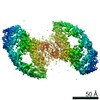
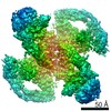
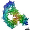
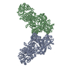
| Title | Cryo-EM structure of RET/GFRa2/NRTN extracellular complex. The 3D refinement was applied with C2 symmetry. | ||||||
 Components Components |
| ||||||
 Keywords Keywords | SIGNALING PROTEIN / RET / receptor tyrosine kinase / cryo-EM | ||||||
| Function / homology |  Function and homology information Function and homology informationglial cell-derived neurotrophic factor receptor activity / glial cell-derived neurotrophic factor receptor binding / Peyer's patch morphogenesis / GDF15-GFRAL signaling pathway / positive regulation of metanephric glomerulus development / ureter maturation / embryonic epithelial tube formation / glial cell-derived neurotrophic factor receptor signaling pathway / lymphocyte migration into lymphoid organs / posterior midgut development ...glial cell-derived neurotrophic factor receptor activity / glial cell-derived neurotrophic factor receptor binding / Peyer's patch morphogenesis / GDF15-GFRAL signaling pathway / positive regulation of metanephric glomerulus development / ureter maturation / embryonic epithelial tube formation / glial cell-derived neurotrophic factor receptor signaling pathway / lymphocyte migration into lymphoid organs / posterior midgut development / Formation of the ureteric bud / membrane protein proteolysis / Formation of the nephric duct / neuron cell-cell adhesion / nerve development / enteric nervous system development / neuron maturation / NCAM1 interactions / plasma membrane protein complex / heparan sulfate binding / positive regulation of extrinsic apoptotic signaling pathway in absence of ligand / cell surface receptor signaling pathway via STAT / positive regulation of cell adhesion mediated by integrin / ureteric bud development / extrinsic component of membrane / neural crest cell migration / response to pain / regulation of axonogenesis / homophilic cell-cell adhesion / RET signaling / positive regulation of cell size / regulation of cell adhesion / cellular response to retinoic acid / NPAS4 regulates expression of target genes / transmembrane receptor protein tyrosine kinase activity / cell surface receptor protein tyrosine kinase signaling pathway / axon guidance / growth factor activity / receptor protein-tyrosine kinase / positive regulation of neuron projection development / receptor tyrosine kinase binding / neuron projection development / MAPK cascade / nervous system development / RAF/MAP kinase cascade / signaling receptor activity / protein tyrosine kinase activity / positive regulation of MAPK cascade / positive regulation of phosphatidylinositol 3-kinase/protein kinase B signal transduction / signaling receptor complex / endosome membrane / positive regulation of cell migration / signaling receptor binding / external side of plasma membrane / axon / calcium ion binding / positive regulation of gene expression / positive regulation of DNA-templated transcription / signal transduction / : / extracellular region / ATP binding / plasma membrane Similarity search - Function | ||||||
| Biological species |  Homo sapiens (human) Homo sapiens (human) | ||||||
| Method | ELECTRON MICROSCOPY / single particle reconstruction / cryo EM / Resolution: 3.65 Å | ||||||
 Authors Authors | Li, J. / Shang, G.J. / Chen, Y.J. / Brautigam, C.A. / Liou, J. / Zhang, X.W. / Bai, X.C. | ||||||
 Citation Citation | Journal: Elife / Year: 2019 Title: Cryo-EM analyses reveal the common mechanism and diversification in the activation of RET by different ligands. Authors: Jie Li / Guijun Shang / Yu-Ju Chen / Chad A Brautigam / Jen Liou / Xuewu Zhang / Xiao-Chen Bai /  Abstract: RET is a receptor tyrosine kinase (RTK) that plays essential roles in development and has been implicated in several human diseases. Different from most of RTKs, RET requires not only its cognate ...RET is a receptor tyrosine kinase (RTK) that plays essential roles in development and has been implicated in several human diseases. Different from most of RTKs, RET requires not only its cognate ligands but also co-receptors for activation, the mechanisms of which remain unclear due to lack of high-resolution structures of the ligand/co-receptor/receptor complexes. Here, we report cryo-EM structures of the extracellular region ternary complexes of GDF15/GFRAL/RET, GDNF/GFRα1/RET, NRTN/GFRα2/RET and ARTN/GFRα3/RET. These structures reveal that all the four ligand/co-receptor pairs, while using different atomic interactions, induce a specific dimerization mode of RET that is poised to bring the two kinase domains into close proximity for cross-phosphorylation. The NRTN/GFRα2/RET dimeric complex further pack into a tetrameric assembly, which is shown by our cell-based assays to regulate the endocytosis of RET. Our analyses therefore reveal both the common mechanism and diversification in the activation of RET by different ligands. | ||||||
| History |
|
- Structure visualization
| Movie |
Movie viewer |
|---|---|
| Structure viewer | Molecule: MolmilJmol/JSmol |
- Downloads & links
-Download
| PDBx/mmCIF format | 6q2o.cif.gz | 353.9 KB | Display | PDBx/mmCIF format |
|---|---|---|---|---|
| PDB format | pdb6q2o.ent.gz | 280.5 KB | Display | PDB format |
| PDBx/mmJSON format | 6q2o.json.gz | Tree view | PDBx/mmJSON format | |
| Others |  Other downloads Other downloads |
-Validation report
| Arichive directory | https://data.pdbj.org/pub/pdb/validation_reports/q2/6q2oftp://data.pdbj.org/pub/pdb/validation_reports/q2/6q2o | HTTPS FTP |
|---|
-Related structure data
| Related structure data |  20576MC  6q2jC  6q2nC  6q2rC  6q2sC M: map data used to model this data C: citing same article ( |
|---|---|
| Similar structure data |


-Links
 PDBj
PDBj


- Assembly
| Deposited unit | 
|
|---|---|
| 1 |
|
-Components
| #1: Protein | Mass: 11706.406 Da / Num. of mol.: 2 Source method: isolated from a genetically manipulated source Source: (gene. exp.) Homo sapiens (human) / Gene: NRTN / Production host:  #2: Protein | Mass: 39572.633 Da / Num. of mol.: 2 Source method: isolated from a genetically manipulated source Source: (gene. exp.) Homo sapiens (human) / Gene: GFRA2, GDNFRB, RETL2, TRNR2 / Cell line (production host): HEK293 / Production host: Homo sapiens (human) / References: UniProt: O00451#3: Protein | Mass: 69100.812 Da / Num. of mol.: 2 Source method: isolated from a genetically manipulated source Source: (gene. exp.) Homo sapiens (human) / Gene: RET, CDHF12, CDHR16, PTC, RET51 / Cell line (production host): HEK293 / Production host: Homo sapiens (human)References: UniProt: P07949, receptor protein-tyrosine kinase #4: Chemical | ChemComp-CA /   Mass: 40.078 Da / Num. of mol.: 8 / Source method: obtained synthetically / Formula: Ca / Feature type: SUBJECT OF INVESTIGATION Mass: 40.078 Da / Num. of mol.: 8 / Source method: obtained synthetically / Formula: Ca / Feature type: SUBJECT OF INVESTIGATION#5: Sugar | ChemComp-NAG /   Type: D-saccharide, beta linking / Mass: 221.208 Da / Num. of mol.: 12 Type: D-saccharide, beta linking / Mass: 221.208 Da / Num. of mol.: 12Source method: isolated from a genetically manipulated source Formula: C8H15NO6 / Feature type: SUBJECT OF INVESTIGATION Has ligand of interest | Y | Has protein modification | Y | |
|---|
-Experimental details
-Experiment
| Experiment | Method: ELECTRON MICROSCOPY |
|---|---|
| EM experiment | Aggregation state: PARTICLE / 3D reconstruction method: single particle reconstruction |
- Sample preparation
| Component | Name: RET, GFRAL and GDF15 extracellular complex / Type: COMPLEX / Entity ID: #1-#3 / Source: RECOMBINANT |
|---|---|
| Molecular weight | Value: 200 kDa/nm / Experimental value: NO |
| Source (natural) | Organism: Homo sapiens (human) |
| Source (recombinant) | Organism: Homo sapiens (human) |
| Buffer solution | pH: 7.4 |
| Specimen | Conc.: 1 mg/ml / Embedding applied: NO / Shadowing applied: NO / Staining applied: NO / Vitrification applied: YES |
| Vitrification | Instrument: FEI VITROBOT MARK IV / Cryogen name: ETHANE / Humidity: 100 % / Chamber temperature: 277 K |
- Electron microscopy imaging
| Experimental equipment |  Model: Titan Krios / Image courtesy: FEI Company |
|---|---|
| Microscopy | Model: FEI TITAN KRIOS |
| Electron gun | Electron source:  FIELD EMISSION GUN / Accelerating voltage: 300 kV / Illumination mode: FLOOD BEAM FIELD EMISSION GUN / Accelerating voltage: 300 kV / Illumination mode: FLOOD BEAM |
| Electron lens | Mode: BRIGHT FIELD / Calibrated magnification: 46729 X / Cs: 2.7 mm / C2 aperture diameter: 70 µm / Alignment procedure: COMA FREE |
| Specimen holder | Cryogen: NITROGEN / Specimen holder model: FEI TITAN KRIOS AUTOGRID HOLDER |
| Image recording | Average exposure time: 15 sec. / Electron dose: 50 e/Å2 / Detector mode: SUPER-RESOLUTION / Film or detector model: GATAN K2 QUANTUM (4k x 4k) / Num. of grids imaged: 1 |
| EM imaging optics | Energyfilter name: GIF Quantum LS / Energyfilter slit width: 20 eV |
| Image scans | Movie frames/image: 30 / Used frames/image: 1-30 |
- Processing
| EM software |
| ||||||||||||||||||||||||||||||
|---|---|---|---|---|---|---|---|---|---|---|---|---|---|---|---|---|---|---|---|---|---|---|---|---|---|---|---|---|---|---|---|
| CTF correction | Type: PHASE FLIPPING AND AMPLITUDE CORRECTION | ||||||||||||||||||||||||||||||
| Symmetry | Point symmetry: C2 (2 fold cyclic) | ||||||||||||||||||||||||||||||
| 3D reconstruction | Resolution: 3.65 Å / Resolution method: FSC 0.143 CUT-OFF / Num. of particles: 247157 / Symmetry type: POINT | ||||||||||||||||||||||||||||||
| Atomic model building | B value: 190 / Protocol: FLEXIBLE FIT / Space: REAL |
