 Movie
Movie Controller

[English] 日本語
 Yorodumi
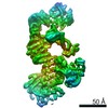
- EMDB-4000: Subtomographic reconstruction of a single VP4 spike from Halorubr... -
+ Open data

- Basic information
| Entry | Database: EMDB / ID: EMD-4000 | |||||||||
|---|---|---|---|---|---|---|---|---|---|---|
| Title | Subtomographic reconstruction of a single VP4 spike from Halorubrum pleomorphic virus 1. | |||||||||
 Map data Map data | None | |||||||||
 Sample Sample |
| |||||||||
| Biological species |  Halorubrum pleomorphic virus 1 Halorubrum pleomorphic virus 1 | |||||||||
| Method | subtomogram averaging / cryo EM / Resolution: 40.0 Å | |||||||||
 Authors Authors | Butcher SJ / Liljeroos L / Manole V | |||||||||
 Citation Citation | Journal: J Virol / Year: 2012 Title: Virion architecture unifies globally distributed pleolipoviruses infecting halophilic archaea. Authors: Maija K Pietilä / Nina S Atanasova / Violeta Manole / Lassi Liljeroos / Sarah J Butcher / Hanna M Oksanen / Dennis H Bamford /  Abstract: Our understanding of the third domain of life, Archaea, has greatly increased since its establishment some 20 years ago. The increasing information on archaea has also brought their viruses into the ...Our understanding of the third domain of life, Archaea, has greatly increased since its establishment some 20 years ago. The increasing information on archaea has also brought their viruses into the limelight. Today, about 100 archaeal viruses are known, which is a low number compared to the numbers of characterized bacterial or eukaryotic viruses. Here, we have performed a comparative biological and structural study of seven pleomorphic viruses infecting extremely halophilic archaea. The pleomorphic nature of this novel virion type was established by sedimentation analysis and cryo-electron microscopy. These nonlytic viruses form virions characterized by a lipid vesicle enclosing the genome, without any nucleoproteins. The viral lipids are unselectively acquired from host cell membranes. The virions contain two to three major structural proteins, which either are embedded in the membrane or form spikes distributed randomly on the external membrane surface. Thus, the most important step during virion assembly is most likely the interaction of the membrane proteins with the genome. The interaction can be driven by single-stranded or double-stranded DNA, resulting in the virions having similar architectures but different genome types. Based on our comparative study, these viruses probably form a novel group, which we define as pleolipoviruses. | |||||||||
| History |
|
- Structure visualization
| Movie |
 Movie viewer Movie viewer |
|---|---|
| Structure viewer | EM map: SurfViewMolmilJmol/JSmol |
| Supplemental images |

- Downloads & links
-EMDB archive
| Map data | emd_4000.map.gz | 89.8 KB | EMDB map data format | |
|---|---|---|---|---|
| Header (meta data) | emd-4000-v30.xmlemd-4000.xml | 13.8 KB 13.8 KB | Display Display | EMDB header |
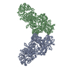
| Images |  emd_4000.png emd_4000.png | 55 KB | ||
| Archive directory |  http://ftp.pdbj.org/pub/emdb/structures/EMD-4000ftp://ftp.pdbj.org/pub/emdb/structures/EMD-4000 http://ftp.pdbj.org/pub/emdb/structures/EMD-4000ftp://ftp.pdbj.org/pub/emdb/structures/EMD-4000 | HTTPS FTP |
-Related structure data
| Similar structure data |
|---|


-Links
| EMDB pages | EMDB (EBI/PDBe) / EMDataResource |
|---|
-Map
| File | Download / File: emd_4000.map.gz / Format: CCP4 / Size: 106.4 KB / Type: IMAGE STORED AS FLOATING POINT NUMBER (4 BYTES) | ||||||||||||||||||||||||||||||||||||||||||||||||||||||||||||
|---|---|---|---|---|---|---|---|---|---|---|---|---|---|---|---|---|---|---|---|---|---|---|---|---|---|---|---|---|---|---|---|---|---|---|---|---|---|---|---|---|---|---|---|---|---|---|---|---|---|---|---|---|---|---|---|---|---|---|---|---|---|
| Annotation | None | ||||||||||||||||||||||||||||||||||||||||||||||||||||||||||||
| Projections & slices | Image control
Images are generated by Spider. | ||||||||||||||||||||||||||||||||||||||||||||||||||||||||||||
| Voxel size | X=Y=Z: 11.5 Å | ||||||||||||||||||||||||||||||||||||||||||||||||||||||||||||
| Density |
| ||||||||||||||||||||||||||||||||||||||||||||||||||||||||||||
| Symmetry | Space group: 1 | ||||||||||||||||||||||||||||||||||||||||||||||||||||||||||||
| Details | EMDB XML:
CCP4 map header:
| ||||||||||||||||||||||||||||||||||||||||||||||||||||||||||||
 Z (Sec.)
 Y (Row.)
 X (Col.)


-Supplemental data
- Sample components
-Entire : Halorubrum pleomorphic virus 1
| Entire | Name: Halorubrum pleomorphic virus 1 |
|---|---|
| Components |
|
-Supramolecule #1: Halorubrum pleomorphic virus 1
| Supramolecule | Name: Halorubrum pleomorphic virus 1 / type: virus / ID: 1 / Parent: 0 / Macromolecule list: all / NCBI-ID: 634168 / Sci species name: Halorubrum pleomorphic virus 1 / Virus type: VIRION / Virus isolate: SPECIES / Virus enveloped: Yes / Virus empty: No |
|---|---|
| Host (natural) | Organism:  Halorubrum sp. PV6 (archaea) Halorubrum sp. PV6 (archaea) |
-Macromolecule #1: VP4
| Macromolecule | Name: VP4 / type: protein_or_peptide / ID: 1 / Enantiomer: LEVO |
|---|---|
| Source (natural) | Organism: Halorubrum pleomorphic virus 1 |
| Sequence | String: msvnrssiks llmvfmivs s sllapvgg aa adefrtp aas dtspea gecs nlddf imfls vgri nadscs rqa yvdaavq dm kdsdanqt k vdiysaaag vkggsetwaa pydnylndt e siawmkae sa iaqsyse ges kteakv aaka aiady ...String: msvnrssiks llmvfmivs s sllapvgg aa adefrtp aas dtspea gecs nlddf imfls vgri nadscs rqa yvdaavq dm kdsdanqt k vdiysaaag vkggsetwaa pydnylndt e siawmkae sa iaqsyse ges kteakv aaka aiady yatkq knli eqwnfa naq mftlreq ar medgisrn y vepayrnve ktnspdysla ysnttveks l vdgttvnt tg vsmdvtv qht tvsdva tvss gpvra gkynn qyne wkatyy sws vepasps qd tlyavhfq p yadrwqriv dmngalqsea dnfvnatwd d ydtgqina sd vlsanta mse ygvrsg sese glwrs taals mmgy dtpnln nsg mmtveyk nv qhtgllma k napngswqv nttyntsnid gpvfmatte g tkldfadg ee ftivgmt akd gtavns tqtt kyryk tantt elle vqnqli elr qeiedre pe aggffgsg s tdtmlvgll alagvlllaq snnrggrr |
-Experimental details
-Structure determination
| Method | cryo EM |
|---|---|
 Processing Processing | subtomogram averaging |
| Aggregation state | particle |
-Sample preparation
| Buffer | pH: 7.5 Component:
| |||||||||||||||
|---|---|---|---|---|---|---|---|---|---|---|---|---|---|---|---|---|
| Grid | Model: Quantifoil R 2/2 / Material: COPPER / Support film - Material: CARBON / Support film - topology: HOLEY ARRAY / Pretreatment - Type: GLOW DISCHARGE / Pretreatment - Atmosphere: AIR / Pretreatment - Pressure: 101.325 kPa / Details: Almost at sea level | |||||||||||||||
| Vitrification | Cryogen name: ETHANE / Instrument: HOMEMADE PLUNGER | |||||||||||||||
| Details | The sample was monodisperse |
- Electron microscopy
| Microscope | FEI TECNAI F20 |
|---|---|
| Temperature | Min: 77.0 K / Max: 77.0 K |
| Image recording | Film or detector model: GATAN ULTRASCAN 4000 (4k x 4k) / Digitization - Dimensions - Width: 4080 pixel / Digitization - Dimensions - Height: 4080 pixel / Digitization - Sampling interval: 15.0 µm / Average electron dose: 2.0 e/Å2 |
| Electron beam | Acceleration voltage: 200 kV / Electron source:  FIELD EMISSION GUN FIELD EMISSION GUN |
| Electron optics | Illumination mode: FLOOD BEAM / Imaging mode: BRIGHT FIELD / Cs: 2.0 mm / Nominal defocus max: 6.0 µm / Nominal defocus min: 6.0 µm / Nominal magnification: 26000 |
| Sample stage | Specimen holder model: GATAN 626 SINGLE TILT LIQUID NITROGEN CRYO TRANSFER HOLDER Cooling holder cryogen: NITROGEN |
| Experimental equipment |  Model: Tecnai F20 / Image courtesy: FEI Company |
-Image processing
| Final reconstruction | Applied symmetry - Point group: C1 (asymmetric) / Algorithm: BACK PROJECTION / Resolution.type: BY AUTHOR / Resolution: 40.0 Å / Resolution method: OTHER / Software - Name: IMOD Details: R-weighted back-projection. The resolution was not estimated quantitatively for this structure, however, I estimate it qualitatively to be around 4 nm. Number subtomograms used: 1019 |
|---|---|
| Extraction | Number tomograms: 17 / Number images used: 3588 / Method: volumes picked interactively / Software - Name: JSUBTOMO Software - details: Particles were iteratively centered, first against a spherical model of a 17.3-nm radius and then against their own 3D radial density profile, without any symmetry imposed. These ...Software - details: Particles were iteratively centered, first against a spherical model of a 17.3-nm radius and then against their own 3D radial density profile, without any symmetry imposed. These particles were all roughly spherical and were assumed to have the membrane at a similar radius in subsequent steps. This enabled the automatic selection of 3,588 membrane subvolumes from all over the surface of the centered particles, on a regular grid, irrespective of whether or not the volume contained spikes, in a box of 30 by 30 by 30 pixels. A surface normal in each location defined the orientation of each subvolume relative to the viral surface. Details: 78 initial subtomograms each contained one virus particle were first extractd. These were further dissected to recover 3588 subtomograms containing spikes. |
| CTF correction | Details: Data were truncated to the first zero of the CTF |
| Final angle assignment | Type: OTHER / Software - Name: IMOD Details: The tilt angles are recorded in the microscope software and utilised in the |
