 Movie
Movie Controller

[English] 日本語
 Yorodumi
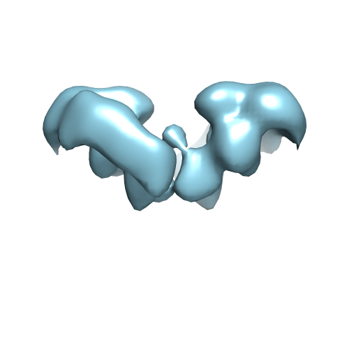
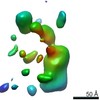
- EMDB-2713: Structure of the zebrafish RET tyrosine kinase extracellular domain -
+ Open data

- Basic information
| Entry | Database: EMDB / ID: EMD-2713 | |||||||||
|---|---|---|---|---|---|---|---|---|---|---|
| Title | Structure of the zebrafish RET tyrosine kinase extracellular domain | |||||||||
 Map data Map data | zTC | |||||||||
 Sample Sample |
| |||||||||
 Keywords Keywords | signal transduction | |||||||||
| Biological species |  | |||||||||
| Method | single particle reconstruction / negative staining / Resolution: 26.0 Å | |||||||||
 Authors Authors | Goodman K / Kjaer S / Beuron F / Knowles P / Nawrotek A / Burns E / Purkiss A / George R / Santoro M / Morris EP / McDonald NQ | |||||||||
 Citation Citation | Journal: Cell Rep / Year: 2014 Title: RET recognition of GDNF-GFRα1 ligand by a composite binding site promotes membrane-proximal self-association. Authors: Kerry M Goodman / Svend Kjær / Fabienne Beuron / Phillip P Knowles / Agata Nawrotek / Emily M Burns / Andrew G Purkiss / Roger George / Massimo Santoro / Edward P Morris / Neil Q McDonald /   Abstract: The RET receptor tyrosine kinase is essential to vertebrate development and implicated in multiple human diseases. RET binds a cell surface bipartite ligand comprising a GDNF family ligand and a ...The RET receptor tyrosine kinase is essential to vertebrate development and implicated in multiple human diseases. RET binds a cell surface bipartite ligand comprising a GDNF family ligand and a GFRα coreceptor, resulting in RET transmembrane signaling. We present a hybrid structural model, derived from electron microscopy (EM) and low-angle X-ray scattering (SAXS) data, of the RET extracellular domain (RET(ECD)), GDNF, and GFRα1 ternary complex, defining the basis for ligand recognition. RET(ECD) envelopes the dimeric ligand complex through a composite binding site comprising four discrete contact sites. The GFRα1-mediated contacts are crucial, particularly close to the invariant RET calcium-binding site, whereas few direct contacts are made by GDNF, explaining how distinct ligand/coreceptor pairs are accommodated. The RET(ECD) cysteine-rich domain (CRD) contacts both ligand components and makes homotypic membrane-proximal interactions occluding three different antibody epitopes. Coupling of these CRD-mediated interactions suggests models for ligand-induced RET activation and ligand-independent oncogenic deregulation. | |||||||||
| History |
|
- Structure visualization
| Movie |
 Movie viewer Movie viewer |
|---|---|
| Structure viewer | EM map: SurfViewMolmilJmol/JSmol |
| Supplemental images |
- Downloads & links
-EMDB archive
| Map data | emd_2713.map.gz | 1.8 MB | EMDB map data format | |
|---|---|---|---|---|
| Header (meta data) | emd-2713-v30.xmlemd-2713.xml | 10.2 KB 10.2 KB | Display Display | EMDB header |
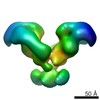
| Images |  EMD-2713.png EMD-2713.png | 54.1 KB | ||
| Archive directory |  http://ftp.pdbj.org/pub/emdb/structures/EMD-2713ftp://ftp.pdbj.org/pub/emdb/structures/EMD-2713 http://ftp.pdbj.org/pub/emdb/structures/EMD-2713ftp://ftp.pdbj.org/pub/emdb/structures/EMD-2713 | HTTPS FTP |
-Related structure data


-Links
| EMDB pages | EMDB (EBI/PDBe) / EMDataResource |
|---|
-Map
| File | Download / File: emd_2713.map.gz / Format: CCP4 / Size: 3.3 MB / Type: IMAGE STORED AS FLOATING POINT NUMBER (4 BYTES) | ||||||||||||||||||||||||||||||||||||||||||||||||||||||||||||||||||||
|---|---|---|---|---|---|---|---|---|---|---|---|---|---|---|---|---|---|---|---|---|---|---|---|---|---|---|---|---|---|---|---|---|---|---|---|---|---|---|---|---|---|---|---|---|---|---|---|---|---|---|---|---|---|---|---|---|---|---|---|---|---|---|---|---|---|---|---|---|---|
| Annotation | zTC | ||||||||||||||||||||||||||||||||||||||||||||||||||||||||||||||||||||
| Projections & slices | Image control
Images are generated by Spider. | ||||||||||||||||||||||||||||||||||||||||||||||||||||||||||||||||||||
| Voxel size | X=Y=Z: 4.34 Å | ||||||||||||||||||||||||||||||||||||||||||||||||||||||||||||||||||||
| Density |
| ||||||||||||||||||||||||||||||||||||||||||||||||||||||||||||||||||||
| Symmetry | Space group: 1 | ||||||||||||||||||||||||||||||||||||||||||||||||||||||||||||||||||||
| Details | EMDB XML:
CCP4 map header:
| ||||||||||||||||||||||||||||||||||||||||||||||||||||||||||||||||||||
 Z (Sec.)
 Y (Row.)
 X (Col.)


-Supplemental data
- Sample components
-Entire : Reconstituted zebrafish RETecd-GDNF-GFRa1 ternary complex
| Entire | Name: Reconstituted zebrafish RETecd-GDNF-GFRa1 ternary complex |
|---|---|
| Components |
|
-Supramolecule #1000: Reconstituted zebrafish RETecd-GDNF-GFRa1 ternary complex
| Supramolecule | Name: Reconstituted zebrafish RETecd-GDNF-GFRa1 ternary complex type: sample / ID: 1000 / Oligomeric state: Hexamer / Number unique components: 3 |
|---|---|
| Molecular weight | Theoretical: 240 KDa |
-Macromolecule #1: RET receptor tyrosine kinase
| Macromolecule | Name: RET receptor tyrosine kinase / type: protein_or_peptide / ID: 1 / Name.synonym: RET / Number of copies: 2 / Recombinant expression: Yes |
|---|---|
| Source (natural) | Organism: |
| Molecular weight | Experimental: 83 KDa |
| Recombinant expression | Organism:  unidentified baculovirus / Recombinant cell: sf9 unidentified baculovirus / Recombinant cell: sf9 |
-Macromolecule #2: glial-cell-line-derived neurotrophic factor
| Macromolecule | Name: glial-cell-line-derived neurotrophic factor / type: protein_or_peptide / ID: 2 / Name.synonym: GDNF / Number of copies: 2 / Recombinant expression: Yes |
|---|---|
| Source (natural) | Organism: |
| Recombinant expression | Organism: unidentified baculovirus / Recombinant cell: sf9 |
-Macromolecule #3: GDNF receptor alpha
| Macromolecule | Name: GDNF receptor alpha / type: protein_or_peptide / ID: 3 / Number of copies: 2 / Recombinant expression: Yes |
|---|---|
| Source (natural) | Organism: |
| Recombinant expression | Organism: unidentified baculovirus / Recombinant cell: sf9 |
-Experimental details
-Structure determination
| Method | negative staining |
|---|---|
 Processing Processing | single particle reconstruction |
| Aggregation state | particle |
-Sample preparation
| Concentration | 0.03 mg/mL |
|---|---|
| Buffer | pH: 8 / Details: 20 mM Tris, 300 mM NaCl, 1 mM Ca++ |
| Staining | Type: NEGATIVE / Details: 2 % uranyle acetate |
| Grid | Details: quantifoil R1.2/R1.3 coated with a thin carbon layer, glow discharge in air |
| Vitrification | Cryogen name: NONE / Instrument: OTHER |
- Electron microscopy
| Microscope | FEI TECNAI F20 |
|---|---|
| Date | May 18, 2012 |
| Image recording | Category: CCD / Film or detector model: TVIPS TEMCAM-F415 (4k x 4k) / Number real images: 800 / Average electron dose: 100 e/Å2 / Bits/pixel: 16 |
| Electron beam | Acceleration voltage: 200 kV / Electron source:  FIELD EMISSION GUN FIELD EMISSION GUN |
| Electron optics | Illumination mode: FLOOD BEAM / Imaging mode: BRIGHT FIELD / Nominal defocus min: 0.9 µm / Nominal magnification: 80000 |
| Sample stage | Specimen holder model: SIDE ENTRY, EUCENTRIC |
| Experimental equipment |  Model: Tecnai F20 / Image courtesy: FEI Company |
-Image processing
| Details | Particles selected manually. 3D starting model consisted of the mTC reconstruction |
|---|---|
| Final reconstruction | Applied symmetry - Point group: C2 (2 fold cyclic) / Algorithm: OTHER / Resolution.type: BY AUTHOR / Resolution: 26.0 Å / Resolution method: OTHER / Software - Name: IMAGIC, SPIDER, in-house-software / Number images used: 7510 |
| Final two d classification | Number classes: 500 |
