 ムービー
ムービー コントローラー


+ データを開く

- 基本情報
基本情報
| 登録情報 | データベース: PDB / ID: 5kg8 | ||||||
|---|---|---|---|---|---|---|---|
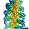
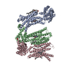
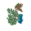
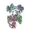
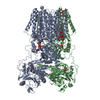
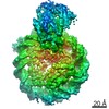
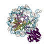
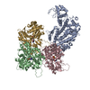
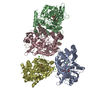
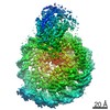
| タイトル | Rigor myosin X co-complexed with an actin filament | ||||||
 要素 要素 |
| ||||||
 キーワード キーワード | MOTOR PROTEIN / myosin molecular motors cytoskeletal motility | ||||||
| 機能・相同性 |  機能・相同性情報 機能・相同性情報plus-end directed microfilament motor activity / Netrin-1 signaling / positive regulation of cell-cell adhesion / cytoskeleton-dependent intracellular transport / filopodium tip / regulation of filopodium assembly / filopodium membrane / myosin complex / cytoskeletal motor activator activity / spectrin binding ...plus-end directed microfilament motor activity / Netrin-1 signaling / positive regulation of cell-cell adhesion / cytoskeleton-dependent intracellular transport / filopodium tip / regulation of filopodium assembly / filopodium membrane / myosin complex / cytoskeletal motor activator activity / spectrin binding / microfilament motor activity / myosin heavy chain binding / tropomyosin binding / actin filament bundle / troponin I binding / filamentous actin / mesenchyme migration / phosphatidylinositol-3,4,5-trisphosphate binding / skeletal muscle myofibril / actin filament bundle assembly / striated muscle thin filament / skeletal muscle thin filament assembly / actin monomer binding / skeletal muscle fiber development / ruffle / stress fiber / titin binding / actin filament polymerization / FCGR3A-mediated phagocytosis / filopodium / actin filament / Regulation of actin dynamics for phagocytic cup formation / 加水分解酵素; 酸無水物に作用; 酸無水物に作用・細胞または細胞小器官の運動に関与 / calcium-dependent protein binding / actin filament binding / regulation of cell shape / lamellipodium / cell body / cell cortex / calmodulin binding / neuron projection / protein domain specific binding / hydrolase activity / neuronal cell body / calcium ion binding / positive regulation of gene expression / nucleolus / magnesium ion binding / signal transduction / ATP binding / identical protein binding / plasma membrane / cytoplasm / cytosol 類似検索 - 分子機能 | ||||||
| 生物種 |  Homo sapiens (ヒト) Homo sapiens (ヒト) | ||||||
| 手法 | 電子顕微鏡法 / らせん対称体再構成法 / クライオ電子顕微鏡法 / 解像度: 9.1 Å | ||||||
 データ登録者 データ登録者 | Sindelar, C.V. / Houdusse, A. / Sweeney, L. | ||||||
| 資金援助 |  米国, 1件 米国, 1件
| ||||||
 引用 引用 | ジャーナル: Nat Commun / 年: 2016 タイトル: The myosin X motor is optimized for movement on actin bundles. 著者: Virginie Ropars / Zhaohui Yang / Tatiana Isabet / Florian Blanc / Kaifeng Zhou / Tianming Lin / Xiaoyan Liu / Pascale Hissier / Frédéric Samazan / Béatrice Amigues / Eric D Yang / Hyokeun ...著者: Virginie Ropars / Zhaohui Yang / Tatiana Isabet / Florian Blanc / Kaifeng Zhou / Tianming Lin / Xiaoyan Liu / Pascale Hissier / Frédéric Samazan / Béatrice Amigues / Eric D Yang / Hyokeun Park / Olena Pylypenko / Marco Cecchini / Charles V Sindelar / H Lee Sweeney / Anne Houdusse /   要旨: Myosin X has features not found in other myosins. Its structure must underlie its unique ability to generate filopodia, which are essential for neuritogenesis, wound healing, cancer metastasis and ...Myosin X has features not found in other myosins. Its structure must underlie its unique ability to generate filopodia, which are essential for neuritogenesis, wound healing, cancer metastasis and some pathogenic infections. By determining high-resolution structures of key components of this motor, and characterizing the in vitro behaviour of the native dimer, we identify the features that explain the myosin X dimer behaviour. Single-molecule studies demonstrate that a native myosin X dimer moves on actin bundles with higher velocities and takes larger steps than on single actin filaments. The largest steps on actin bundles are larger than previously reported for artificially dimerized myosin X constructs or any other myosin. Our model and kinetic data explain why these large steps and high velocities can only occur on bundled filaments. Thus, myosin X functions as an antiparallel dimer in cells with a unique geometry optimized for movement on actin bundles. | ||||||
| 履歴 |
|
- 構造の表示
構造の表示
| ムービー |
ムービービューア |
|---|---|
| 構造ビューア | 分子: MolmilJmol/JSmol |
- ダウンロードとリンク
-ダウンロード
| PDBx/mmCIF形式 | 5kg8.cif.gz | 229 KB | 表示 | PDBx/mmCIF形式 |
|---|---|---|---|---|
| PDB形式 | pdb5kg8.ent.gz | 140.4 KB | 表示 | PDB形式 |
| PDBx/mmJSON形式 | 5kg8.json.gz | ツリー表示 | PDBx/mmJSON形式 | |
| その他 |  その他のダウンロード その他のダウンロード |
-検証レポート
| アーカイブディレクトリ | https://data.pdbj.org/pub/pdb/validation_reports/kg/5kg8ftp://data.pdbj.org/pub/pdb/validation_reports/kg/5kg8 | HTTPS FTP |
|---|
-関連構造データ


-リンク
 PDBj
PDBj


- 集合体
集合体
| 登録構造単位 | 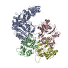
|
|---|---|
| 1 |
|
-要素
| #1: タンパク質 | 分子量: 85083.086 Da / 分子数: 1 / 断片: motor domain (UNP residues 3-741) / 由来タイプ: 組換発現 / 由来: (組換発現) Homo sapiens (ヒト) / 遺伝子: MYO10, KIAA0799 / 発現宿主:  |
|---|---|
| #2: タンパク質 | 分子量: 41827.609 Da / 分子数: 3 / 由来タイプ: 天然 / 由来: (天然) |
-実験情報
-実験
| 実験 | 手法: 電子顕微鏡法 |
|---|---|
| EM実験 | 試料の集合状態: FILAMENT / 3次元再構成法: らせん対称体再構成法 |
- 試料調製
試料調製
| 構成要素 | 名称: Actin filament decorated by the myosin X motor domain タイプ: ORGANELLE OR CELLULAR COMPONENT / Entity ID: all / 由来: MULTIPLE SOURCES |
|---|---|
| 緩衝液 | pH: 6.8 |
| 緩衝液成分 | 濃度: 5 mM / 名称: MOPS |
| 試料 | 濃度: 1.7 mg/ml / 包埋: NO / シャドウイング: NO / 染色: NO / 凍結: YES |
| 試料支持 | グリッドの材料: COPPER / グリッドのサイズ: 300 divisions/in. / グリッドのタイプ: Quantifoil |
| 急速凍結 | 装置: HOMEMADE PLUNGER / 凍結剤: ETHANE |
- 電子顕微鏡撮影
| 実験機器 |  モデル: Tecnai F20 / 画像提供: FEI Company |
|---|---|
| 顕微鏡 | モデル: FEI TECNAI F20 |
| 電子銃 | 電子線源:  FIELD EMISSION GUN / 加速電圧: 200 kV / 照射モード: FLOOD BEAM FIELD EMISSION GUN / 加速電圧: 200 kV / 照射モード: FLOOD BEAM |
| 電子レンズ | モード: BRIGHT FIELD / 倍率(補正後): 26780 X / 最大 デフォーカス(公称値): 5000 nm / 最小 デフォーカス(公称値): 1000 nm / Calibrated defocus min: 1438 nm / 最大 デフォーカス(補正後): 5251 nm / Cs: 2 mm / C2レンズ絞り径: 70 µm / アライメント法: COMA FREE |
| 試料ホルダ | 凍結剤: NITROGEN 試料ホルダーモデル: GATAN 626 SINGLE TILT LIQUID NITROGEN CRYO TRANSFER HOLDER |
| 撮影 | 平均露光時間: 13 sec. / 電子線照射量: 50 e/Å2 / 検出モード: COUNTING フィルム・検出器のモデル: GATAN K2 SUMMIT (4k x 4k) 撮影したグリッド数: 2 / 実像数: 154 |
- 解析
解析
| EMソフトウェア |
| |||||||||||||||||||||||||||||||||||
|---|---|---|---|---|---|---|---|---|---|---|---|---|---|---|---|---|---|---|---|---|---|---|---|---|---|---|---|---|---|---|---|---|---|---|---|---|
| CTF補正 | タイプ: PHASE FLIPPING AND AMPLITUDE CORRECTION | |||||||||||||||||||||||||||||||||||
| らせん対称 | 回転角度/サブユニット: 167.1 ° / 軸方向距離/サブユニット: 27.44 Å / らせん対称軸の対称性: C1 / 詳細: Not directly applicable to the modeled coordinates | |||||||||||||||||||||||||||||||||||
| 3次元再構成 | 解像度: 9.1 Å / 解像度の算出法: FSC 0.143 CUT-OFF / 粒子像の数: 57927 / アルゴリズム: FOURIER SPACE 詳細: Symmetry was not applied in this reconstruction. The provided symmetry parameters are nominal, and were not actually used. クラス平均像の数: 1 / 対称性のタイプ: HELICAL |
