 Movie
Movie Controller

[English] 日本語
 Yorodumi
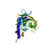
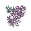
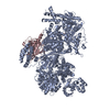
- PDB-4bgd: Crystal structure of Brr2 in complex with the Jab1/MPN domain of Prp8 -
+ Open data

- Basic information
| Entry | Database: PDB / ID: 4bgd | ||||||
|---|---|---|---|---|---|---|---|
| Title | Crystal structure of Brr2 in complex with the Jab1/MPN domain of Prp8 | ||||||
 Components Components | (PRE-MRNA-SPLICING ...) x 2 | ||||||
 Keywords Keywords | TRANSCRIPTION / SPLICEOSOME / RNA HELICASE / U5 SNRNP / RETINITIS PIGMENTOSA | ||||||
| Function / homology |  Function and homology information Function and homology informationspliceosome conformational change to release U4 (or U4atac) and U1 (or U11) / generation of catalytic spliceosome for second transesterification step / mRNA 5'-splice site recognition / mRNA 3'-splice site recognition / spliceosomal tri-snRNP complex assembly / Prp19 complex / U5 snRNP / U5 snRNA binding / pre-mRNA intronic binding / spliceosomal snRNP assembly ...spliceosome conformational change to release U4 (or U4atac) and U1 (or U11) / generation of catalytic spliceosome for second transesterification step / mRNA 5'-splice site recognition / mRNA 3'-splice site recognition / spliceosomal tri-snRNP complex assembly / Prp19 complex / U5 snRNP / U5 snRNA binding / pre-mRNA intronic binding / spliceosomal snRNP assembly / U2 snRNA binding / U6 snRNA binding / U1 snRNA binding / U4/U6 x U5 tri-snRNP complex / catalytic step 2 spliceosome / spliceosomal complex / mRNA splicing, via spliceosome / metallopeptidase activity / nucleic acid binding / RNA helicase activity / RNA helicase / mRNA binding / ATP hydrolysis activity / ATP binding / identical protein binding / nucleus / cytoplasm Similarity search - Function | ||||||
| Biological species |  | ||||||
| Method |  X-RAY DIFFRACTION / SYNCHROTRON / SIRAS / Resolution: 3.1 Å X-RAY DIFFRACTION / SYNCHROTRON / SIRAS / Resolution: 3.1 Å | ||||||
 Authors Authors | Nguyen, T.H.D. / Li, J. / Nagai, K. | ||||||
 Citation Citation | Journal: Structure / Year: 2013 Title: Structural Basis of Brr2-Prp8 Interactions and Implications for U5 Snrnp Biogenesis and the Spliceosome Active Site Authors: Nguyen, T.H.D. / Li, J. / Galej, W.P. / Oshikane, H. / Newman, A.J. / Nagai, K. | ||||||
| History |
|
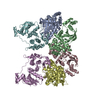
- Structure visualization
| Structure viewer | Molecule: MolmilJmol/JSmol |
|---|
- Downloads & links
-Download
| PDBx/mmCIF format | 4bgd.cif.gz | 410.9 KB | Display | PDBx/mmCIF format |
|---|---|---|---|---|
| PDB format | pdb4bgd.ent.gz | 323.2 KB | Display | PDB format |
| PDBx/mmJSON format | 4bgd.json.gz | Tree view | PDBx/mmJSON format | |
| Others |  Other downloads Other downloads |
-Validation report
| Arichive directory | https://data.pdbj.org/pub/pdb/validation_reports/bg/4bgdftp://data.pdbj.org/pub/pdb/validation_reports/bg/4bgd | HTTPS FTP |
|---|
-Related structure data


-Links
 PDBj
PDBj


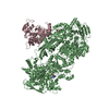
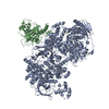
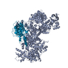
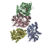
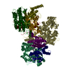
- Assembly
| Deposited unit | 
| ||||||||
|---|---|---|---|---|---|---|---|---|---|
| 1 |
| ||||||||
| Unit cell |
|
-Components
-PRE-MRNA-SPLICING ... , 2 types, 2 molecules AC
| #1: Protein | Mass: 195729.453 Da / Num. of mol.: 1 / Fragment: RESIDUES 442-2163 Source method: isolated from a genetically manipulated source Source: (gene. exp.) Strain: S288C / Production host: |
|---|---|
| #2: Protein | Mass: 28180.924 Da / Num. of mol.: 1 / Fragment: RESIDUES 2148-2395 Source method: isolated from a genetically manipulated source Source: (gene. exp.) Strain: S288C / Production host: |
-Non-polymers , 4 types, 18 molecules 

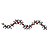

| #3: Chemical | ChemComp-ADP /  Mass: 427.201 Da / Num. of mol.: 1 / Source method: obtained synthetically / Formula: C10H15N5O10P2 / Comment: ADP, energy-carrying molecule*YM Mass: 427.201 Da / Num. of mol.: 1 / Source method: obtained synthetically / Formula: C10H15N5O10P2 / Comment: ADP, energy-carrying molecule*YM |
|---|---|
| #4: Chemical | ChemComp-MG /  Mass: 24.305 Da / Num. of mol.: 1 / Source method: obtained synthetically / Formula: Mg Mass: 24.305 Da / Num. of mol.: 1 / Source method: obtained synthetically / Formula: Mg |
| #5: Chemical | ChemComp-PE5 /  Mass: 398.489 Da / Num. of mol.: 1 / Source method: obtained synthetically / Formula: C18H38O9 / Comment: precipitant*YM Mass: 398.489 Da / Num. of mol.: 1 / Source method: obtained synthetically / Formula: C18H38O9 / Comment: precipitant*YM |
| #6: Water | ChemComp-HOH / Mass: 18.015 Da / Num. of mol.: 15 / Source method: isolated from a natural source / Formula: H2O |
-Experimental details
-Experiment
| Experiment | Method: X-RAY DIFFRACTION / Number of used crystals: 1 |
|---|
- Sample preparation
| Crystal | Density Matthews: 3.77 Å3/Da / Density % sol: 67 % Description: THE TWO KNOWN FRAGMENTS COULD ALSO BE FOUND BY MOLECULAR REPLACEMENT, BUT IT WAS NOT SUFFICIENT FOR STRUCTURE DETERMINATION. THE STRUCTURE WAS SOLVED BY SIRAS. |
|---|---|
| Crystal grow | pH: 7.5 / Details: 100 MM HEPESNA PH 7.5, 200 MM MGCL2, 32-40% PEG400 |
-Data collection
| Diffraction | Mean temperature: 77 K |
|---|---|
| Diffraction source | Source: SYNCHROTRON / Site: Diamond  / Beamline: I02 / Wavelength: 0.9795 / Beamline: I02 / Wavelength: 0.9795 |
| Detector | Type: DECTRIS PILATUS 6M / Detector: PIXEL / Date: May 17, 2012 |
| Radiation | Protocol: SINGLE WAVELENGTH / Monochromatic (M) / Laue (L): M / Scattering type: x-ray |
| Radiation wavelength | Wavelength: 0.9795 Å / Relative weight: 1 |
| Reflection | Resolution: 3.1→92.2 Å / Num. obs: 63788 / % possible obs: 99.9 % / Observed criterion σ(I): 6 / Redundancy: 4.2 % / Rmerge(I) obs: 0.08 / Net I/σ(I): 9.8 |
| Reflection shell | Resolution: 3.1→3.18 Å / Redundancy: 4 % / Rmerge(I) obs: 0.88 / Mean I/σ(I) obs: 1.4 / % possible all: 100 |
- Processing
| Software |
| ||||||||||||||||||||||||||||||||||||||||||||||||||||||||||||||||||||||||||||||||||||||||||||||||||||||||||||||||||||||||||||||||||||||||||||||||||||||||||||||||||||||||||||||||||||||
|---|---|---|---|---|---|---|---|---|---|---|---|---|---|---|---|---|---|---|---|---|---|---|---|---|---|---|---|---|---|---|---|---|---|---|---|---|---|---|---|---|---|---|---|---|---|---|---|---|---|---|---|---|---|---|---|---|---|---|---|---|---|---|---|---|---|---|---|---|---|---|---|---|---|---|---|---|---|---|---|---|---|---|---|---|---|---|---|---|---|---|---|---|---|---|---|---|---|---|---|---|---|---|---|---|---|---|---|---|---|---|---|---|---|---|---|---|---|---|---|---|---|---|---|---|---|---|---|---|---|---|---|---|---|---|---|---|---|---|---|---|---|---|---|---|---|---|---|---|---|---|---|---|---|---|---|---|---|---|---|---|---|---|---|---|---|---|---|---|---|---|---|---|---|---|---|---|---|---|---|---|---|---|---|
| Refinement | Method to determine structure: SIRAS Starting model: PDB ENTRIES 3HIB AND 2OG4 Resolution: 3.1→92.37 Å / Cor.coef. Fo:Fc: 0.934 / Cor.coef. Fo:Fc free: 0.903 / SU B: 19.956 / SU ML: 0.347 / Cross valid method: THROUGHOUT / ESU R Free: 0.436 Stereochemistry target values: MAXIMUM LIKELIHOOD WITH PHASES Details: HYDROGENS HAVE BEEN ADDED IN THE RIDING POSITIONS. U VALUES REFINED INDIVIDUALLY
| ||||||||||||||||||||||||||||||||||||||||||||||||||||||||||||||||||||||||||||||||||||||||||||||||||||||||||||||||||||||||||||||||||||||||||||||||||||||||||||||||||||||||||||||||||||||
| Solvent computation | Ion probe radii: 0.8 Å / Shrinkage radii: 0.8 Å / VDW probe radii: 1.2 Å / Solvent model: MASK | ||||||||||||||||||||||||||||||||||||||||||||||||||||||||||||||||||||||||||||||||||||||||||||||||||||||||||||||||||||||||||||||||||||||||||||||||||||||||||||||||||||||||||||||||||||||
| Displacement parameters | Biso mean: 121.282 Å2
| ||||||||||||||||||||||||||||||||||||||||||||||||||||||||||||||||||||||||||||||||||||||||||||||||||||||||||||||||||||||||||||||||||||||||||||||||||||||||||||||||||||||||||||||||||||||
| Refinement step | Cycle: LAST / Resolution: 3.1→92.37 Å
| ||||||||||||||||||||||||||||||||||||||||||||||||||||||||||||||||||||||||||||||||||||||||||||||||||||||||||||||||||||||||||||||||||||||||||||||||||||||||||||||||||||||||||||||||||||||
| Refine LS restraints |
|