 Movie
Movie Controller

[English] 日本語
 Yorodumi
- PDB-5g5z: S.pneumoniae ABC-transporter substrate binding protein FusA in co... -
+ Open data

- Basic information
| Entry | Database: PDB / ID: 5g5z | |||||||||
|---|---|---|---|---|---|---|---|---|---|---|
| Title | S.pneumoniae ABC-transporter substrate binding protein FusA in complex with kestose | |||||||||
 Components Components | ABC TRANSPORTER, SUBSTRATE-BINDING PROTEIN | |||||||||
 Keywords Keywords | TRANSPORT PROTEIN / FUSA / SUBSTRATE-BINDING-PROTEIN / ABC-TRANSPORTER / TRANSPORTER / FRUCTOOLIGOSACCHARIDES / KESTOSE / NYSTOSE / FRUCTO-NYSTOSE / CARBOHYDRATE / SUGAR / TRANSPORT / PNEUMONIAE | |||||||||
| Function / homology | polysaccharide transport / : / Bacterial extracellular solute-binding protein / Prokaryotic membrane lipoprotein lipid attachment site profile. / metal ion binding / plasma membrane / 1-kestose / Fructooligosaccharide ABC transporter substrate-binding protein FusA Function and homology information Function and homology information | |||||||||
| Biological species |   STREPTOCOCCUS PNEUMONIAE (bacteria) STREPTOCOCCUS PNEUMONIAE (bacteria) | |||||||||
| Method |  X-RAY DIFFRACTION / SYNCHROTRON / MOLECULAR REPLACEMENT / Resolution: 2.01 Å X-RAY DIFFRACTION / SYNCHROTRON / MOLECULAR REPLACEMENT / Resolution: 2.01 Å | |||||||||
 Authors Authors | Culurgioni, S. / Harris, G. / Singh, A.K. / King, S.J. / Walsh, M.A. | |||||||||
 Citation Citation | Journal: Structure / Year: 2017 Title: Structural Basis for Regulation and Specificity of Fructooligosaccharide Import in Streptococcus pneumoniae. Authors: Culurgioni, S. / Harris, G. / Singh, A.K. / King, S.J. / Walsh, M.A. | |||||||||
| History |
|
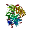
- Structure visualization
| Structure viewer | Molecule: MolmilJmol/JSmol |
|---|
- Downloads & links
-Download
| PDBx/mmCIF format | 5g5z.cif.gz | 803.4 KB | Display | PDBx/mmCIF format |
|---|---|---|---|---|
| PDB format | pdb5g5z.ent.gz | 665.8 KB | Display | PDB format |
| PDBx/mmJSON format | 5g5z.json.gz | Tree view | PDBx/mmJSON format | |
| Others |  Other downloads Other downloads |
-Validation report
| Arichive directory | https://data.pdbj.org/pub/pdb/validation_reports/g5/5g5zftp://data.pdbj.org/pub/pdb/validation_reports/g5/5g5z | HTTPS FTP |
|---|
-Related structure data
| Related structure data |  5g5ySC  5g60C  5g61C  5g62C S: Starting model for refinement C: citing same article ( |
|---|---|
| Similar structure data |


-Links
 PDBj
PDBj


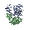
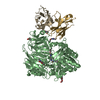
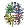
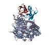
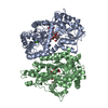
- Assembly
| Deposited unit | 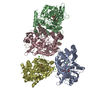
| ||||||||||||||||
|---|---|---|---|---|---|---|---|---|---|---|---|---|---|---|---|---|---|
| 1 | 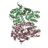
| ||||||||||||||||
| 2 | 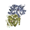
| ||||||||||||||||
| 3 | 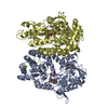
| ||||||||||||||||
| Unit cell |
| ||||||||||||||||
| Noncrystallographic symmetry (NCS) | NCS oper:
|
-Components
| #1: Protein | Mass: 56236.637 Da / Num. of mol.: 4 Source method: isolated from a genetically manipulated source Source: (gene. exp.) STREPTOCOCCUS PNEUMONIAE (bacteria) / Strain: TIGR4 / Plasmid: POPINF / Production host: #2: Polysaccharide | beta-D-fructofuranose-(2-1)-beta-D-fructofuranose-(2-1)-alpha-D-glucopyranose / 1-kestose 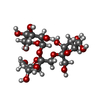  Source method: isolated from a genetically manipulated source Details: oligosaccharide with reducing-end-to-reducing-end glycosidic bond References: 1-kestose #3: Chemical | ChemComp-CA /   Mass: 40.078 Da / Num. of mol.: 9 / Source method: obtained synthetically / Formula: Ca Mass: 40.078 Da / Num. of mol.: 9 / Source method: obtained synthetically / Formula: Ca#4: Water | ChemComp-HOH / |  Mass: 18.015 Da / Num. of mol.: 1156 / Source method: isolated from a natural source / Formula: H2O Mass: 18.015 Da / Num. of mol.: 1156 / Source method: isolated from a natural source / Formula: H2OSequence details | LACKING THE FIRST 46 N-TERMINAL RESIDUES | |
|---|
-Experimental details
-Experiment
| Experiment | Method: X-RAY DIFFRACTION / Number of used crystals: 1 |
|---|
- Sample preparation
| Crystal | Density Matthews: 2.75 Å3/Da / Density % sol: 49.76 % / Description: NONE |
|---|---|
| Crystal grow | pH: 5 Details: 20 % PEG6000, 0.1M SODIUM ACETATE PH 5.0, 0.02 M CALCIUM CHLORIDE |
-Data collection
| Diffraction | Mean temperature: 100 K |
|---|---|
| Diffraction source | Source: SYNCHROTRON / Site: Diamond  / Beamline: I03 / Wavelength: 0.9763 / Beamline: I03 / Wavelength: 0.9763 |
| Detector | Type: DECTRIS PILATUS 6M / Detector: PIXEL / Date: Apr 23, 2015 / Details: MIRRORS |
| Radiation | Protocol: SINGLE WAVELENGTH / Monochromatic (M) / Laue (L): M / Scattering type: x-ray |
| Radiation wavelength | Wavelength: 0.9763 Å / Relative weight: 1 |
| Reflection | Resolution: 2.01→70.14 Å / Num. obs: 140542 / % possible obs: 97.7 % / Observed criterion σ(I): 2 / Redundancy: 5.7 % / Biso Wilson estimate: 31 Å2 / Rmerge(I) obs: 0.13 / Net I/σ(I): 8 |
| Reflection shell | Resolution: 2.01→2.06 Å / Redundancy: 5.8 % / Rmerge(I) obs: 1.21 / Mean I/σ(I) obs: 1.4 / % possible all: 99.4 |
- Processing
| Software |
| |||||||||||||||||||||||||||||||||||||||||||||||||||||||||||||||||||||||||||||||||||||||||||||||||||||||||||||||||||||||||||||||||||||||||||||||||||||||||||||||||||||||||||||||||||||||||||||||||||||||||||||||||||||||||
|---|---|---|---|---|---|---|---|---|---|---|---|---|---|---|---|---|---|---|---|---|---|---|---|---|---|---|---|---|---|---|---|---|---|---|---|---|---|---|---|---|---|---|---|---|---|---|---|---|---|---|---|---|---|---|---|---|---|---|---|---|---|---|---|---|---|---|---|---|---|---|---|---|---|---|---|---|---|---|---|---|---|---|---|---|---|---|---|---|---|---|---|---|---|---|---|---|---|---|---|---|---|---|---|---|---|---|---|---|---|---|---|---|---|---|---|---|---|---|---|---|---|---|---|---|---|---|---|---|---|---|---|---|---|---|---|---|---|---|---|---|---|---|---|---|---|---|---|---|---|---|---|---|---|---|---|---|---|---|---|---|---|---|---|---|---|---|---|---|---|---|---|---|---|---|---|---|---|---|---|---|---|---|---|---|---|---|---|---|---|---|---|---|---|---|---|---|---|---|---|---|---|---|---|---|---|---|---|---|---|---|---|---|---|---|---|---|---|---|
| Refinement | Method to determine structure: MOLECULAR REPLACEMENT Starting model: PDB ENTRY 5G5Y Resolution: 2.01→70.145 Å / SU ML: 0.25 / σ(F): 1.34 / Phase error: 23.96 / Stereochemistry target values: ML
| |||||||||||||||||||||||||||||||||||||||||||||||||||||||||||||||||||||||||||||||||||||||||||||||||||||||||||||||||||||||||||||||||||||||||||||||||||||||||||||||||||||||||||||||||||||||||||||||||||||||||||||||||||||||||
| Solvent computation | Shrinkage radii: 0.9 Å / VDW probe radii: 1.11 Å / Solvent model: FLAT BULK SOLVENT MODEL | |||||||||||||||||||||||||||||||||||||||||||||||||||||||||||||||||||||||||||||||||||||||||||||||||||||||||||||||||||||||||||||||||||||||||||||||||||||||||||||||||||||||||||||||||||||||||||||||||||||||||||||||||||||||||
| Displacement parameters | Biso mean: 38.71 Å2 | |||||||||||||||||||||||||||||||||||||||||||||||||||||||||||||||||||||||||||||||||||||||||||||||||||||||||||||||||||||||||||||||||||||||||||||||||||||||||||||||||||||||||||||||||||||||||||||||||||||||||||||||||||||||||
| Refinement step | Cycle: LAST / Resolution: 2.01→70.145 Å
| |||||||||||||||||||||||||||||||||||||||||||||||||||||||||||||||||||||||||||||||||||||||||||||||||||||||||||||||||||||||||||||||||||||||||||||||||||||||||||||||||||||||||||||||||||||||||||||||||||||||||||||||||||||||||
| Refine LS restraints |
| |||||||||||||||||||||||||||||||||||||||||||||||||||||||||||||||||||||||||||||||||||||||||||||||||||||||||||||||||||||||||||||||||||||||||||||||||||||||||||||||||||||||||||||||||||||||||||||||||||||||||||||||||||||||||
| LS refinement shell |
|
