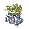
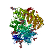
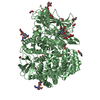
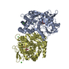
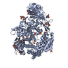
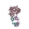
A: Dipeptidyl peptidase 4 B: Dipeptidyl peptidase 4 C: Dipeptidyl peptidase 4 D: Dipeptidyl peptidase 4 L: Fab light chain E: Fab light chain H: Fab heavy chain F: Fab heavy chain G: Fab light chain I: Fab heavy chain J: Fab light chain K: Fab heavy chain hetero molecules
Mass: 23031.551 Da / Num. of mol.: 4 Source method: isolated from a genetically manipulated source Source: (gene. exp.) Mus musculus (house mouse) / Production host: Homo sapiens (human)
#3: Antibody
Fabheavychain
Mass: 23267.041 Da / Num. of mol.: 4 Source method: isolated from a genetically manipulated source Source: (gene. exp.) Mus musculus (house mouse) / Production host: Homo sapiens (human)
-
Protein / Sugars , 2 types, 25 molecules ABCD
#1: Protein
Dipeptidylpeptidase4 / Bile canaliculus domain-specific membrane glycoprotein / Dipeptidyl peptidase IV / DPP IV / GP110 ...Bile canaliculus domain-specific membrane glycoprotein / Dipeptidyl peptidase IV / DPP IV / GP110 glycoprotein / T-cell activation antigen CD26
Mass: 85529.836 Da / Num. of mol.: 4 / Fragment: residues 37-767 Source method: isolated from a genetically manipulated source Source: (gene. exp.) Rattus norvegicus (Norway rat) / Gene: Dpp4, Cd26 / Production host: Homo sapiens (human) / References: UniProt: P14740, dipeptidyl-peptidase IV
Method to determine structure: MOLECULAR REPLACEMENT / Resolution: 2.8→30 Å / Cor.coef. Fo:Fc: 0.886 / Cor.coef. Fo:Fc free: 0.824 / SU B: 23.751 / SU ML: 0.44 / Cross valid method: THROUGHOUT / σ(F): 0 / ESU R Free: 0.453 / Stereochemistry target values: MAXIMUM LIKELIHOOD Details: HYDROGENS HAVE BEEN ADDED IN THE RIDING POSITIONS U VALUES : REFINED INDIVIDUALLY
Rfactor
Num. reflection
% reflection
Selection details
Rfree
0.2998
6869
5 %
RANDOM
Rwork
0.2497
-
-
-
obs
0.2522
130806
88.5 %
-
Solvent computation
Ion probe radii: 0.8 Å / Shrinkage radii: 0.8 Å / VDW probe radii: 1.2 Å / Solvent model: MASK
In the structure databanks used in Yorodumi, some data are registered as the other names, "COVID-19 virus" and "2019-nCoV". Here are the details of the virus and the list of structure data.
Jan 31, 2019. EMDB accession codes are about to change! (news from PDBe EMDB page)
EMDB accession codes are about to change! (news from PDBe EMDB page)
The allocation of 4 digits for EMDB accession codes will soon come to an end. Whilst these codes will remain in use, new EMDB accession codes will include an additional digit and will expand incrementally as the available range of codes is exhausted. The current 4-digit format prefixed with “EMD-” (i.e. EMD-XXXX) will advance to a 5-digit format (i.e. EMD-XXXXX), and so on. It is currently estimated that the 4-digit codes will be depleted around Spring 2019, at which point the 5-digit format will come into force.
The EM Navigator/Yorodumi systems omit the EMD- prefix.
Related info.:Q: What is EMD? / ID/Accession-code notation in Yorodumi/EM Navigator
Yorodumi is a browser for structure data from EMDB, PDB, SASBDB, etc.
This page is also the successor to EM Navigator detail page, and also detail information page/front-end page for Omokage search.
The word "yorodu" (or yorozu) is an old Japanese word meaning "ten thousand". "mi" (miru) is to see.
Related info.:EMDB / PDB / SASBDB / Comparison of 3 databanks / Yorodumi Search / Aug 31, 2016. New EM Navigator & Yorodumi / Yorodumi Papers / Jmol/JSmol / Function and homology information / Changes in new EM Navigator and Yorodumi
 Movie
Movie Controller


 Open data
 Basic information
 Components
 Keywords
 Function and homology information
 X-RAY DIFFRACTION /
  Authors
 Citation
 Structure visualization
 Downloads & links
 Other downloads


 PDBj
PDBj


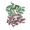
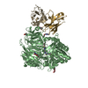
 Assembly

 Homo sapiens (human)
 Type: D-saccharide, beta linking / Mass: 221.208 Da / Num. of mol.: 21


 Mass: 194.226 Da / Num. of mol.: 1 / Source method: obtained synthetically / Formula: C8H18O5 / Comment: precipitant*YM
 Mass: 106.120 Da / Num. of mol.: 3 / Source method: obtained synthetically / Formula: C4H10O3
 Mass: 62.068 Da / Num. of mol.: 10 / Source method: obtained synthetically / Formula: C2H6O2
 Mass: 707.779 Da / Num. of mol.: 4 / Source method: obtained synthetically / Formula: C32H52F3N5O9
 Sample preparation
 / Beamline: 5.0.2 / Wavelength: 1 Å
 Processing