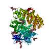
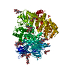
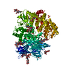
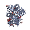
[English] 日本語
Yorodumi
- PDB-4hom: Crystal structure of porcine aminopeptidase-N complexed with subs... -

+
Open data


ID or keywords:

Loading...

-
Basic information

Entry
Database: PDB / ID: 4hom
TitleCrystal structure of porcine aminopeptidase-N complexed with substance P
Components
  • Aminopeptidase N
  • substance P
KeywordsHYDROLASE / zinc aminopeptidase
Function / homology
Function and homology information


substance P receptor binding / insemination / Tachykinin receptors bind tachykinins / detection of abiotic stimulus / tachykinin receptor signaling pathway / membrane alanyl aminopeptidase / response to pain / peptide catabolic process / metalloaminopeptidase activity / neuropeptide signaling pathway ...substance P receptor binding / insemination / Tachykinin receptors bind tachykinins / detection of abiotic stimulus / tachykinin receptor signaling pathway / membrane alanyl aminopeptidase / response to pain / peptide catabolic process / metalloaminopeptidase activity / neuropeptide signaling pathway / sensory perception of pain / cellular response to nerve growth factor stimulus / peptide binding / cell-cell signaling / virus receptor activity / positive regulation of cytosolic calcium ion concentration / G alpha (q) signalling events / chemical synaptic transmission / angiogenesis / cell differentiation / inflammatory response / axon / neuronal cell body / synapse / proteolysis / extracellular space / zinc ion binding / extracellular region / plasma membrane / cytoplasm
Similarity search - Function
Tachykinin family / Tachykinin family / Tachykinin domain / Tachykinin family / Tachykinin/Neurokinin-like, conserved site / Tachykinin family signature. / Zincin-like fold - #20 / Immunoglobulin-like - #1910 / Aminopeptidase N-type / ERAP1-like C-terminal domain ...Tachykinin family / Tachykinin family / Tachykinin domain / Tachykinin family / Tachykinin/Neurokinin-like, conserved site / Tachykinin family signature. / Zincin-like fold - #20 / Immunoglobulin-like - #1910 / Aminopeptidase N-type / ERAP1-like C-terminal domain / ERAP1-like C-terminal domain / Zincin-like fold / tricorn interacting facor f3 domain / Aminopeptidase N-like , N-terminal domain / Peptidase M1, alanine aminopeptidase/leukotriene A4 hydrolase / Peptidase M1, membrane alanine aminopeptidase / Peptidase family M1 domain / Peptidase M1 N-terminal domain / Aminopeptidase N-like , N-terminal domain superfamliy / Neutral Protease Domain 2 / Neutral Protease; domain 2 / Peptidase M4/M1, CTD superfamily / Neutral zinc metallopeptidases, zinc-binding region signature. / Alpha Horseshoe / Immunoglobulin-like / Sandwich / Orthogonal Bundle / Mainly Beta / Mainly Alpha
Similarity search - Domain/homology
triacetyl-beta-chitotriose / Aminopeptidase N / Protachykinin-1
Similarity search - Component
Biological speciesSus scrofa (pig)
MethodX-RAY DIFFRACTION / SYNCHROTRON / MOLECULAR REPLACEMENT / Resolution: 1.9 Å
AuthorsChen, L. / Lin, Y.L. / Peng, G. / Li, F.
CitationJournal: Proc.Natl.Acad.Sci.USA / Year: 2012
Title: Structural basis for multifunctional roles of mammalian aminopeptidase N.
Authors: Chen, L. / Lin, Y.L. / Peng, G. / Li, F.
History
DepositionOct 22, 2012Deposition site: RCSB / Processing site: RCSB
SupersessionOct 31, 2012ID: 4FKN
Revision 1.0Oct 31, 2012Provider: repository / Type: Initial release
Revision 1.1Nov 14, 2012Group: Database references
Revision 2.0Jul 29, 2020Group: Advisory / Atomic model ...Advisory / Atomic model / Data collection / Database references / Derived calculations / Structure summary
Category: atom_site / atom_site_anisotrop ...atom_site / atom_site_anisotrop / chem_comp / entity / entity_name_com / pdbx_branch_scheme / pdbx_chem_comp_identifier / pdbx_entity_branch / pdbx_entity_branch_descriptor / pdbx_entity_branch_link / pdbx_entity_branch_list / pdbx_entity_nonpoly / pdbx_molecule_features / pdbx_nonpoly_scheme / pdbx_struct_assembly_gen / pdbx_struct_conn_angle / pdbx_validate_close_contact / struct_asym / struct_conn / struct_ref_seq_dif / struct_site / struct_site_gen
Item: _atom_site.B_iso_or_equiv / _atom_site.Cartn_x ..._atom_site.B_iso_or_equiv / _atom_site.Cartn_x / _atom_site.Cartn_y / _atom_site.Cartn_z / _atom_site.auth_asym_id / _atom_site.auth_seq_id / _atom_site.label_asym_id / _atom_site.label_entity_id / _atom_site_anisotrop.U[1][1] / _atom_site_anisotrop.U[1][2] / _atom_site_anisotrop.U[1][3] / _atom_site_anisotrop.U[2][2] / _atom_site_anisotrop.U[2][3] / _atom_site_anisotrop.U[3][3] / _atom_site_anisotrop.pdbx_auth_asym_id / _atom_site_anisotrop.pdbx_auth_seq_id / _atom_site_anisotrop.pdbx_label_asym_id / _chem_comp.name / _chem_comp.type / _pdbx_entity_nonpoly.entity_id / _pdbx_entity_nonpoly.name / _pdbx_struct_assembly_gen.asym_id_list / _pdbx_struct_conn_angle.ptnr1_auth_comp_id / _pdbx_struct_conn_angle.ptnr1_auth_seq_id / _pdbx_struct_conn_angle.ptnr1_label_atom_id / _pdbx_struct_conn_angle.ptnr1_label_comp_id / _pdbx_struct_conn_angle.ptnr1_label_seq_id / _pdbx_struct_conn_angle.ptnr2_label_asym_id / _pdbx_struct_conn_angle.ptnr3_auth_comp_id / _pdbx_struct_conn_angle.ptnr3_auth_seq_id / _pdbx_struct_conn_angle.ptnr3_label_asym_id / _pdbx_struct_conn_angle.ptnr3_label_atom_id / _pdbx_struct_conn_angle.ptnr3_label_comp_id / _pdbx_struct_conn_angle.ptnr3_label_seq_id / _pdbx_struct_conn_angle.value / _pdbx_validate_close_contact.auth_asym_id_2 / _pdbx_validate_close_contact.auth_seq_id_2 / _struct_conn.pdbx_dist_value / _struct_conn.pdbx_leaving_atom_flag / _struct_conn.pdbx_role / _struct_conn.ptnr1_auth_asym_id / _struct_conn.ptnr1_auth_comp_id / _struct_conn.ptnr1_auth_seq_id / _struct_conn.ptnr1_label_asym_id / _struct_conn.ptnr1_label_atom_id / _struct_conn.ptnr1_label_comp_id / _struct_conn.ptnr1_label_seq_id / _struct_conn.ptnr2_auth_asym_id / _struct_conn.ptnr2_auth_seq_id / _struct_conn.ptnr2_label_asym_id / _struct_ref_seq_dif.details
Description: Carbohydrate remediation / Provider: repository / Type: Remediation

-
Structure visualization

Structure viewerMolecule:
MolmilJmol/JSmol

Downloads & links

-
Assembly

Deposited unit
A: Aminopeptidase N
B: substance P
hetero molecules


Theoretical massNumber of molelcules
Total (without water)109,80914
Polymers104,8722
Non-polymers4,93712
Water25,8341434
1


  • Idetical with deposited unit
  • defined by author&software
TypeNameSymmetry operationNumber
identity operation1_555x,y,z1
Buried area7140 Å2
ΔGint23 kcal/mol
Surface area39110 Å2
MethodPISA
Unit cell
Length a, b, c (Å)261.573, 62.598, 81.553
Angle α, β, γ (deg.)90.00, 100.27, 90.00
Int Tables number5
Space group name H-MC121

-
Components

-
Protein / Protein/peptide , 2 types, 2 molecules AB

#1: Protein Aminopeptidase N / AP-N / pAPN / Alanyl aminopeptidase / Aminopeptidase M / AP-M / Microsomal aminopeptidase / gp130


Mass: 103521.156 Da / Num. of mol.: 1 / Fragment: UNP residues 62-963
Source method: isolated from a genetically manipulated source
Source: (gene. exp.) Sus scrofa (pig) / Gene: ANPEP / Plasmid: pFastbac / Production host: 
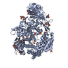
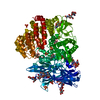
Spodoptera frugiperda (fall armyworm) / Strain (production host): SF9 / References: UniProt: P15145, membrane alanyl aminopeptidase
#2: Protein/peptide substance P


Mass: 1350.629 Da / Num. of mol.: 1 / Source method: obtained synthetically / References: UniProt: P20366*PLUS

-
Sugars , 3 types, 11 molecules

#3: Polysaccharide 2-acetamido-2-deoxy-beta-D-glucopyranose-(1-4)-2-acetamido-2-deoxy-beta-D-glucopyranose-(1-4)-2- ...2-acetamido-2-deoxy-beta-D-glucopyranose-(1-4)-2-acetamido-2-deoxy-beta-D-glucopyranose-(1-4)-2-acetamido-2-deoxy-beta-D-glucopyranose / triacetyl-beta-chitotriose


Type: oligosaccharide, Oligosaccharide / Class: Inhibitor / Mass: 627.594 Da / Num. of mol.: 3
Source method: isolated from a genetically manipulated source
Details: oligosaccharide / References: triacetyl-beta-chitotriose
DescriptorTypeProgram
DGlcpNAcb1-4DGlcpNAcb1-4DGlcpNAcb1-Glycam Condensed SequenceGMML 1.0
WURCS=2.0/1,3,2/[a2122h-1b_1-5_2*NCC/3=O]/1-1-1/a4-b1_b4-c1WURCSPDB2Glycan 1.1.0
[]{[(4+1)][b-D-GlcpNAc]{[(4+1)][b-D-GlcpNAc]{[(4+1)][b-D-GlcpNAc]{}}}}LINUCSPDB-CARE
#4: Polysaccharide
2-acetamido-2-deoxy-beta-D-glucopyranose-(1-4)-2-acetamido-2-deoxy-beta-D-glucopyranose


Type: oligosaccharide / Mass: 424.401 Da / Num. of mol.: 6
Source method: isolated from a genetically manipulated source
DescriptorTypeProgram
DGlcpNAcb1-4DGlcpNAcb1-Glycam Condensed SequenceGMML 1.0
WURCS=2.0/1,2,1/[a2122h-1b_1-5_2*NCC/3=O]/1-1/a4-b1WURCSPDB2Glycan 1.1.0
[]{[(4+1)][b-D-GlcpNAc]{[(4+1)][b-D-GlcpNAc]{}}}LINUCSPDB-CARE
#5: Sugar ChemComp-NAG / 2-acetamido-2-deoxy-beta-D-glucopyranose


Type: D-saccharide, beta linking / Mass: 221.208 Da / Num. of mol.: 2
Source method: isolated from a genetically manipulated source
Formula: C8H15NO6
IdentifierTypeProgram
DGlcpNAcbCONDENSED IUPAC CARBOHYDRATE SYMBOLGMML 1.0
N-acetyl-b-D-glucopyranosamineCOMMON NAMEGMML 1.0
b-D-GlcpNAcIUPAC CARBOHYDRATE SYMBOLPDB-CARE 1.0
GlcNAcSNFG CARBOHYDRATE SYMBOLGMML 1.0

-
Non-polymers , 2 types, 1435 molecules

#6: Chemical ChemComp-ZN / ZINC ION


Mass: 65.409 Da / Num. of mol.: 1 / Source method: obtained synthetically / Formula: Zn
#7: Water ChemComp-HOH / water


Mass: 18.015 Da / Num. of mol.: 1434 / Source method: isolated from a natural source / Formula: H2O

-
Experimental details

-
Experiment

ExperimentMethod: X-RAY DIFFRACTION / Number of used crystals: 1

-
Sample preparation

CrystalDensity Matthews: 3.13 Å3/Da / Density % sol: 60.73 %
Crystal growTemperature: 277 K / Method: vapor diffusion, sitting drop / pH: 7.2
Details: 18% PEG3350, 200 mM lithium sulfate, 100 mM HEPES, pH 7.2, VAPOR DIFFUSION, SITTING DROP, temperature 277K

-
Data collection

Diffraction sourceSource: SYNCHROTRON / Site: APS / Beamline: 19-ID
DetectorType: ADSC QUANTUM 315 / Detector: CCD
RadiationMonochromator: Rosenbaum-Rock high resolution double crystal Si(111)
Protocol: SINGLE WAVELENGTH / Monochromatic (M) / Laue (L): M / Scattering type: x-ray
Radiation wavelengthRelative weight: 1
ReflectionResolution: 1.85→50 Å / Num. all: 102618 / Num. obs: 100774 / % possible obs: 98.2 % / Observed criterion σ(F): 2 / Observed criterion σ(I): 2

-
Processing

Software
NameVersionClassification
HKL-2000data collection
SOLVEphasing
REFMAC5.7.0029refinement
HKL-2000data reduction
HKL-2000data scaling
RefinementMethod to determine structure: MOLECULAR REPLACEMENT / Resolution: 1.9→37.18 Å / Cor.coef. Fo:Fc: 0.974 / Cor.coef. Fo:Fc free: 0.954 / SU B: 6.228 / SU ML: 0.082 / Cross valid method: THROUGHOUT / ESU R: 0.233 / ESU R Free: 0.121 / Stereochemistry target values: MAXIMUM LIKELIHOOD
RfactorNum. reflection% reflectionSelection details
Rfree0.20123 5027 5 %RANDOM
Rwork0.1471 ---
obs0.14979 95596 98.05 %-
Solvent computationIon probe radii: 0.8 Å / Shrinkage radii: 0.8 Å / VDW probe radii: 1.4 Å / Solvent model: BABINET MODEL WITH MASK
Displacement parametersBiso mean: 33.54 Å2
Baniso -1Baniso -2Baniso -3
1-0.83 Å2-0 Å20.86 Å2
2---0.29 Å20 Å2
3----0.66 Å2
Refinement stepCycle: LAST / Resolution: 1.9→37.18 Å
ProteinNucleic acidLigandSolventTotal
Num. atoms7336 0 323 1434 9093
Refine LS restraints
Refine-IDTypeDev idealDev ideal targetNumber
X-RAY DIFFRACTIONr_bond_refined_d0.0060.027875
X-RAY DIFFRACTIONr_bond_other_d
X-RAY DIFFRACTIONr_angle_refined_deg1.1271.98710755
X-RAY DIFFRACTIONr_angle_other_deg
X-RAY DIFFRACTIONr_dihedral_angle_1_deg6.365911
X-RAY DIFFRACTIONr_dihedral_angle_2_deg39.11824.933371
X-RAY DIFFRACTIONr_dihedral_angle_3_deg14.42151235
X-RAY DIFFRACTIONr_dihedral_angle_4_deg16.0121532
X-RAY DIFFRACTIONr_chiral_restr0.080.21227
X-RAY DIFFRACTIONr_gen_planes_refined0.0050.0215977
X-RAY DIFFRACTIONr_gen_planes_other
X-RAY DIFFRACTIONr_nbd_refined
X-RAY DIFFRACTIONr_nbd_other
X-RAY DIFFRACTIONr_nbtor_refined
X-RAY DIFFRACTIONr_nbtor_other
X-RAY DIFFRACTIONr_xyhbond_nbd_refined
X-RAY DIFFRACTIONr_xyhbond_nbd_other
X-RAY DIFFRACTIONr_metal_ion_refined
X-RAY DIFFRACTIONr_metal_ion_other
X-RAY DIFFRACTIONr_symmetry_vdw_refined
X-RAY DIFFRACTIONr_symmetry_vdw_other
X-RAY DIFFRACTIONr_symmetry_hbond_refined
X-RAY DIFFRACTIONr_symmetry_hbond_other
X-RAY DIFFRACTIONr_symmetry_metal_ion_refined
X-RAY DIFFRACTIONr_symmetry_metal_ion_other
X-RAY DIFFRACTIONr_mcbond_it
X-RAY DIFFRACTIONr_mcbond_other
X-RAY DIFFRACTIONr_mcangle_it
X-RAY DIFFRACTIONr_scbond_it
X-RAY DIFFRACTIONr_scangle_it
X-RAY DIFFRACTIONr_rigid_bond_restr2.16937874
X-RAY DIFFRACTIONr_sphericity_free26.2125561
X-RAY DIFFRACTIONr_sphericity_bonded16.9858533
LS refinement shellResolution: 1.9→2.003 Å / Total num. of bins used: 10
RfactorNum. reflection% reflection
Rfree0.283 716 -
Rwork0.219 13946 -
obs--98.89 %
Refinement TLS params.Method: refined / Origin x: 22.1893 Å / Origin y: 18.3368 Å / Origin z: 59.669 Å
111213212223313233
T0.1093 Å2-0.0322 Å2-0.0445 Å2-0.1145 Å2-0.0463 Å2--0.1635 Å2
L1.5446 °2-0.3189 °2-0.3913 °2-0.715 °2-0.0955 °2--1.0435 °2
S0.035 Å °0.0748 Å °0.1681 Å °-0.104 Å °-0.0458 Å °0.2452 Å °0.009 Å °-0.2753 Å °0.0108 Å °
Refinement TLS group
IDRefine-IDRefine TLS-IDAuth asym-IDAuth seq-ID
1X-RAY DIFFRACTION1A63 - 281
2X-RAY DIFFRACTION1A282 - 543
3X-RAY DIFFRACTION1A544 - 632
4X-RAY DIFFRACTION1A633 - 964

+
About Yorodumi

-
News

-
Feb 9, 2022. New format data for meta-information of EMDB entries

New format data for meta-information of EMDB entries

  • Version 3 of the EMDB header file is now the official format.
  • The previous official version 1.9 will be removed from the archive.

Related info.:EMDB header

External links:wwPDB to switch to version 3 of the EMDB data model

-
Aug 12, 2020. Covid-19 info

Covid-19 info

URL: https://pdbjlvh1.pdbj.org/emnavi/covid19.php

New page: Covid-19 featured information page in EM Navigator.

Related info.:Covid-19 info / Mar 5, 2020. Novel coronavirus structure data

+
Mar 5, 2020. Novel coronavirus structure data

Novel coronavirus structure data

Related info.:Yorodumi Speices / Aug 12, 2020. Covid-19 info

External links:COVID-19 featured content - PDBj / Molecule of the Month (242):Coronavirus Proteases

+
Jan 31, 2019. EMDB accession codes are about to change! (news from PDBe EMDB page)

EMDB accession codes are about to change! (news from PDBe EMDB page)

  • The allocation of 4 digits for EMDB accession codes will soon come to an end. Whilst these codes will remain in use, new EMDB accession codes will include an additional digit and will expand incrementally as the available range of codes is exhausted. The current 4-digit format prefixed with “EMD-” (i.e. EMD-XXXX) will advance to a 5-digit format (i.e. EMD-XXXXX), and so on. It is currently estimated that the 4-digit codes will be depleted around Spring 2019, at which point the 5-digit format will come into force.
  • The EM Navigator/Yorodumi systems omit the EMD- prefix.

Related info.:Q: What is EMD? / ID/Accession-code notation in Yorodumi/EM Navigator

External links:EMDB Accession Codes are Changing Soon! / Contact to PDBj

+
Jul 12, 2017. Major update of PDB

Major update of PDB

  • wwPDB released updated PDB data conforming to the new PDBx/mmCIF dictionary.
  • This is a major update changing the version number from 4 to 5, and with Remediation, in which all the entries are updated.
  • In this update, many items about electron microscopy experimental information are reorganized (e.g. em_software).
  • Now, EM Navigator and Yorodumi are based on the updated data.

External links:wwPDB Remediation / Enriched Model Files Conforming to OneDep Data Standards Now Available in the PDB FTP Archive

-
Yorodumi

Thousand views of thousand structures

  • Yorodumi is a browser for structure data from EMDB, PDB, SASBDB, etc.
  • This page is also the successor to EM Navigator detail page, and also detail information page/front-end page for Omokage search.
  • The word "yorodu" (or yorozu) is an old Japanese word meaning "ten thousand". "mi" (miru) is to see.

Related info.:EMDB / PDB / SASBDB / Comparison of 3 databanks / Yorodumi Search / Aug 31, 2016. New EM Navigator & Yorodumi / Yorodumi Papers / Jmol/JSmol / Function and homology information / Changes in new EM Navigator and Yorodumi

Read more