 Movie
Movie Controller


+ Open data

- Basic information
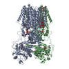
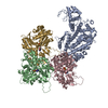
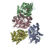
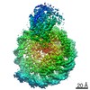
| Entry | Database: PDB / ID: 5kg8 | ||||||
|---|---|---|---|---|---|---|---|
| Title | Rigor myosin X co-complexed with an actin filament | ||||||
 Components Components |
| ||||||
 Keywords Keywords | MOTOR PROTEIN / myosin molecular motors cytoskeletal motility | ||||||
| Function / homology |  Function and homology information Function and homology informationplus-end directed microfilament motor activity / Netrin-1 signaling / positive regulation of cell-cell adhesion / cytoskeleton-dependent intracellular transport / filopodium tip / regulation of filopodium assembly / filopodium membrane / myosin complex / cytoskeletal motor activator activity / spectrin binding ...plus-end directed microfilament motor activity / Netrin-1 signaling / positive regulation of cell-cell adhesion / cytoskeleton-dependent intracellular transport / filopodium tip / regulation of filopodium assembly / filopodium membrane / myosin complex / cytoskeletal motor activator activity / spectrin binding / microfilament motor activity / myosin heavy chain binding / tropomyosin binding / actin filament bundle / troponin I binding / filamentous actin / mesenchyme migration / phosphatidylinositol-3,4,5-trisphosphate binding / skeletal muscle myofibril / actin filament bundle assembly / striated muscle thin filament / skeletal muscle thin filament assembly / actin monomer binding / skeletal muscle fiber development / ruffle / stress fiber / titin binding / actin filament polymerization / FCGR3A-mediated phagocytosis / filopodium / actin filament / Regulation of actin dynamics for phagocytic cup formation / Hydrolases; Acting on acid anhydrides; Acting on acid anhydrides to facilitate cellular and subcellular movement / calcium-dependent protein binding / actin filament binding / regulation of cell shape / lamellipodium / cell body / cell cortex / calmodulin binding / neuron projection / protein domain specific binding / hydrolase activity / neuronal cell body / calcium ion binding / positive regulation of gene expression / nucleolus / magnesium ion binding / signal transduction / ATP binding / identical protein binding / plasma membrane / cytoplasm / cytosol Similarity search - Function | ||||||
| Biological species |  Homo sapiens (human) Homo sapiens (human) | ||||||
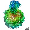
| Method | ELECTRON MICROSCOPY / helical reconstruction / cryo EM / Resolution: 9.1 Å | ||||||
 Authors Authors | Sindelar, C.V. / Houdusse, A. / Sweeney, L. | ||||||
| Funding support |  United States, 1items United States, 1items
| ||||||
 Citation Citation | Journal: Nat Commun / Year: 2016 Title: The myosin X motor is optimized for movement on actin bundles. Authors: Virginie Ropars / Zhaohui Yang / Tatiana Isabet / Florian Blanc / Kaifeng Zhou / Tianming Lin / Xiaoyan Liu / Pascale Hissier / Frédéric Samazan / Béatrice Amigues / Eric D Yang / Hyokeun ...Authors: Virginie Ropars / Zhaohui Yang / Tatiana Isabet / Florian Blanc / Kaifeng Zhou / Tianming Lin / Xiaoyan Liu / Pascale Hissier / Frédéric Samazan / Béatrice Amigues / Eric D Yang / Hyokeun Park / Olena Pylypenko / Marco Cecchini / Charles V Sindelar / H Lee Sweeney / Anne Houdusse /   Abstract: Myosin X has features not found in other myosins. Its structure must underlie its unique ability to generate filopodia, which are essential for neuritogenesis, wound healing, cancer metastasis and ...Myosin X has features not found in other myosins. Its structure must underlie its unique ability to generate filopodia, which are essential for neuritogenesis, wound healing, cancer metastasis and some pathogenic infections. By determining high-resolution structures of key components of this motor, and characterizing the in vitro behaviour of the native dimer, we identify the features that explain the myosin X dimer behaviour. Single-molecule studies demonstrate that a native myosin X dimer moves on actin bundles with higher velocities and takes larger steps than on single actin filaments. The largest steps on actin bundles are larger than previously reported for artificially dimerized myosin X constructs or any other myosin. Our model and kinetic data explain why these large steps and high velocities can only occur on bundled filaments. Thus, myosin X functions as an antiparallel dimer in cells with a unique geometry optimized for movement on actin bundles. | ||||||
| History |
|
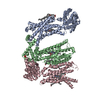
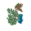
- Structure visualization
| Movie |
Movie viewer |
|---|---|
| Structure viewer | Molecule: MolmilJmol/JSmol |
- Downloads & links
-Download
| PDBx/mmCIF format | 5kg8.cif.gz | 229 KB | Display | PDBx/mmCIF format |
|---|---|---|---|---|
| PDB format | pdb5kg8.ent.gz | 140.4 KB | Display | PDB format |
| PDBx/mmJSON format | 5kg8.json.gz | Tree view | PDBx/mmJSON format | |
| Others |  Other downloads Other downloads |
-Validation report
| Arichive directory | https://data.pdbj.org/pub/pdb/validation_reports/kg/5kg8ftp://data.pdbj.org/pub/pdb/validation_reports/kg/5kg8 | HTTPS FTP |
|---|
-Related structure data
| Related structure data | 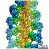 8244MC  5hmoC  5hmpC  5i0hC  5i0iC M: map data used to model this data C: citing same article ( |
|---|---|
| Similar structure data |

-Links
 PDBj
PDBj


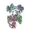
- Assembly
| Deposited unit | 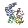
|
|---|---|
| 1 |
|
-Components
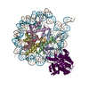
| #1: Protein | Mass: 85083.086 Da / Num. of mol.: 1 / Fragment: motor domain (UNP residues 3-741) Source method: isolated from a genetically manipulated source Source: (gene. exp.) Homo sapiens (human) / Gene: MYO10, KIAA0799 / Production host:  |
|---|---|
| #2: Protein | Mass: 41827.609 Da / Num. of mol.: 3 / Source method: isolated from a natural source / Source: (natural) |
-Experimental details
-Experiment
| Experiment | Method: ELECTRON MICROSCOPY |
|---|---|
| EM experiment | Aggregation state: FILAMENT / 3D reconstruction method: helical reconstruction |
- Sample preparation
| Component | Name: Actin filament decorated by the myosin X motor domain / Type: ORGANELLE OR CELLULAR COMPONENT / Entity ID: all / Source: MULTIPLE SOURCES |
|---|---|
| Buffer solution | pH: 6.8 |
| Buffer component | Conc.: 5 mM / Name: MOPS |
| Specimen | Conc.: 1.7 mg/ml / Embedding applied: NO / Shadowing applied: NO / Staining applied: NO / Vitrification applied: YES |
| Specimen support | Grid material: COPPER / Grid mesh size: 300 divisions/in. / Grid type: Quantifoil |
| Vitrification | Instrument: HOMEMADE PLUNGER / Cryogen name: ETHANE |
- Electron microscopy imaging
| Experimental equipment |  Model: Tecnai F20 / Image courtesy: FEI Company |
|---|---|
| Microscopy | Model: FEI TECNAI F20 |
| Electron gun | Electron source:  FIELD EMISSION GUN / Accelerating voltage: 200 kV / Illumination mode: FLOOD BEAM FIELD EMISSION GUN / Accelerating voltage: 200 kV / Illumination mode: FLOOD BEAM |
| Electron lens | Mode: BRIGHT FIELD / Calibrated magnification: 26780 X / Nominal defocus max: 5000 nm / Nominal defocus min: 1000 nm / Calibrated defocus min: 1438 nm / Calibrated defocus max: 5251 nm / Cs: 2 mm / C2 aperture diameter: 70 µm / Alignment procedure: COMA FREE |
| Specimen holder | Cryogen: NITROGEN Specimen holder model: GATAN 626 SINGLE TILT LIQUID NITROGEN CRYO TRANSFER HOLDER |
| Image recording | Average exposure time: 13 sec. / Electron dose: 50 e/Å2 / Detector mode: COUNTING / Film or detector model: GATAN K2 SUMMIT (4k x 4k) / Num. of grids imaged: 2 / Num. of real images: 154 |
- Processing
| EM software |
| |||||||||||||||||||||||||||||||||||
|---|---|---|---|---|---|---|---|---|---|---|---|---|---|---|---|---|---|---|---|---|---|---|---|---|---|---|---|---|---|---|---|---|---|---|---|---|
| CTF correction | Type: PHASE FLIPPING AND AMPLITUDE CORRECTION | |||||||||||||||||||||||||||||||||||
| Helical symmerty | Angular rotation/subunit: 167.1 ° / Axial rise/subunit: 27.44 Å / Axial symmetry: C1 / Details: Not directly applicable to the modeled coordinates | |||||||||||||||||||||||||||||||||||
| 3D reconstruction | Resolution: 9.1 Å / Resolution method: FSC 0.143 CUT-OFF / Num. of particles: 57927 / Algorithm: FOURIER SPACE Details: Symmetry was not applied in this reconstruction. The provided symmetry parameters are nominal, and were not actually used. Num. of class averages: 1 / Symmetry type: HELICAL |
