[English] 日本語
Yorodumi
- EMDB-0977: Cyanobacterial PSI Monomer from T. elongatus by Single Particle C... -

+
Open data


ID or keywords:

Loading...

-
Basic information

Entry
Database: EMDB / ID: EMD-0977
TitleCyanobacterial PSI Monomer from T. elongatus by Single Particle CRYO-EM at 3.2 A Resolution
Map data
Sample
  • Complex: Cyanobacterial PSI Monomer from T. elongatus
    • Protein or peptide: x 10 types
  • Ligand: x 7 types
Keywordsmonomer / complex / photosystem / photosynthesis / MEMBRANE PROTEIN
Function / homology
Function and homology information


photosystem I reaction center / photosystem I / photosynthetic electron transport in photosystem I / photosystem I / chlorophyll binding / plasma membrane-derived thylakoid membrane / photosynthesis / 4 iron, 4 sulfur cluster binding / electron transfer activity / magnesium ion binding / metal ion binding
Similarity search - Function
Photosystem I PsaM, reaction centre superfamily / Photosystem I PsaM, reaction centre / Photosystem I protein M (PsaM) / Photosystem I PsaL, reaction centre subunit XI / Photosystem I, reaction centre subunit XI / Photosystem I PsaL, reaction centre subunit XI superfamily / Photosystem I reaction centre subunit XI / Photosystem I reaction centre subunit VIII / Photosystem I reaction centre subunit VIII / Photosystem I reaction centre subunit VIII superfamily ...Photosystem I PsaM, reaction centre superfamily / Photosystem I PsaM, reaction centre / Photosystem I protein M (PsaM) / Photosystem I PsaL, reaction centre subunit XI / Photosystem I, reaction centre subunit XI / Photosystem I PsaL, reaction centre subunit XI superfamily / Photosystem I reaction centre subunit XI / Photosystem I reaction centre subunit VIII / Photosystem I reaction centre subunit VIII / Photosystem I reaction centre subunit VIII superfamily / Photosystem I PsaF, reaction centre subunit III / Photosystem I PsaF, reaction centre subunit III superfamily / Photosystem I reaction centre subunit III / Photosystem I PsaJ, reaction centre subunit IX / Photosystem I PsaD / Photosystem I PsaJ, reaction centre subunit IX superfamily / Photosystem I, reaction centre subunit PsaD superfamily / Photosystem I reaction centre subunit IX / PsaJ / PsaD / Photosystem I PsaE, reaction centre subunit IV / Photosystem I reaction centre subunit IV / PsaE / Photosystem I protein PsaC / Photosystem I PsaA / Photosystem I PsaB / Photosystem I PsaA/PsaB, conserved site / Photosystem I psaA and psaB proteins signature. / Photosystem I PsaA/PsaB / Photosystem I PsaA/PsaB superfamily / Photosystem I psaA/psaB protein / Electron transport accessory-like domain superfamily / 4Fe-4S dicluster domain / 4Fe-4S ferredoxin, iron-sulphur binding, conserved site / 4Fe-4S ferredoxin-type iron-sulfur binding region signature. / 4Fe-4S ferredoxin-type iron-sulfur binding domain profile. / 4Fe-4S ferredoxin-type, iron-sulphur binding domain
Similarity search - Domain/homology
Photosystem I reaction center subunit III / Photosystem I reaction center subunit XII / Photosystem I P700 chlorophyll a apoprotein A1 / Photosystem I P700 chlorophyll a apoprotein A2 / Photosystem I iron-sulfur center / Photosystem I reaction center subunit II / Photosystem I reaction center subunit IV / Photosystem I reaction center subunit VIII / Photosystem I reaction center subunit IX / Photosystem I reaction center subunit XI
Similarity search - Component
Biological speciesThermosynechococcus elongatus BP-1 (bacteria) / Thermosynechococcus elongatus (strain BP-1) (bacteria)
Methodsingle particle reconstruction / cryo EM / Resolution: 3.2 Å
AuthorsKurisu G / Coruh O
Funding support Japan, 2 items
OrganizationGrant numberCountry
Japan Science and TechnologyJPMJCR13M4 Japan
Ministry of Education, Culture, Sports, Science and Technology (Japan)16H06560 Japan
CitationJournal: Commun Biol / Year: 2021
Title: Cryo-EM structure of a functional monomeric Photosystem I from Thermosynechococcus elongatus reveals red chlorophyll cluster.
Authors: Orkun Çoruh / Anna Frank / Hideaki Tanaka / Akihiro Kawamoto / Eithar El-Mohsnawy / Takayuki Kato / Keiichi Namba / Christoph Gerle / Marc M Nowaczyk / Genji Kurisu /
Abstract: A high-resolution structure of trimeric cyanobacterial Photosystem I (PSI) from Thermosynechococcus elongatus was reported as the first atomic model of PSI almost 20 years ago. However, the monomeric ...A high-resolution structure of trimeric cyanobacterial Photosystem I (PSI) from Thermosynechococcus elongatus was reported as the first atomic model of PSI almost 20 years ago. However, the monomeric PSI structure has not yet been reported despite long-standing interest in its structure and extensive spectroscopic characterization of the loss of red chlorophylls upon monomerization. Here, we describe the structure of monomeric PSI from Thermosynechococcus elongatus BP-1. Comparison with the trimer structure gave detailed insights into monomerization-induced changes in both the central trimerization domain and the peripheral regions of the complex. Monomerization-induced loss of red chlorophylls is assigned to a cluster of chlorophylls adjacent to PsaX. Based on our findings, we propose a role of PsaX in the stabilization of red chlorophylls and that lipids of the surrounding membrane present a major source of thermal energy for uphill excitation energy transfer from red chlorophylls to P700.
History
DepositionJan 24, 2020-
Header (metadata) releaseMar 17, 2021-
Map releaseMar 17, 2021-
UpdateMar 27, 2024-
Current statusMar 27, 2024Processing site: PDBj / Status: Released

-
Structure visualization

Movie
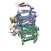
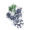
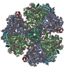
  • Surface view with section colored by density value
  • Surface level: 0.0416
  • Imaged by UCSF Chimera
  • Download
  • Surface view colored by height
  • Surface level: 0.0416
  • Imaged by UCSF Chimera
  • Download
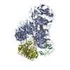
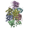
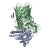
  • Surface view with fitted model
  • Atomic models: PDB-6lu1
  • Surface level: 0.0416
  • Imaged by UCSF Chimera
  • Download
Movie viewer
Structure viewerEM map:
SurfViewMolmilJmol/JSmol
Supplemental images

Downloads & links

-
Map

FileDownload / File: emd_0977.map.gz / Format: CCP4 / Size: 64 MB / Type: IMAGE STORED AS FLOATING POINT NUMBER (4 BYTES)
Voxel sizeX=Y=Z: 0.89 Å
Density
Contour LevelBy AUTHOR: 0.0416 / Movie #1: 0.0416
Minimum - Maximum-0.106297866 - 0.18965338
Average (Standard dev.)-0.0000073780034 (±0.007665535)
SymmetrySpace group: 1
Details

EMDB XML:

Map geometry
Axis orderXYZ
Origin000
Dimensions256256256
Spacing256256256
CellA=B=C: 227.84 Å
α=β=γ: 90.0 °

CCP4 map header:

modeImage stored as Reals
Å/pix. X/Y/Z0.890.890.89
M x/y/z256256256
origin x/y/z0.0000.0000.000
length x/y/z227.840227.840227.840
α/β/γ90.00090.00090.000
MAP C/R/S123
start NC/NR/NS000
NC/NR/NS256256256
D min/max/mean-0.1060.190-0.000

-
Supplemental data

-
Half map: #1

Fileemd_0977_half_map_1.map
Projections & Slices
AxesZYX

Projections

Slices (1/2)
Density Histograms

-
Half map: #2

Fileemd_0977_half_map_2.map
Projections & Slices
AxesZYX

Projections

Slices (1/2)
Density Histograms

-
Sample components

+
Entire : Cyanobacterial PSI Monomer from T. elongatus

EntireName: Cyanobacterial PSI Monomer from T. elongatus
Components
  • Complex: Cyanobacterial PSI Monomer from T. elongatus
    • Protein or peptide: Photosystem I P700 chlorophyll a apoprotein A1
    • Protein or peptide: Photosystem I P700 chlorophyll a apoprotein A2
    • Protein or peptide: Photosystem I iron-sulfur center
    • Protein or peptide: Photosystem I reaction center subunit II
    • Protein or peptide: Photosystem I reaction center subunit IV
    • Protein or peptide: Photosystem I reaction center subunit III
    • Protein or peptide: Photosystem I reaction center subunit VIII
    • Protein or peptide: Photosystem I reaction center subunit IX
    • Protein or peptide: Photosystem I reaction center subunit XI
    • Protein or peptide: Photosystem I reaction center subunit XII
  • Ligand: CHLOROPHYLL A
  • Ligand: PHYLLOQUINONEPhytomenadione
  • Ligand: BETA-CAROTENEΒ-Carotene
  • Ligand: 1,2-DIPALMITOYL-PHOSPHATIDYL-GLYCEROLE
  • Ligand: IRON/SULFUR CLUSTERIron–sulfur cluster
  • Ligand: 1,2-DISTEAROYL-MONOGALACTOSYL-DIGLYCERIDE
  • Ligand: water

+
Supramolecule #1: Cyanobacterial PSI Monomer from T. elongatus

SupramoleculeName: Cyanobacterial PSI Monomer from T. elongatus / type: complex / ID: 1 / Parent: 0 / Macromolecule list: #1-#10
Details: The monomers in this sample are generated by the monomerization of trimers of PSI during the purification process.
Source (natural)Organism: Thermosynechococcus elongatus BP-1 (bacteria)
Molecular weightTheoretical: 350 kDa/nm

+
Macromolecule #1: Photosystem I P700 chlorophyll a apoprotein A1

MacromoleculeName: Photosystem I P700 chlorophyll a apoprotein A1 / type: protein_or_peptide / ID: 1 / Number of copies: 1 / Enantiomer: LEVO / EC number: photosystem I
Source (natural)Organism: Thermosynechococcus elongatus BP-1 (bacteria) / Strain: BP-1
Molecular weightTheoretical: 83.267773 KDa
SequenceString: MTISPPEREP KVRVVVDNDP VPTSFEKWAK PGHFDRTLAR GPQTTTWIWN LHALAHDFDT HTSDLEDISR KIFSAHFGHL AVVFIWLSG MYFHGAKFSN YEAWLADPTG IKPSAQVVWP IVGQGILNGD VGGGFHGIQI TSGLFQLWRA SGITNEFQLY C TAIGGLVM ...String:
MTISPPEREP KVRVVVDNDP VPTSFEKWAK PGHFDRTLAR GPQTTTWIWN LHALAHDFDT HTSDLEDISR KIFSAHFGHL AVVFIWLSG MYFHGAKFSN YEAWLADPTG IKPSAQVVWP IVGQGILNGD VGGGFHGIQI TSGLFQLWRA SGITNEFQLY C TAIGGLVM AGLMLFAGWF HYHKRAPKLE WFQNVESMLN HHLAGLLGLG SLAWAGHQIH VSLPINKLLD AGVAAKDIPL PH EFILNPS LMAELYPKVD WGFFSGVIPF FTFNWAAYSD FLTFNGGLNP VTGGLWLSDT AHHHLAIAVL FIIAGHMYRT NWG IGHSLK EILEAHKGPF TGAGHKGLYE VLTTSWHAQL AINLAMMGSL SIIVAQHMYA MPPYPYLATD YPTQLSLFTH HMWI GGFLV VGGAAHGAIF MVRDYDPAMN QNNVLDRVLR HRDAIISHLN WVCIFLGFHS FGLYVHNDTM RAFGRPQDMF SDTGI QLQP VFAQWVQNLH TLAPGGTAPN AAATASVAFG GDVVAVGGKV AMMPIVLGTA DFMVHHIHAF TIHVTVLILL KGVLFA RSS RLIPDKANLG FRFPCDGPGR GGTCQVSGWD HVFLGLFWMY NCISVVIFHF SWKMQSDVWG TVAPDGTVSH ITGGNFA QS AITINGWLRD FLWAQASQVI GSYGSALSAY GLLFLGAHFI WAFSLMFLFS GRGYWQELIE SIVWAHNKLK VAPAIQPR A LSIIQGRAVG VAHYLLGGIA TTWAFFLARI ISVG

UniProtKB: Photosystem I P700 chlorophyll a apoprotein A1

+
Macromolecule #2: Photosystem I P700 chlorophyll a apoprotein A2

MacromoleculeName: Photosystem I P700 chlorophyll a apoprotein A2 / type: protein_or_peptide / ID: 2 / Number of copies: 1 / Enantiomer: LEVO / EC number: photosystem I
Source (natural)Organism: Thermosynechococcus elongatus (strain BP-1) (bacteria)
Strain: BP-1
Molecular weightTheoretical: 83.123648 KDa
SequenceString: MATKFPKFSQ DLAQDPTTRR IWYAIAMAHD FESHDGMTEE NLYQKIFASH FGHLAIIFLW VSGSLFHVAW QGNFEQWVQD PVNTRPIAH AIWDPQFGKA AVDAFTQAGA SNPVDIAYSG VYHWWYTIGM RTNGDLYQGA IFLLILASLA LFAGWLHLQP K FRPSLSWF ...String:
MATKFPKFSQ DLAQDPTTRR IWYAIAMAHD FESHDGMTEE NLYQKIFASH FGHLAIIFLW VSGSLFHVAW QGNFEQWVQD PVNTRPIAH AIWDPQFGKA AVDAFTQAGA SNPVDIAYSG VYHWWYTIGM RTNGDLYQGA IFLLILASLA LFAGWLHLQP K FRPSLSWF KNAESRLNHH LAGLFGVSSL AWAGHLIHVA IPESRGQHVG WDNFLSTMPH PAGLAPFFTG NWGVYAQNPD TA SHVFGTA QGAGTAILTF LGGFHPQTES LWLTDMAHHH LAIAVLFIVA GHMYRTQFGI GHSIKEMMDA KDFFGTKVEG PFN MPHQGI YETYNNSLHF QLGWHLACLG VITSLVAQHM YSLPPYAFIA QDHTTMAALY THHQYIAGFL MVGAFAHGAI FLVR DYDPA QNKGNVLDRV LQHKEAIISH LSWVSLFLGF HTLGLYVHND VVVAFGTPEK QILIEPVFAQ FIQAAHGKLL YGFDT LLSN PDSIASTAWP NYGNVWLPGW LDAINSGTNS LFLTIGPGDF LVHHAIALGL HTTTLILVKG ALDARGSKLM PDKKDF GYA FPCDGPGRGG TCDISAWDAF YLAMFWMLNT IGWVTFYWHW KHLGVWEGNV AQFNESSTYL MGWLRDYLWL NSSQLIN GY NPFGTNNLSV WAWMFLFGHL VWATGFMFLI SWRGYWQELI ETLVWAHERT PLANLVRWKD KPVALSIVQA RLVGLAHF S VGYILTYAAF LIASTAAKFG

UniProtKB: Photosystem I P700 chlorophyll a apoprotein A2

+
Macromolecule #3: Photosystem I iron-sulfur center

MacromoleculeName: Photosystem I iron-sulfur center / type: protein_or_peptide / ID: 3 / Number of copies: 1 / Enantiomer: LEVO / EC number: photosystem I
Source (natural)Organism: Thermosynechococcus elongatus (strain BP-1) (bacteria)
Strain: BP-1
Molecular weightTheoretical: 8.809207 KDa
SequenceString:
MAHTVKIYDT CIGCTQCVRA CPTDVLEMVP WDGCKAGQIA SSPRTEDCVG CKRCETACPT DFLSIRVYLG AETTRSMGLA Y

UniProtKB: Photosystem I iron-sulfur center

+
Macromolecule #4: Photosystem I reaction center subunit II

MacromoleculeName: Photosystem I reaction center subunit II / type: protein_or_peptide / ID: 4 / Number of copies: 1 / Enantiomer: LEVO
Source (natural)Organism: Thermosynechococcus elongatus (strain BP-1) (bacteria)
Strain: BP-1
Molecular weightTheoretical: 15.389494 KDa
SequenceString:
MTTLTGQPPL YGGSTGGLLS AADTEEKYAI TWTSPKEQVF EMPTAGAAVM REGENLVYFA RKEQCLALAA QQLRPRKIND YKIYRIFPD GETVLIHPKD GVFPEKVNKG REAVNSVPRS IGQNPNPSQL KFTGKKPYDP

UniProtKB: Photosystem I reaction center subunit II

+
Macromolecule #5: Photosystem I reaction center subunit IV

MacromoleculeName: Photosystem I reaction center subunit IV / type: protein_or_peptide / ID: 5 / Number of copies: 1 / Enantiomer: LEVO
Source (natural)Organism: Thermosynechococcus elongatus (strain BP-1) (bacteria)
Strain: BP-1
Molecular weightTheoretical: 8.399485 KDa
SequenceString:
MVQRGSKVKI LRPESYWYNE VGTVASVDQT PGVKYPVIVR FDKVNYTGYS GSASGVNTNN FALHEVQEVA PPKKGK

UniProtKB: Photosystem I reaction center subunit IV

+
Macromolecule #6: Photosystem I reaction center subunit III

MacromoleculeName: Photosystem I reaction center subunit III / type: protein_or_peptide / ID: 6 / Number of copies: 1 / Enantiomer: LEVO
Source (natural)Organism: Thermosynechococcus elongatus BP-1 (bacteria) / Strain: BP-1
Molecular weightTheoretical: 17.716586 KDa
SequenceString:
MRRFLALLLV LTLWLGFTPL ASADVAGLVP CKDSPAFQKR AAAAVNTTAD PASGQKRFER YSQALCGEDG LPHLVVDGRL SRAGDFLIP SVLFLYIAGW IGWVGRAYLI AVRNSGEANE KEIIIDVPLA IKCMLTGFAW PLAALKELAS GELTAKDNEI T VSPR

UniProtKB: Photosystem I reaction center subunit III

+
Macromolecule #7: Photosystem I reaction center subunit VIII

MacromoleculeName: Photosystem I reaction center subunit VIII / type: protein_or_peptide / ID: 7 / Number of copies: 1 / Enantiomer: LEVO
Source (natural)Organism: Thermosynechococcus elongatus BP-1 (bacteria) / Strain: BP-1
Molecular weightTheoretical: 4.297234 KDa
SequenceString:
MMGSYAASFL PWIFIPVVCW LMPTVVMGLL FLYIEGEA

UniProtKB: Photosystem I reaction center subunit VIII

+
Macromolecule #8: Photosystem I reaction center subunit IX

MacromoleculeName: Photosystem I reaction center subunit IX / type: protein_or_peptide / ID: 8 / Number of copies: 1 / Enantiomer: LEVO
Source (natural)Organism: Thermosynechococcus elongatus BP-1 (bacteria) / Strain: BP-1
Molecular weightTheoretical: 4.770698 KDa
SequenceString:
MKHFLTYLST APVLAAIWMT ITAGILIEFN RFYPDLLFHP L

UniProtKB: Photosystem I reaction center subunit IX

+
Macromolecule #9: Photosystem I reaction center subunit XI

MacromoleculeName: Photosystem I reaction center subunit XI / type: protein_or_peptide / ID: 9 / Number of copies: 1 / Enantiomer: LEVO
Source (natural)Organism: Thermosynechococcus elongatus (strain BP-1) (bacteria)
Strain: BP-1
Molecular weightTheoretical: 16.261685 KDa
SequenceString:
MAEELVKPYN GDPFVGHLST PISDSGLVKT FIGNLPAYRQ GLSPILRGLE VGMAHGYFLI GPWVKLGPLR DSDVANLGGL ISGIALILV ATACLAAYGL VSFQKGGSSS DPLKTSEGWS QFTAGFFVGA MGSAFVAFFL LENFSVVDGI MTGLFN

UniProtKB: Photosystem I reaction center subunit XI

+
Macromolecule #10: Photosystem I reaction center subunit XII

MacromoleculeName: Photosystem I reaction center subunit XII / type: protein_or_peptide / ID: 10 / Number of copies: 1 / Enantiomer: LEVO
Source (natural)Organism: Thermosynechococcus elongatus (strain BP-1) (bacteria)
Strain: BP-1
Molecular weightTheoretical: 3.426115 KDa
SequenceString:
MALTDTQVYV ALVIALLPAV LAFRLSTELY K

UniProtKB: Photosystem I reaction center subunit XII

+
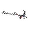
Macromolecule #11: CHLOROPHYLL A

MacromoleculeName: CHLOROPHYLL A / type: ligand / ID: 11 / Number of copies: 82 / Formula: CLA
Molecular weightTheoretical: 893.489 Da
Chemical component information

ChemComp-CLA:
CHLOROPHYLL A / Chlorophyll a

+
Macromolecule #12: PHYLLOQUINONE

MacromoleculeName: PHYLLOQUINONE / type: ligand / ID: 12 / Number of copies: 2 / Formula: PQN
Molecular weightTheoretical: 450.696 Da
Chemical component information

ChemComp-PQN:
PHYLLOQUINONE / Phytomenadione

+
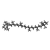
Macromolecule #13: BETA-CAROTENE

MacromoleculeName: BETA-CAROTENE / type: ligand / ID: 13 / Number of copies: 26 / Formula: BCR
Molecular weightTheoretical: 536.873 Da
Chemical component information

ChemComp-BCR:
BETA-CAROTENE / Β-Carotene

+
Macromolecule #14: 1,2-DIPALMITOYL-PHOSPHATIDYL-GLYCEROLE

MacromoleculeName: 1,2-DIPALMITOYL-PHOSPHATIDYL-GLYCEROLE / type: ligand / ID: 14 / Number of copies: 2 / Formula: LHG
Molecular weightTheoretical: 722.97 Da
Chemical component information

ChemComp-LHG:
1,2-DIPALMITOYL-PHOSPHATIDYL-GLYCEROLE / phospholipid*YM / Phosphatidylglycerol

+
Macromolecule #15: IRON/SULFUR CLUSTER

MacromoleculeName: IRON/SULFUR CLUSTER / type: ligand / ID: 15 / Number of copies: 3 / Formula: SF4
Molecular weightTheoretical: 351.64 Da
Chemical component information

ChemComp-FS1:
IRON/SULFUR CLUSTER / Iron–sulfur cluster

+
Macromolecule #16: 1,2-DISTEAROYL-MONOGALACTOSYL-DIGLYCERIDE

MacromoleculeName: 1,2-DISTEAROYL-MONOGALACTOSYL-DIGLYCERIDE / type: ligand / ID: 16 / Number of copies: 1 / Formula: LMG
Molecular weightTheoretical: 787.158 Da
Chemical component information

ChemComp-LMG:
1,2-DISTEAROYL-MONOGALACTOSYL-DIGLYCERIDE

+
Macromolecule #17: water

MacromoleculeName: water / type: ligand / ID: 17 / Number of copies: 5 / Formula: HOH
Molecular weightTheoretical: 18.015 Da
Chemical component information

ChemComp-HOH:
WATER / Water

-
Experimental details

-
Structure determination

Methodcryo EM
Processingsingle particle reconstruction
Aggregation stateparticle

-
Sample preparation

Concentration5 mg/mL
BufferpH: 7.5
Component:
ConcentrationFormulaName
20.0 mMHEPES2-[4-(2-hydroxyethyl)piperazin-1-yl]ethanesulfonic acid
10.0 mMMgCl2Magnesium Chloride
0.0005 %C56H92O25Glyco-diosgenin (GDN)
GridModel: Quantifoil R1.2/1.3 / Material: COPPER / Mesh: 300 / Support film - Material: CARBON / Support film - topology: HOLEY / Support film - Film thickness: 20 / Pretreatment - Type: GLOW DISCHARGE / Pretreatment - Time: 30 sec.
VitrificationCryogen name: ETHANE / Chamber humidity: 95 % / Chamber temperature: 277.15 K / Instrument: FEI VITROBOT MARK IV
DetailsSample was monodisperse.

-
Electron microscopy

MicroscopeJEOL CRYO ARM 200
Electron beamAcceleration voltage: 200 kV / Electron source: FIELD EMISSION GUN
Electron opticsCalibrated defocus max: 3.5 µm / Calibrated defocus min: 0.5 µm / Calibrated magnification: 56497 / Illumination mode: FLOOD BEAM / Imaging mode: BRIGHT FIELDBright-field microscopy / Cs: 1.4 mm / Nominal magnification: 60000
Sample stageSpecimen holder model: JEOL / Cooling holder cryogen: NITROGEN
TemperatureMin: 100.4 K / Max: 100.4 K
Image recordingFilm or detector model: GATAN K2 SUMMIT (4k x 4k) / Detector mode: COUNTING / Digitization - Dimensions - Width: 3710 pixel / Digitization - Dimensions - Height: 3838 pixel / Digitization - Frames/image: 1-60 / Number grids imaged: 1 / Average exposure time: 12.0 sec. / Average electron dose: 1.34 e/Å2
-
Image processing

Particle selectionNumber selected: 182018
Startup modelType of model: OTHER / Details: Initial model by stochastic gradient descent.
Initial angle assignmentType: MAXIMUM LIKELIHOOD / Software - Name: RELION (ver. 3.0)
Final 3D classificationNumber classes: 1 / Avg.num./class: 46105 / Software - Name: RELION (ver. 3.0) / Details: Only one class selected out of four classes.
Final angle assignmentType: MAXIMUM LIKELIHOOD / Software - Name: RELION (ver. 3.0)
Final reconstructionNumber classes used: 1 / Applied symmetry - Point group: C1 (asymmetric) / Algorithm: BACK PROJECTION / Resolution.type: BY AUTHOR / Resolution: 3.2 Å / Resolution method: FSC 0.143 CUT-OFF / Software - Name: RELION (ver. 3.0) / Number images used: 46105
FSC plot (resolution estimation)

-
Atomic model buiding 1

Initial modelPDB ID:

Chain - Source name: PDB / Chain - Initial model type: experimental model
RefinementSpace: REAL / Protocol: OTHER / Overall B value: 45
Output model

PDB-6lu1:
Cyanobacterial PSI Monomer from T. elongatus by Single Particle CRYO-EM at 3.2 A Resolution

+
About Yorodumi

-
News

-
Feb 9, 2022. New format data for meta-information of EMDB entries

New format data for meta-information of EMDB entries

  • Version 3 of the EMDB header file is now the official format.
  • The previous official version 1.9 will be removed from the archive.

Related info.:EMDB header

External links:wwPDB to switch to version 3 of the EMDB data model

-
Aug 12, 2020. Covid-19 info

Covid-19 info

URL: https://pdbj.org/emnavi/covid19.php

New page: Covid-19 featured information page in EM Navigator.

Related info.:Covid-19 info / Mar 5, 2020. Novel coronavirus structure data

+
Mar 5, 2020. Novel coronavirus structure data

Novel coronavirus structure data

Related info.:Yorodumi Speices / Aug 12, 2020. Covid-19 info

External links:COVID-19 featured content - PDBj / Molecule of the Month (242):Coronavirus Proteases

+
Jan 31, 2019. EMDB accession codes are about to change! (news from PDBe EMDB page)

EMDB accession codes are about to change! (news from PDBe EMDB page)

  • The allocation of 4 digits for EMDB accession codes will soon come to an end. Whilst these codes will remain in use, new EMDB accession codes will include an additional digit and will expand incrementally as the available range of codes is exhausted. The current 4-digit format prefixed with “EMD-” (i.e. EMD-XXXX) will advance to a 5-digit format (i.e. EMD-XXXXX), and so on. It is currently estimated that the 4-digit codes will be depleted around Spring 2019, at which point the 5-digit format will come into force.
  • The EM Navigator/Yorodumi systems omit the EMD- prefix.

Related info.:Q: What is EMD? / ID/Accession-code notation in Yorodumi/EM Navigator

External links:EMDB Accession Codes are Changing Soon! / Contact to PDBj

+
Jul 12, 2017. Major update of PDB

Major update of PDB

  • wwPDB released updated PDB data conforming to the new PDBx/mmCIF dictionary.
  • This is a major update changing the version number from 4 to 5, and with Remediation, in which all the entries are updated.
  • In this update, many items about electron microscopy experimental information are reorganized (e.g. em_software).
  • Now, EM Navigator and Yorodumi are based on the updated data.

External links:wwPDB Remediation / Enriched Model Files Conforming to OneDep Data Standards Now Available in the PDB FTP Archive

-
Yorodumi

Thousand views of thousand structures

  • Yorodumi is a browser for structure data from EMDB, PDB, SASBDB, etc.
  • This page is also the successor to EM Navigator detail page, and also detail information page/front-end page for Omokage search.
  • The word "yorodu" (or yorozu) is an old Japanese word meaning "ten thousand". "mi" (miru) is to see.

Related info.:EMDB / PDB / SASBDB / Comparison of 3 databanks / Yorodumi Search / Aug 31, 2016. New EM Navigator & Yorodumi / Yorodumi Papers / Jmol/JSmol / Function and homology information / Changes in new EM Navigator and Yorodumi

Read more