 Movie
Movie Controller

[English] 日本語
 Yorodumi
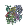
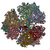
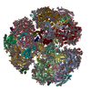
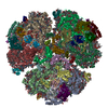
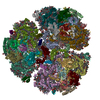
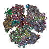
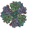
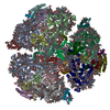
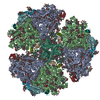
- PDB-1jb0: Crystal Structure of Photosystem I: a Photosynthetic Reaction Cen... -
+ Open data

- Basic information
| Entry | Database: PDB / ID: 1jb0 | |||||||||
|---|---|---|---|---|---|---|---|---|---|---|
| Title | Crystal Structure of Photosystem I: a Photosynthetic Reaction Center and Core Antenna System from Cyanobacteria | |||||||||
 Components Components |
| |||||||||
 Keywords Keywords | PHOTOSYNTHESIS / MEMBRANE PROTEIN / MULTIPROTEIN-PIGMENT COMPLEX | |||||||||
| Function / homology |  Function and homology information Function and homology information: / thylakoid membrane / photosystem I reaction center / photosystem I / photosynthetic electron transport in photosystem I / photosystem I / plasma membrane-derived thylakoid membrane / chlorophyll binding / membrane => GO:0016020 / photosynthesis ...: / thylakoid membrane / photosystem I reaction center / photosystem I / photosynthetic electron transport in photosystem I / photosystem I / plasma membrane-derived thylakoid membrane / chlorophyll binding / membrane => GO:0016020 / photosynthesis / 4 iron, 4 sulfur cluster binding / electron transfer activity / oxidoreductase activity / magnesium ion binding / metal ion binding Similarity search - Function | |||||||||
| Biological species |  Synechococcus elongatus (bacteria) Synechococcus elongatus (bacteria) | |||||||||
| Method |  X-RAY DIFFRACTION / SYNCHROTRON / MIR / Resolution: 2.5 Å X-RAY DIFFRACTION / SYNCHROTRON / MIR / Resolution: 2.5 Å | |||||||||
 Authors Authors | Jordan, P. / Fromme, P. / Witt, H.T. / Klukas, O. / Saenger, W. / Krauss, N. | |||||||||
 Citation Citation | Journal: NATURE / Year: 2001 Title: Three-dimensional Structure of Cyanobacterial Photosystem I at 2.5 A Resolution Authors: Jordan, P. / Fromme, P. / Witt, H.T. / Klukas, O. / Saenger, W. / Krauss, N. #1: Journal: J.Biol.Chem. / Year: 1999Title: PHOTOSYSTEM I, AN IMPROVED MODEL OF THE STROMAL SUBUNITS PSAC, PSAD AND PSAE Authors: Klukas, O. / Schubert, W.D. / Jordan, P. / Krauss, N. / Fromme, P. / Witt, H.T. / Saenger, W. #2: Journal: J.Biol.Chem. / Year: 1999Title: LOCALISATION OF TWO PHYLLOQUINONES, QK AND QK', IN AN IMPROVED ELECTRON DENSITY MAP OF PHOTOSYSTEM I AT 4-A RESOLUTION Authors: Klukas, O. / Schubert, W.D. / Jordan, P. / Krau, N. / Fromme, P. / Witt, H.T. / Saenger, W. #3: Journal: J.Mol.Biol. / Year: 1997Title: PHOTOSYSTEM I OF SYNECHOCOCCUS ELONGATUS AT 4 A RESOLUTION: COMPREHENSIVE STRUCTURE ANALYSIS Authors: Schubert, W.D. / Klukas, O. / Krauss, N. / Saenger, W. / Fromme, P. / Witt, H.T. #4: Journal: Nat.Struct.Biol. / Year: 1996Title: PHOTOSYSTEM I AT 4 A RESOLUTION REPRESENTS THE FIRST STRUCTURAL MODEL OF A JOINT PHOTOSYNTHETIC REACTION CENTRE AND CORE ANTENNA SYSTEM Authors: Krauss, N. / Schubert, W.D. / Klukas, O. / Fromme, P. / Witt, H.T. / Saenger, W. | |||||||||
| History |
|
- Structure visualization
| Structure viewer | Molecule: MolmilJmol/JSmol |
|---|
- Downloads & links
-Download
| PDBx/mmCIF format | 1jb0.cif.gz | 628.8 KB | Display | PDBx/mmCIF format |
|---|---|---|---|---|
| PDB format | pdb1jb0.ent.gz | 537.1 KB | Display | PDB format |
| PDBx/mmJSON format | 1jb0.json.gz | Tree view | PDBx/mmJSON format | |
| Others |  Other downloads Other downloads |
-Validation report
| Arichive directory | https://data.pdbj.org/pub/pdb/validation_reports/jb/1jb0ftp://data.pdbj.org/pub/pdb/validation_reports/jb/1jb0 | HTTPS FTP |
|---|
-Related structure data
| Related structure data | |
|---|---|
| Similar structure data |

-Links
 PDBj
PDBj


- Assembly
| Deposited unit | 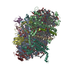
| ||||||||
|---|---|---|---|---|---|---|---|---|---|
| 1 | 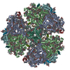
| ||||||||
| Unit cell |
| ||||||||
| Components on special symmetry positions |
|
-Components
-PHOTOSYSTEM I ... , 4 types, 4 molecules ABCX
| #1: Protein | Mass: 83267.773 Da / Num. of mol.: 1 / Source method: isolated from a natural source / Source: (natural) Synechococcus elongatus (bacteria) / References: UniProt: P25896, UniProt: P0A405*PLUS |
|---|---|
| #2: Protein | Mass: 82992.453 Da / Num. of mol.: 1 / Source method: isolated from a natural source / Source: (natural) Synechococcus elongatus (bacteria) / References: UniProt: P25897, UniProt: P0A407*PLUS |
| #3: Protein | Mass: 8678.011 Da / Num. of mol.: 1 / Source method: isolated from a natural source / Source: (natural) Synechococcus elongatus (bacteria) / References: UniProt: P18083, UniProt: P0A415*PLUS |
| #12: Protein/peptide | Mass: 3845.508 Da / Num. of mol.: 1 / Source method: isolated from a natural source / Source: (natural) Synechococcus elongatus (bacteria) / References: UniProt: Q8DKP6*PLUS |
-PHOTOSYSTEM 1 REACTION CENTRE SUBUNIT ... , 8 types, 8 molecules DEFIJKLM
| #4: Protein | Mass: 15258.297 Da / Num. of mol.: 1 / Source method: isolated from a natural source / Source: (natural) Synechococcus elongatus (bacteria) / References: UniProt: P20452, UniProt: P0A420*PLUS |
|---|---|
| #5: Protein | Mass: 8268.290 Da / Num. of mol.: 1 / Source method: isolated from a natural source / Source: (natural) Synechococcus elongatus (bacteria) / References: UniProt: P25898, UniProt: P0A423*PLUS |
| #6: Protein | Mass: 17716.586 Da / Num. of mol.: 1 / Source method: isolated from a natural source / Source: (natural) Synechococcus elongatus (bacteria) / References: UniProt: P25899, UniProt: P0A401*PLUS |
| #7: Protein/peptide | Mass: 4297.234 Da / Num. of mol.: 1 / Source method: isolated from a natural source / Source: (natural) Synechococcus elongatus (bacteria) / References: UniProt: P25900, UniProt: P0A427*PLUS |
| #8: Protein/peptide | Mass: 4770.698 Da / Num. of mol.: 1 / Source method: isolated from a natural source / Source: (natural) Synechococcus elongatus (bacteria) / References: UniProt: P25901, UniProt: P0A429*PLUS |
| #9: Protein | Mass: 8483.983 Da / Num. of mol.: 1 / Source method: isolated from a natural source / Source: (natural) Synechococcus elongatus (bacteria) / References: UniProt: P20453, UniProt: P0A425*PLUS |
| #10: Protein | Mass: 16156.569 Da / Num. of mol.: 1 / Source method: isolated from a natural source / Source: (natural) Synechococcus elongatus (bacteria) / References: UniProt: P25902, UniProt: Q8DGB4*PLUS |
| #11: Protein/peptide | Mass: 3426.115 Da / Num. of mol.: 1 / Source method: isolated from a natural source / Source: (natural) Synechococcus elongatus (bacteria) / References: UniProt: P25903, UniProt: P0A403*PLUS |
-Non-polymers , 8 types, 329 molecules 


| #13: Chemical | ChemComp-CLA /  Mass: 893.489 Da / Num. of mol.: 96 / Source method: obtained synthetically / Formula: C55H72MgN4O5 Mass: 893.489 Da / Num. of mol.: 96 / Source method: obtained synthetically / Formula: C55H72MgN4O5#14: Chemical |  Mass: 450.696 Da / Num. of mol.: 2 / Source method: obtained synthetically / Formula: C31H46O2 Mass: 450.696 Da / Num. of mol.: 2 / Source method: obtained synthetically / Formula: C31H46O2#15: Chemical |  Mass: 351.640 Da / Num. of mol.: 3 / Source method: obtained synthetically / Formula: Fe4S4 Mass: 351.640 Da / Num. of mol.: 3 / Source method: obtained synthetically / Formula: Fe4S4#16: Chemical | ChemComp-BCR /  Mass: 536.873 Da / Num. of mol.: 22 / Source method: obtained synthetically / Formula: C40H56 Mass: 536.873 Da / Num. of mol.: 22 / Source method: obtained synthetically / Formula: C40H56#17: Chemical |  Mass: 722.970 Da / Num. of mol.: 3 / Source method: obtained synthetically / Formula: C38H75O10P / Comment: phospholipid*YM Mass: 722.970 Da / Num. of mol.: 3 / Source method: obtained synthetically / Formula: C38H75O10P / Comment: phospholipid*YM#18: Chemical | ChemComp-LMG / |  Mass: 787.158 Da / Num. of mol.: 1 / Source method: obtained synthetically / Formula: C45H86O10 Mass: 787.158 Da / Num. of mol.: 1 / Source method: obtained synthetically / Formula: C45H86O10#19: Chemical | ChemComp-CA / |  Mass: 40.078 Da / Num. of mol.: 1 / Source method: obtained synthetically / Formula: Ca Mass: 40.078 Da / Num. of mol.: 1 / Source method: obtained synthetically / Formula: Ca#20: Water | ChemComp-HOH / | Mass: 18.015 Da / Num. of mol.: 201 / Source method: isolated from a natural source / Formula: H2O |
|---|
-Details
| Has protein modification | Y |
|---|
-Experimental details
-Experiment
| Experiment | Method: X-RAY DIFFRACTION / Number of used crystals: 2 |
|---|
- Sample preparation
| Crystal | Density Matthews: 7.32 Å3/Da / Density % sol: 83.2 % | ||||||||||||||||||
|---|---|---|---|---|---|---|---|---|---|---|---|---|---|---|---|---|---|---|---|
| Crystal grow | Temperature: 277 K / Method: microdialysis / pH: 6.4 Details: beta-dodecylmaltoside, magnesium sulfate, pH 6.4, MICRODIALYSIS, temperature 277K | ||||||||||||||||||
| Crystal grow | *PLUS Method: batch methodDetails: Fromme, P., (1998) Biochim. Biophys. Acta, 1365, 175. | ||||||||||||||||||
| Components of the solutions | *PLUS
|
-Data collection
| Diffraction |
| ||||||||||||||||||
|---|---|---|---|---|---|---|---|---|---|---|---|---|---|---|---|---|---|---|---|
| Diffraction source |
| ||||||||||||||||||
| Detector |
| ||||||||||||||||||
| Radiation |
| ||||||||||||||||||
| Radiation wavelength | Wavelength: 0.99 Å / Relative weight: 1 | ||||||||||||||||||
| Reflection | Resolution: 2.5→30 Å / Num. all: 246961 / Num. obs: 246961 / % possible obs: 97.1 % / Observed criterion σ(F): 0 / Observed criterion σ(I): -3 / Redundancy: 3.6 % / Biso Wilson estimate: 48 Å2 / Rmerge(I) obs: 0.064 / Net I/σ(I): 14 | ||||||||||||||||||
| Reflection shell | Resolution: 2.5→2.59 Å / Redundancy: 2.2 % / Rmerge(I) obs: 0.248 / Mean I/σ(I) obs: 2.9 / Num. unique all: 22495 / % possible all: 88.9 | ||||||||||||||||||
| Reflection | *PLUS | ||||||||||||||||||
| Reflection shell | *PLUS % possible obs: 88.9 % |

- Processing
| Software |
| |||||||||||||||||||||||||
|---|---|---|---|---|---|---|---|---|---|---|---|---|---|---|---|---|---|---|---|---|---|---|---|---|---|---|
| Refinement | Method to determine structure: MIR / Resolution: 2.5→30 Å / Isotropic thermal model: isotropic / Cross valid method: THROUGHOUT / σ(F): 0 / σ(I): 0 / Stereochemistry target values: Engh & Huber
| |||||||||||||||||||||||||
| Solvent computation | Solvent model: BULK SOLVENT CORRECTION HAS BEEN APPLIED IN CNS | |||||||||||||||||||||||||
| Displacement parameters | Biso mean: 46.7 Å2 | |||||||||||||||||||||||||
| Refine analyze |
| |||||||||||||||||||||||||
| Refinement step | Cycle: LAST / Resolution: 2.5→30 Å
| |||||||||||||||||||||||||
| Refine LS restraints |
| |||||||||||||||||||||||||
| LS refinement shell |
| |||||||||||||||||||||||||
| Software | *PLUS Name: CNS / Version: 0.9 / Classification: refinement | |||||||||||||||||||||||||
| Refinement | *PLUS Highest resolution: 2.5 Å / Lowest resolution: 30 Å / σ(F): 0 | |||||||||||||||||||||||||
| Solvent computation | *PLUS | |||||||||||||||||||||||||
| Displacement parameters | *PLUS | |||||||||||||||||||||||||
| Refine LS restraints | *PLUS
| |||||||||||||||||||||||||
| LS refinement shell | *PLUS Rfactor Rfree: 0.287 / Rfactor Rwork: 0.296 |
