 Movie
Movie Controller

[English] 日本語
 Yorodumi
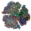
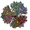
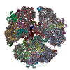
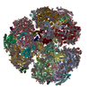
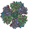
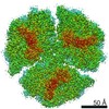
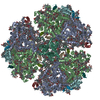
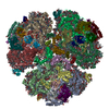
- PDB-1c51: PHOTOSYNTHETIC REACTION CENTER AND CORE ANTENNA SYSTEM (TRIMERIC)... -
+ Open data

- Basic information
| Entry | Database: PDB / ID: 1c51 | |||||||||
|---|---|---|---|---|---|---|---|---|---|---|
| Title | PHOTOSYNTHETIC REACTION CENTER AND CORE ANTENNA SYSTEM (TRIMERIC), ALPHA CARBON ONLY | |||||||||
 Components Components | (PROTEIN (PHOTOSYSTEM I: SUBUNIT ...) x 8 | |||||||||
 Keywords Keywords | PHOTOSYNTHESIS/ELECTRON TRANSPORT / PHOTOSYNTHESIS / PHOTOSYNTHETIC REACTION CENTER / OXYGENIC PHOTOSYNTHESIS / CORE- ANTENNA LIGHT-HARVESTING SYSTEM / THERMOPHILIC CYANOBACTERIUM / HELIX-BUNDLE MEMBRANE PROTEIN / PHOTOSYNTHESIS-ELECTRON TRANSPORT COMPLEX | |||||||||
| Function / homology | CHLOROPHYLL A / PHYLLOQUINONE / IRON/SULFUR CLUSTER Function and homology information Function and homology information | |||||||||
| Biological species |  Synechococcus elongatus (bacteria) Synechococcus elongatus (bacteria) | |||||||||
| Method |  X-RAY DIFFRACTION / SYNCHROTRON / MIRAS / Resolution: 4 Å X-RAY DIFFRACTION / SYNCHROTRON / MIRAS / Resolution: 4 Å | |||||||||
 Authors Authors | Klukas, O. / Schubert, W.D. / Jordan, P. / Krauss, N. / Fromme, P. / Witt, H.T. / Saenger, W. | |||||||||
 Citation Citation | Journal: J.Biol.Chem. / Year: 1999 Title: Photosystem I, an improved model of the stromal subunits PsaC, PsaD, and PsaE. Authors: Klukas, O. / Schubert, W.D. / Jordan, P. / Krauss, N. / Fromme, P. / Witt, H.T. / Saenger, W. #1: Journal: J.Biol.Chem. / Year: 1999Title: Localisation of Two Phylloquinones, Qk and Qk', in an Improved Electron Density Map of Photosystem I at 4-A Resolution Authors: Klukas, O. / Schubert, W.-D. / Jordan, P. / Krauss, N. / Fromme, P. / Witt, H.T. / Saenger, W. #2: Journal: J.Mol.Biol. / Year: 1997Title: Photosystem I of Synechococcus Elongatus at 4 A Resolution: Comprehensive Structure Analysis Authors: Schubert, W.-D. / Klukas, O. / Krauss, N. / Saenger, W. / Fromme, P. / Witt, H.T. #3: Journal: Nat.Struct.Biol. / Year: 1996Title: Photosystem I at 4 A Resolution Represents the First Structural Model of a Joint Photosynthetic Reaction Centre and Core Antenna System Authors: Krauss, N. / Schubert, W.-D. / Klukas, O. / Fromme, P. / Witt, H.T. / Saenger, W. #4: Journal: Nature / Year: 1993Title: Three-Dimensional Structure of System I of Photosynthesis at 6A Resolution Authors: Krauss, N. / Hinrichs, W. / Witt, I. / Fromme, P. / Pritzkow, W. / Dauter, Z. / Betzel, C. / Wilson, K.S. / Witt, H.T. / Saenger, W. #5: Journal: Ber.Bunsenges.Phys.Chem. / Year: 1988Title: X-Ray Characterization of Single Crystals of the Reaction Center I of Water Splitting Photosynthesis Authors: Witt, I. / Witt, H.T. / Di Fiore, D. / Rogner, M. / Hinrichs, W. / Saenger, W. / Granzin, J. / Betzel, C. / Dauter, Z. | |||||||||
| History |
|
- Structure visualization
| Structure viewer | Molecule: MolmilJmol/JSmol |
|---|
- Downloads & links
-Download
| PDBx/mmCIF format | 1c51.cif.gz | 120.6 KB | Display | PDBx/mmCIF format |
|---|---|---|---|---|
| PDB format | pdb1c51.ent.gz | 81.7 KB | Display | PDB format |
| PDBx/mmJSON format | 1c51.json.gz | Tree view | PDBx/mmJSON format | |
| Others |  Other downloads Other downloads |
-Validation report
| Arichive directory | https://data.pdbj.org/pub/pdb/validation_reports/c5/1c51ftp://data.pdbj.org/pub/pdb/validation_reports/c5/1c51 | HTTPS FTP |
|---|
-Related structure data
| Similar structure data |
|---|
-Links
 PDBj
PDBj


- Assembly
| Deposited unit | 
| ||||||||
|---|---|---|---|---|---|---|---|---|---|
| 1 | 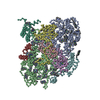
| ||||||||
| Unit cell |
|
-Components
-PROTEIN (PHOTOSYSTEM I: SUBUNIT ... , 8 types, 8 molecules ABCDEFKL
| #1: Protein | Mass: 50825.766 Da / Num. of mol.: 1 / Source method: isolated from a natural source / Source: (natural) Synechococcus elongatus (bacteria) / Cellular location: THYLAKOID MEMBRANE |
|---|---|
| #2: Protein | Mass: 52698.094 Da / Num. of mol.: 1 / Source method: isolated from a natural source / Source: (natural) Synechococcus elongatus (bacteria) / Cellular location: THYLAKOID MEMBRANE |
| #3: Protein | Mass: 6571.091 Da / Num. of mol.: 1 / Source method: isolated from a natural source / Source: (natural) Synechococcus elongatus (bacteria) / Cellular location: THYLAKOID MEMBRANE |
| #4: Protein | Mass: 10656.127 Da / Num. of mol.: 1 / Source method: isolated from a natural source / Source: (natural) Synechococcus elongatus (bacteria) / Cellular location: THYLAKOID MEMBRANE |
| #5: Protein | Mass: 6400.881 Da / Num. of mol.: 1 / Source method: isolated from a natural source / Source: (natural) Synechococcus elongatus (bacteria) / Cellular location: THYLAKOID MEMBRANE |
| #6: Protein | Mass: 13039.064 Da / Num. of mol.: 1 / Source method: isolated from a natural source / Source: (natural) Synechococcus elongatus (bacteria) / Cellular location: THYLAKOID MEMBRANE |
| #7: Protein | Mass: 6656.196 Da / Num. of mol.: 1 / Source method: isolated from a natural source / Source: (natural) Synechococcus elongatus (bacteria) / Cellular location: THYLAKOID MEMBRANE |
| #8: Protein | Mass: 10230.603 Da / Num. of mol.: 1 / Source method: isolated from a natural source / Source: (natural) Synechococcus elongatus (bacteria) / Cellular location: THYLAKOID MEMBRANE |
-Non-polymers , 3 types, 77 molecules 


| #9: Chemical | ChemComp-CLA /  Mass: 893.489 Da / Num. of mol.: 72 / Source method: obtained synthetically / Formula: C55H72MgN4O5 Mass: 893.489 Da / Num. of mol.: 72 / Source method: obtained synthetically / Formula: C55H72MgN4O5#10: Chemical |  Mass: 450.696 Da / Num. of mol.: 2 / Source method: obtained synthetically / Formula: C31H46O2 Mass: 450.696 Da / Num. of mol.: 2 / Source method: obtained synthetically / Formula: C31H46O2#11: Chemical |  Mass: 351.640 Da / Num. of mol.: 3 / Source method: obtained synthetically / Formula: Fe4S4 Mass: 351.640 Da / Num. of mol.: 3 / Source method: obtained synthetically / Formula: Fe4S4 |
|---|
-Details
| Compound details | CHAIN A COMPRISES ONE OF THE TWO LARGE, CENTRAL, MEMBRANE INTEGRAL SUBUNITS OF THE REACTION CENTER ...CHAIN A COMPRISES ONE OF THE TWO LARGE, CENTRAL, MEMBRANE INTEGRAL SUBUNITS OF THE REACTION CENTER OF PSI - EITHER PSAA OR PSAB. CHAIN B COMPRISES THE SECOND OF THE TWO LARGE, CENTRAL, MEMBRANE INTEGRAL SUBUNITS OF THE REACTION CENTER OF PSI - EITHER PSAB OR PSAA. CHAIN C COMPRISES THE MODEL OF THE STROMAL EXTRINSIC SUBUNIT OF PSI - PSAC. CHAIN F COMPRISES THOSE SECTIONS OF THE PRESENT MODEL LOCATED DISTAL TO THE TRIMERIZAT |
|---|---|
| Nonpolymer details | THE ATOM NAMING OF THE CHLOROPHYLL MOLECULES IS ARBITRARY IN THE SENSE THAT THE AUTHORS DO NOT WISH ...THE ATOM NAMING OF THE CHLOROPHYL |
-Experimental details
-Experiment
| Experiment | Method: X-RAY DIFFRACTION / Number of used crystals: 6 |
|---|
- Sample preparation
| Crystal | Density Matthews: 5.8 Å3/Da / Density % sol: 80 % Description: DATA WERE COLLECTED AT DIFFERENT SYNCHROTRONS, SEE FIELD 'BEAMLINE' | ||||||||||||||||||
|---|---|---|---|---|---|---|---|---|---|---|---|---|---|---|---|---|---|---|---|
| Crystal grow | pH: 6.4 / Details: pH 6.4 | ||||||||||||||||||
| Crystal grow | *PLUS Method: batch methodDetails: Fromme, P., (1998) Biochim. Biophys. Acta, 1365, 175. | ||||||||||||||||||
| Components of the solutions | *PLUS
|
-Data collection
| Diffraction | Mean temperature: 277 K |
|---|---|
| Diffraction source | Source: SYNCHROTRON / Site: MPG/DESY, HAMBURG  / Beamline: BW6 / Wavelength: 0.9 / Beamline: BW6 / Wavelength: 0.9 |
| Detector | Type: MAR scanner 300 mm plate / Detector: IMAGE PLATE / Date: Jul 1, 1996 |
| Radiation | Protocol: SINGLE WAVELENGTH / Monochromatic (M) / Laue (L): M / Scattering type: x-ray |
| Radiation wavelength | Wavelength: 0.9 Å / Relative weight: 1 |
| Reflection | Resolution: 3.5→60 Å / Num. obs: 89999 / % possible obs: 93.4 % / Observed criterion σ(I): -3 / Redundancy: 4.8 % / Rmerge(I) obs: 0.107 / Net I/σ(I): 11.2 |
| Reflection shell | Resolution: 3.5→3.56 Å / Redundancy: 2.1 % / Rmerge(I) obs: 0.243 / Mean I/σ(I) obs: 4.2 / % possible all: 78.9 |
- Processing
| Software |
| ||||||||||||
|---|---|---|---|---|---|---|---|---|---|---|---|---|---|
| Refinement | Method to determine structure: MIRAS / Resolution: 4→60 Å / Details: NO REFINEMENT WAS CARRIED OUT ON THE MODEL | ||||||||||||
| Refinement step | Cycle: LAST / Resolution: 4→60 Å
| ||||||||||||
| Refinement | *PLUS Highest resolution: 4 Å / Lowest resolution: 60 Å | ||||||||||||
| Solvent computation | *PLUS | ||||||||||||
| Displacement parameters | *PLUS |
