 Movie
Movie Controller

[English] 日本語
 Yorodumi
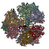
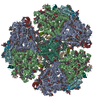
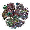
- PDB-6pfy: Membrane Protein Megahertz Crystallography at the European XFEL, ... -
+ Open data

- Basic information
| Entry | Database: PDB / ID: 6pfy | |||||||||
|---|---|---|---|---|---|---|---|---|---|---|
| Title | Membrane Protein Megahertz Crystallography at the European XFEL, Photosystem I at synchrotron to 2.9 A | |||||||||
 Components Components | (Photosystem I ...) x 12 | |||||||||
 Keywords Keywords | PHOTOSYNTHESIS / Light Harvesting / photosystem I | |||||||||
| Function / homology |  Function and homology information Function and homology informationphotosystem I reaction center / photosystem I / photosynthetic electron transport in photosystem I / photosystem I / plasma membrane-derived thylakoid membrane / chlorophyll binding / photosynthesis / 4 iron, 4 sulfur cluster binding / electron transfer activity / oxidoreductase activity ...photosystem I reaction center / photosystem I / photosynthetic electron transport in photosystem I / photosystem I / plasma membrane-derived thylakoid membrane / chlorophyll binding / photosynthesis / 4 iron, 4 sulfur cluster binding / electron transfer activity / oxidoreductase activity / magnesium ion binding / metal ion binding Similarity search - Function | |||||||||
| Biological species |   Thermosynechococcus elongatus (bacteria) Thermosynechococcus elongatus (bacteria) | |||||||||
| Method |  X-RAY DIFFRACTION / SYNCHROTRON / MOLECULAR REPLACEMENT / Resolution: 2.9 Å X-RAY DIFFRACTION / SYNCHROTRON / MOLECULAR REPLACEMENT / Resolution: 2.9 Å | |||||||||
 Authors Authors | Fromme, R. | |||||||||
| Funding support |  United States, 1items United States, 1items
| |||||||||
 Citation Citation | Journal: Nat Commun / Year: 2019 Title: Membrane protein megahertz crystallography at the European XFEL. Authors: Gisriel, C. / Coe, J. / Letrun, R. / Yefanov, O.M. / Luna-Chavez, C. / Stander, N.E. / Lisova, S. / Mariani, V. / Kuhn, M. / Aplin, S. / Grant, T.D. / Dorner, K. / Sato, T. / Echelmeier, A. ...Authors: Gisriel, C. / Coe, J. / Letrun, R. / Yefanov, O.M. / Luna-Chavez, C. / Stander, N.E. / Lisova, S. / Mariani, V. / Kuhn, M. / Aplin, S. / Grant, T.D. / Dorner, K. / Sato, T. / Echelmeier, A. / Cruz Villarreal, J. / Hunter, M.S. / Wiedorn, M.O. / Knoska, J. / Mazalova, V. / Roy-Chowdhury, S. / Yang, J.H. / Jones, A. / Bean, R. / Bielecki, J. / Kim, Y. / Mills, G. / Weinhausen, B. / Meza, J.D. / Al-Qudami, N. / Bajt, S. / Brehm, G. / Botha, S. / Boukhelef, D. / Brockhauser, S. / Bruce, B.D. / Coleman, M.A. / Danilevski, C. / Discianno, E. / Dobson, Z. / Fangohr, H. / Martin-Garcia, J.M. / Gevorkov, Y. / Hauf, S. / Hosseinizadeh, A. / Januschek, F. / Ketawala, G.K. / Kupitz, C. / Maia, L. / Manetti, M. / Messerschmidt, M. / Michelat, T. / Mondal, J. / Ourmazd, A. / Previtali, G. / Sarrou, I. / Schon, S. / Schwander, P. / Shelby, M.L. / Silenzi, A. / Sztuk-Dambietz, J. / Szuba, J. / Turcato, M. / White, T.A. / Wrona, K. / Xu, C. / Abdellatif, M.H. / Zook, J.D. / Spence, J.C.H. / Chapman, H.N. / Barty, A. / Kirian, R.A. / Frank, M. / Ros, A. / Schmidt, M. / Fromme, R. / Mancuso, A.P. / Fromme, P. / Zatsepin, N.A. #1: Journal: Nat Commun / Year: 2019Title: Membrane protein megahertz crystallography at the European XFEL Authors: Gisriel, C. / Coe, J. / Letrun, R. / Luna-Chavez, C. / Stander, N.E. / Roy-Chauhundry, S. / Lisova, S. / Mariani, V. / Kuhn, M. / Aplin, S. / Grant, T.D. / Doerner, K. / Sato, T. / ...Authors: Gisriel, C. / Coe, J. / Letrun, R. / Luna-Chavez, C. / Stander, N.E. / Roy-Chauhundry, S. / Lisova, S. / Mariani, V. / Kuhn, M. / Aplin, S. / Grant, T.D. / Doerner, K. / Sato, T. / Echelmeier, A. / Villareal, J. / Hunter, M.S. / Wiedorn, M. / Knoska, J. / Mazalova, V. / Yang, J.-H. / Jones, A. / Bean, R. / Bielecki, J. / Kim, Y. / Mills, G. / Weinhausen, B. / Meza, J.D. / Al-Quadami, N. / Bajt, S. / Brehm, G. / Botha, S. / Boukhelef, D. / Brockhauser, S. / Bruse, B.D. / Coleman, M.A. / Danilevski, C. / Discianno, E. / Dobson, Z. / Hosseinizadeh, H. / Januschek, F. / Ketawala, G. / Kupitz, C. / Maia, L. / Manetti, M. / Messerschmidt, M. / Michalat, T. / Mondal, J. / Ourmazd, A. / Previtali, G. / Sarrou, I. / Schoen, S. / Schwander, P. / Shelby, M.L. / Silenzi, A. / Sztuk-Dambietz, J. / Szuba, J. / Turcato, M. / White, T.A. / Wrona, K. / Xu, C. / Abdellatif, M.H. / Zook, J.D. / Spence, J.C.H. / Chapman, H.N. / Barty, A. / Kirian, R.A. / Frank, M. / Ros, A. / Schmidt, M. / Fromme, R. / Manusco, A.P. / Fromme, P. / Zatsepin, N.A. | |||||||||
| History |
|
- Structure visualization
| Structure viewer | Molecule: MolmilJmol/JSmol |
|---|
- Downloads & links
-Download
| PDBx/mmCIF format | 6pfy.cif.gz | 1.8 MB | Display | PDBx/mmCIF format |
|---|---|---|---|---|
| PDB format | pdb6pfy.ent.gz | 1.5 MB | Display | PDB format |
| PDBx/mmJSON format | 6pfy.json.gz | Tree view | PDBx/mmJSON format | |
| Others |  Other downloads Other downloads |
-Validation report
| Arichive directory | https://data.pdbj.org/pub/pdb/validation_reports/pf/6pfyftp://data.pdbj.org/pub/pdb/validation_reports/pf/6pfy | HTTPS FTP |
|---|
-Related structure data
| Related structure data |  6pgkC 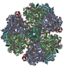 1jb0S S: Starting model for refinement C: citing same article ( |
|---|---|
| Similar structure data |

-Links
 PDBj
PDBj


- Assembly
| Deposited unit | 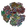
| ||||||||
|---|---|---|---|---|---|---|---|---|---|
| 1 |
| ||||||||
| Unit cell |
|
-Components
-Photosystem I ... , 12 types, 36 molecules AGYBHZCNaDObEPcFQdIReJSfKTgLUh...
| #1: Protein | Mass: 83267.773 Da / Num. of mol.: 3 / Source method: isolated from a natural source Source: (natural) Thermosynechococcus elongatus (strain BP-1) (bacteria)Strain: BP-1 / References: UniProt: P0A405, photosystem I #2: Protein | Mass: 83123.648 Da / Num. of mol.: 3 / Source method: isolated from a natural source Source: (natural) Thermosynechococcus elongatus (strain BP-1) (bacteria)Strain: BP-1 / References: UniProt: P0A407, photosystem I #3: Protein | Mass: 8809.207 Da / Num. of mol.: 3 / Source method: isolated from a natural source Source: (natural) Thermosynechococcus elongatus (strain BP-1) (bacteria)Strain: BP-1 / References: UniProt: P0A415, photosystem I #4: Protein | Mass: 15389.494 Da / Num. of mol.: 3 / Source method: isolated from a natural source Source: (natural) Thermosynechococcus elongatus (strain BP-1) (bacteria)Strain: BP-1 / References: UniProt: P0A420 #5: Protein | Mass: 8399.485 Da / Num. of mol.: 3 / Source method: isolated from a natural source Source: (natural) Thermosynechococcus elongatus (strain BP-1) (bacteria)Strain: BP-1 / References: UniProt: P0A423 #6: Protein | Mass: 17716.586 Da / Num. of mol.: 3 / Source method: isolated from a natural source Source: (natural) Thermosynechococcus elongatus (strain BP-1) (bacteria)Strain: BP-1 / References: UniProt: P0A401 #7: Protein/peptide | Mass: 4297.234 Da / Num. of mol.: 3 / Source method: isolated from a natural source Source: (natural) Thermosynechococcus elongatus (strain BP-1) (bacteria)Strain: BP-1 / References: UniProt: P0A427 #8: Protein/peptide | Mass: 4770.698 Da / Num. of mol.: 3 / Source method: isolated from a natural source Source: (natural) Thermosynechococcus elongatus (strain BP-1) (bacteria)Strain: BP-1 / References: UniProt: P0A429 #9: Protein | Mass: 8483.983 Da / Num. of mol.: 3 / Source method: isolated from a natural source Source: (natural) Thermosynechococcus elongatus (strain BP-1) (bacteria)Strain: BP-1 / References: UniProt: P0A425 #10: Protein | Mass: 16287.765 Da / Num. of mol.: 3 / Source method: isolated from a natural source Source: (natural) Thermosynechococcus elongatus (strain BP-1) (bacteria)Strain: BP-1 / References: UniProt: Q8DGB4 #11: Protein/peptide | Mass: 3426.115 Da / Num. of mol.: 3 / Source method: isolated from a natural source Source: (natural) Thermosynechococcus elongatus (strain BP-1) (bacteria)Strain: BP-1 / References: UniProt: P0A403 #12: Protein/peptide | Mass: 4424.317 Da / Num. of mol.: 3 / Source method: isolated from a natural source Source: (natural) Thermosynechococcus elongatus (strain BP-1) (bacteria)Strain: BP-1 / References: UniProt: Q8DKP6 |
|---|
-Non-polymers , 9 types, 657 molecules 


| #13: Chemical |  Mass: 893.489 Da / Num. of mol.: 3 / Source method: obtained synthetically / Formula: C55H72MgN4O5 / Feature type: SUBJECT OF INVESTIGATION Mass: 893.489 Da / Num. of mol.: 3 / Source method: obtained synthetically / Formula: C55H72MgN4O5 / Feature type: SUBJECT OF INVESTIGATION#14: Chemical | ChemComp-CLA /  Mass: 893.489 Da / Num. of mol.: 282 / Source method: obtained synthetically / Formula: C55H72MgN4O5 / Feature type: SUBJECT OF INVESTIGATION Mass: 893.489 Da / Num. of mol.: 282 / Source method: obtained synthetically / Formula: C55H72MgN4O5 / Feature type: SUBJECT OF INVESTIGATION#15: Chemical | ChemComp-PQN /  Mass: 450.696 Da / Num. of mol.: 6 / Source method: obtained synthetically / Formula: C31H46O2 / Feature type: SUBJECT OF INVESTIGATION Mass: 450.696 Da / Num. of mol.: 6 / Source method: obtained synthetically / Formula: C31H46O2 / Feature type: SUBJECT OF INVESTIGATION#16: Chemical | ChemComp-SF4 /  Mass: 351.640 Da / Num. of mol.: 9 / Source method: obtained synthetically / Formula: Fe4S4 / Feature type: SUBJECT OF INVESTIGATION Mass: 351.640 Da / Num. of mol.: 9 / Source method: obtained synthetically / Formula: Fe4S4 / Feature type: SUBJECT OF INVESTIGATION#17: Chemical | ChemComp-BCR /  Mass: 536.873 Da / Num. of mol.: 66 / Source method: obtained synthetically / Formula: C40H56 / Feature type: SUBJECT OF INVESTIGATION Mass: 536.873 Da / Num. of mol.: 66 / Source method: obtained synthetically / Formula: C40H56 / Feature type: SUBJECT OF INVESTIGATION#18: Chemical | ChemComp-LHG /  Mass: 722.970 Da / Num. of mol.: 9 / Source method: obtained synthetically / Formula: C38H75O10P / Feature type: SUBJECT OF INVESTIGATION / Comment: phospholipid*YM Mass: 722.970 Da / Num. of mol.: 9 / Source method: obtained synthetically / Formula: C38H75O10P / Feature type: SUBJECT OF INVESTIGATION / Comment: phospholipid*YM#19: Chemical |  Mass: 787.158 Da / Num. of mol.: 3 / Source method: obtained synthetically / Formula: C45H86O10 / Feature type: SUBJECT OF INVESTIGATION Mass: 787.158 Da / Num. of mol.: 3 / Source method: obtained synthetically / Formula: C45H86O10 / Feature type: SUBJECT OF INVESTIGATION#20: Chemical |  Mass: 40.078 Da / Num. of mol.: 3 / Source method: obtained synthetically / Formula: Ca / Feature type: SUBJECT OF INVESTIGATION Mass: 40.078 Da / Num. of mol.: 3 / Source method: obtained synthetically / Formula: Ca / Feature type: SUBJECT OF INVESTIGATION#21: Water | ChemComp-HOH / | Mass: 18.015 Da / Num. of mol.: 276 / Source method: isolated from a natural source / Formula: H2O |
|---|
-Details
| Has ligand of interest | Y |
|---|---|
| Has protein modification | Y |
-Experimental details
-Experiment
| Experiment | Method: X-RAY DIFFRACTION / Number of used crystals: 1 |
|---|
- Sample preparation
| Crystal | Density % sol: 79 % / Description: Rod shape 200 x 200 by 400 um |
|---|---|
| Crystal grow | Temperature: 283 K / Method: microdialysis / pH: 6.4 / Details: MES 5 mM 5- 30 mM MgSO4 0.02 % beta-DDM |
-Data collection
| Diffraction | Mean temperature: 100 K / Serial crystal experiment: N |
|---|---|
| Diffraction source | Source: SYNCHROTRON / Site: ALS / Beamline: 5.0.2 / Wavelength: 0.987 Å |
| Detector | Type: ADSC QUANTUM 315 / Detector: CCD / Date: Apr 8, 2005 |
| Radiation | Protocol: SINGLE WAVELENGTH / Monochromatic (M) / Laue (L): M / Scattering type: x-ray |
| Radiation wavelength | Wavelength: 0.987 Å / Relative weight: 1 |
| Reflection | Resolution: 2.9→48.87 Å / Num. obs: 492002 / % possible obs: 99.7 % / Redundancy: 3.6 % / CC1/2: 0.966 / Rmerge(I) obs: 0.136 / Χ2: 1.06 / Net I/σ(I): 7.1 |
| Reflection shell | Resolution: 2.9→2.95 Å / Redundancy: 3.5 % / Rmerge(I) obs: 0.481 / Mean I/σ(I) obs: 2.5 / Num. unique obs: 24360 / CC1/2: 0.684 / Χ2: 0.84 / % possible all: 100 |
- Processing
| Software |
| ||||||||||||||||||||||||||||||||||||||||||||||||||||||||||||||||||||||||||||||||||||||||||||||||||||||||||||||||||||||||||||||||||||||||||||||||||||||||||||||||||||||||||||||||||||||
|---|---|---|---|---|---|---|---|---|---|---|---|---|---|---|---|---|---|---|---|---|---|---|---|---|---|---|---|---|---|---|---|---|---|---|---|---|---|---|---|---|---|---|---|---|---|---|---|---|---|---|---|---|---|---|---|---|---|---|---|---|---|---|---|---|---|---|---|---|---|---|---|---|---|---|---|---|---|---|---|---|---|---|---|---|---|---|---|---|---|---|---|---|---|---|---|---|---|---|---|---|---|---|---|---|---|---|---|---|---|---|---|---|---|---|---|---|---|---|---|---|---|---|---|---|---|---|---|---|---|---|---|---|---|---|---|---|---|---|---|---|---|---|---|---|---|---|---|---|---|---|---|---|---|---|---|---|---|---|---|---|---|---|---|---|---|---|---|---|---|---|---|---|---|---|---|---|---|---|---|---|---|---|---|
| Refinement | Method to determine structure: MOLECULAR REPLACEMENT Starting model: 1jb0 Resolution: 2.9→48.87 Å / Cor.coef. Fo:Fc: 0.765 / Cor.coef. Fo:Fc free: 0.698 / SU B: 25.1 / SU ML: 0.48 / Cross valid method: THROUGHOUT / ESU R: 0.576 / ESU R Free: 0.4 / Details: HYDROGENS HAVE BEEN ADDED IN THE RIDING POSITIONS
| ||||||||||||||||||||||||||||||||||||||||||||||||||||||||||||||||||||||||||||||||||||||||||||||||||||||||||||||||||||||||||||||||||||||||||||||||||||||||||||||||||||||||||||||||||||||
| Solvent computation | Ion probe radii: 0.8 Å / Shrinkage radii: 0.8 Å / VDW probe radii: 1.2 Å | ||||||||||||||||||||||||||||||||||||||||||||||||||||||||||||||||||||||||||||||||||||||||||||||||||||||||||||||||||||||||||||||||||||||||||||||||||||||||||||||||||||||||||||||||||||||
| Displacement parameters | Biso mean: 43.362 Å2
| ||||||||||||||||||||||||||||||||||||||||||||||||||||||||||||||||||||||||||||||||||||||||||||||||||||||||||||||||||||||||||||||||||||||||||||||||||||||||||||||||||||||||||||||||||||||
| Refinement step | Cycle: 1 / Resolution: 2.9→48.87 Å
| ||||||||||||||||||||||||||||||||||||||||||||||||||||||||||||||||||||||||||||||||||||||||||||||||||||||||||||||||||||||||||||||||||||||||||||||||||||||||||||||||||||||||||||||||||||||
| Refine LS restraints |
|