 Movie
Movie Controller

[English] 日本語
 Yorodumi
- PDB-5zf0: X-ray Structure of the Electron Transfer Complex between Ferredox... -
+ Open data

- Basic information
| Entry | Database: PDB / ID: 5zf0 | ||||||||||||
|---|---|---|---|---|---|---|---|---|---|---|---|---|---|
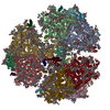
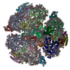
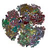
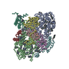
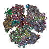
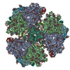
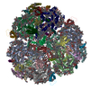
| Title | X-ray Structure of the Electron Transfer Complex between Ferredoxin and Photosystem I | ||||||||||||
 Components Components |
| ||||||||||||
 Keywords Keywords | PHOTOSYNTHESIS/ELECTRON TRANSPORT / Photosystem I / Ferredoxin / PHOTOSYNTHESIS-ELECTRON TRANSPORT complex | ||||||||||||
| Function / homology |  Function and homology information Function and homology informationphotosystem I reaction center / photosystem I / photosynthetic electron transport in photosystem I / photosystem I / plasma membrane-derived thylakoid membrane / chlorophyll binding / photosynthesis / electron transport chain / 2 iron, 2 sulfur cluster binding / 4 iron, 4 sulfur cluster binding ...photosystem I reaction center / photosystem I / photosynthetic electron transport in photosystem I / photosystem I / plasma membrane-derived thylakoid membrane / chlorophyll binding / photosynthesis / electron transport chain / 2 iron, 2 sulfur cluster binding / 4 iron, 4 sulfur cluster binding / electron transfer activity / oxidoreductase activity / magnesium ion binding / metal ion binding Similarity search - Function | ||||||||||||
| Biological species |   Thermosynechococcus elongatus BP-1 (bacteria)Thermosynechococcus elongatus (bacteria) Thermosynechococcus elongatus BP-1 (bacteria)Thermosynechococcus elongatus (bacteria) | ||||||||||||
| Method |  X-RAY DIFFRACTION / SYNCHROTRON / MOLECULAR REPLACEMENT / Resolution: 4.2 Å X-RAY DIFFRACTION / SYNCHROTRON / MOLECULAR REPLACEMENT / Resolution: 4.2 Å | ||||||||||||
 Authors Authors | Kubota-Kawai, H. / Mutoh, R. / Shinmura, K. / Setif, P. / Nowaczyk, M. / Roegner, M. / Ikegami, T. / Tanaka, T. / Kurisu, G. | ||||||||||||
| Funding support |  Japan, 3items Japan, 3items
| ||||||||||||
 Citation Citation | Journal: Nat Plants / Year: 2018 Title: X-ray structure of an asymmetrical trimeric ferredoxin-photosystem I complex Authors: Kubota-Kawai, H. / Mutoh, R. / Shinmura, K. / Setif, P. / Nowaczyk, M.M. / Rogner, M. / Ikegami, T. / Tanaka, H. / Kurisu, G. #1: Journal: Biochemistry / Year: 2015Title: X-ray Structure and Nuclear Magnetic Resonance Analysis of the Interaction Sites of the Ga-Substituted Cyanobacterial Ferredoxin Authors: Mutoh, R. / Muraki, N. / Shinmura, K. / Kubota-Kawai, H. / Lee, Y.H. / Nowaczyk, M.M. / Roegner, M. / Hase, T. / Ikegami, T. / Kurisu, G. | ||||||||||||
| History |
|
- Structure visualization
| Structure viewer | Molecule: MolmilJmol/JSmol |
|---|
- Downloads & links
-Download
| PDBx/mmCIF format | 5zf0.cif.gz | 3.6 MB | Display | PDBx/mmCIF format |
|---|---|---|---|---|
| PDB format | pdb5zf0.ent.gz | Display | PDB format | |
| PDBx/mmJSON format | 5zf0.json.gz | Tree view | PDBx/mmJSON format | |
| Others |  Other downloads Other downloads |
-Validation report
| Arichive directory | https://data.pdbj.org/pub/pdb/validation_reports/zf/5zf0ftp://data.pdbj.org/pub/pdb/validation_reports/zf/5zf0 | HTTPS FTP |
|---|
-Related structure data
| Related structure data | 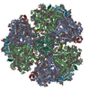 1jb0S S: Starting model for refinement |
|---|---|
| Similar structure data |

-Links
 PDBj
PDBj


- Assembly
| Deposited unit | 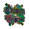
| ||||||||
|---|---|---|---|---|---|---|---|---|---|
| 1 | 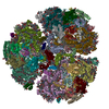
| ||||||||
| 2 | 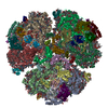
| ||||||||
| Unit cell |
|
-Components
-Photosystem I P700 chlorophyll a apoprotein ... , 2 types, 12 molecules A1A2A3A4A6A5B1B2B3B4B6B5
| #1: Protein | Mass: 83267.773 Da / Num. of mol.: 6 / Source method: isolated from a natural source Source: (natural) Thermosynechococcus elongatus BP-1 (bacteria)Strain: BP-1 / References: UniProt: P0A405, photosystem I #2: Protein | Mass: 82992.453 Da / Num. of mol.: 6 / Fragment: UNP residues 2-741 / Source method: isolated from a natural source Source: (natural) Thermosynechococcus elongatus BP-1 (bacteria)Strain: BP-1 / References: UniProt: P0A407, photosystem I |
|---|
-Protein , 2 types, 12 molecules C1C2C3C4C6C5P1P2P3P4P6P5
| #3: Protein | Mass: 8678.011 Da / Num. of mol.: 6 / Fragment: UNP residues 2-81 / Source method: isolated from a natural source Source: (natural) Thermosynechococcus elongatus BP-1 (bacteria)Strain: BP-1 / References: UniProt: P0A415, photosystem I #13: Protein | Mass: 10722.764 Da / Num. of mol.: 6 / Fragment: UNP residues 2-98 Source method: isolated from a genetically manipulated source Source: (gene. exp.) Thermosynechococcus elongatus BP-1 (bacteria)Strain: BP-1 / Gene: petF1, petF, tsl1009 / Production host: |
|---|
-Photosystem I reaction center subunit ... , 8 types, 48 molecules D1D2D3D4D6D5E1E2E3E4E6E5F1F2F3F4F6F5I1I2I3I4I6I5J1J2J3J4J6J5...
| #4: Protein | Mass: 15258.297 Da / Num. of mol.: 6 / Fragment: UNP residues 2-139 / Source method: isolated from a natural source Source: (natural) Thermosynechococcus elongatus BP-1 (bacteria)Strain: BP-1 / References: UniProt: P0A420 #5: Protein | Mass: 8268.290 Da / Num. of mol.: 6 / Fragment: UNP residues 2-76 / Source method: isolated from a natural source Source: (natural) Thermosynechococcus elongatus BP-1 (bacteria)Strain: BP-1 / References: UniProt: P0A423 #6: Protein | Mass: 17716.586 Da / Num. of mol.: 6 / Source method: isolated from a natural source Source: (natural) Thermosynechococcus elongatus BP-1 (bacteria)Strain: BP-1 / References: UniProt: P0A401 #7: Protein/peptide | Mass: 4297.234 Da / Num. of mol.: 6 / Source method: isolated from a natural source Source: (natural) Thermosynechococcus elongatus BP-1 (bacteria)Strain: BP-1 / References: UniProt: P0A427 #8: Protein/peptide | Mass: 4770.698 Da / Num. of mol.: 6 / Source method: isolated from a natural source Source: (natural) Thermosynechococcus elongatus BP-1 (bacteria)Strain: BP-1 / References: UniProt: P0A429 #9: Protein | Mass: 8483.983 Da / Num. of mol.: 6 / Source method: isolated from a natural source Source: (natural) Thermosynechococcus elongatus BP-1 (bacteria)Strain: BP-1 / References: UniProt: P0A425 #10: Protein | Mass: 16156.569 Da / Num. of mol.: 6 / Source method: isolated from a natural source Source: (natural) Thermosynechococcus elongatus BP-1 (bacteria)Strain: BP-1 / References: UniProt: Q8DGB4 #11: Protein/peptide | Mass: 3426.115 Da / Num. of mol.: 6 / Source method: isolated from a natural source Source: (natural) Thermosynechococcus elongatus BP-1 (bacteria)Strain: BP-1 / References: UniProt: P0A403 |
|---|
-Protein/peptide , 1 types, 6 molecules X1X2X3X4X6X5
| #12: Protein/peptide | Mass: 3845.508 Da / Num. of mol.: 6 / Source method: isolated from a natural source / Source: (natural) Thermosynechococcus elongatus (bacteria) |
|---|
-Non-polymers , 8 types, 774 molecules 


| #14: Chemical | ChemComp-CLA /  Mass: 893.489 Da / Num. of mol.: 576 / Source method: obtained synthetically / Formula: C55H72MgN4O5 Mass: 893.489 Da / Num. of mol.: 576 / Source method: obtained synthetically / Formula: C55H72MgN4O5#15: Chemical | ChemComp-PQN /  Mass: 450.696 Da / Num. of mol.: 12 / Source method: obtained synthetically / Formula: C31H46O2 Mass: 450.696 Da / Num. of mol.: 12 / Source method: obtained synthetically / Formula: C31H46O2#16: Chemical | ChemComp-BCR /  Mass: 536.873 Da / Num. of mol.: 132 / Source method: obtained synthetically / Formula: C40H56 Mass: 536.873 Da / Num. of mol.: 132 / Source method: obtained synthetically / Formula: C40H56#17: Chemical | ChemComp-LHG /  Mass: 722.970 Da / Num. of mol.: 18 / Source method: obtained synthetically / Formula: C38H75O10P / Comment: phospholipid*YM Mass: 722.970 Da / Num. of mol.: 18 / Source method: obtained synthetically / Formula: C38H75O10P / Comment: phospholipid*YM#18: Chemical | ChemComp-SF4 /  Mass: 351.640 Da / Num. of mol.: 18 / Source method: obtained synthetically / Formula: Fe4S4 Mass: 351.640 Da / Num. of mol.: 18 / Source method: obtained synthetically / Formula: Fe4S4#19: Chemical | ChemComp-LMG /  Mass: 787.158 Da / Num. of mol.: 6 / Source method: obtained synthetically / Formula: C45H86O10 Mass: 787.158 Da / Num. of mol.: 6 / Source method: obtained synthetically / Formula: C45H86O10#20: Chemical | ChemComp-CA /  Mass: 40.078 Da / Num. of mol.: 6 / Source method: obtained synthetically / Formula: Ca Mass: 40.078 Da / Num. of mol.: 6 / Source method: obtained synthetically / Formula: Ca#21: Chemical | ChemComp-FES /  Mass: 175.820 Da / Num. of mol.: 6 / Source method: obtained synthetically / Formula: Fe2S2 Mass: 175.820 Da / Num. of mol.: 6 / Source method: obtained synthetically / Formula: Fe2S2 |
|---|
-Details
| Has protein modification | Y |
|---|
-Experimental details
-Experiment
| Experiment | Method: X-RAY DIFFRACTION / Number of used crystals: 1 |
|---|
- Sample preparation
| Crystal | Density Matthews: 4.32 Å3/Da / Density % sol: 71.5 % |
|---|---|
| Crystal grow | Temperature: 277 K / Method: microbatch / pH: 4.6 / Details: 41% PEG200, 100 mM acetate buffer, 100 mM NaCl |
-Data collection
| Diffraction | Mean temperature: 100 K |
|---|---|
| Diffraction source | Source: SYNCHROTRON / Site: SPring-8 / Beamline: BL44XU / Wavelength: 0.9 Å |
| Detector | Type: RAYONIX MX300HE / Detector: CCD / Date: May 21, 2013 |
| Radiation | Protocol: SINGLE WAVELENGTH / Monochromatic (M) / Laue (L): M / Scattering type: x-ray |
| Radiation wavelength | Wavelength: 0.9 Å / Relative weight: 1 |
| Reflection | Resolution: 4.2→158.04 Å / Num. obs: 191480 / % possible obs: 100 % / Redundancy: 7.7 % / Rmerge(I) obs: 0.159 / Net I/σ(I): 8.7 |
| Reflection shell | Resolution: 4.2→4.43 Å / Redundancy: 7.6 % / Rmerge(I) obs: 0.868 / Mean I/σ(I) obs: 2.3 / Num. unique all: 27882 / Num. unique obs: 212827 / % possible all: 100 |
- Processing
| Software |
| ||||||||||||||||||||
|---|---|---|---|---|---|---|---|---|---|---|---|---|---|---|---|---|---|---|---|---|---|
| Refinement | Method to determine structure: MOLECULAR REPLACEMENT Starting model: 1JB0 Resolution: 4.2→158.04 Å / Cross valid method: FREE R-VALUE
| ||||||||||||||||||||
| Displacement parameters | Biso mean: 158.055 Å2 | ||||||||||||||||||||
| Refinement step | Cycle: LAST / Resolution: 4.2→158.04 Å
| ||||||||||||||||||||
| LS refinement shell | Resolution: 4.199→4.308 Å / Total num. of bins used: 20
|
