 Movie
Movie Controller

[English] 日本語
 Yorodumi
- PDB-7bw2: Crystal Structure of Cyanobacterial PSI Monomer from T.elongatus ... -
+ Open data

- Basic information
| Entry | Database: PDB / ID: 7bw2 | |||||||||
|---|---|---|---|---|---|---|---|---|---|---|
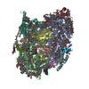
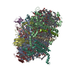
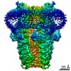
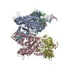
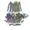
| Title | Crystal Structure of Cyanobacterial PSI Monomer from T.elongatus at 6.5 A Resolution | |||||||||
 Components Components | (Photosystem I ...) x 12 | |||||||||
 Keywords Keywords | MEMBRANE PROTEIN / monomer / complex / photosystem / photosynthesis | |||||||||
| Function / homology |  Function and homology information Function and homology informationphotosystem I reaction center / photosystem I / photosynthetic electron transport in photosystem I / photosystem I / plasma membrane-derived thylakoid membrane / chlorophyll binding / photosynthesis / 4 iron, 4 sulfur cluster binding / electron transfer activity / oxidoreductase activity ...photosystem I reaction center / photosystem I / photosynthetic electron transport in photosystem I / photosystem I / plasma membrane-derived thylakoid membrane / chlorophyll binding / photosynthesis / 4 iron, 4 sulfur cluster binding / electron transfer activity / oxidoreductase activity / magnesium ion binding / metal ion binding Similarity search - Function | |||||||||
| Biological species |   Thermosynechococcus elongatus BP-1 (bacteria)Thermosynechococcus elongatus (bacteria) Thermosynechococcus elongatus BP-1 (bacteria)Thermosynechococcus elongatus (bacteria) | |||||||||
| Method |  X-RAY DIFFRACTION / SYNCHROTRON / MOLECULAR REPLACEMENT / Resolution: 6.5 Å X-RAY DIFFRACTION / SYNCHROTRON / MOLECULAR REPLACEMENT / Resolution: 6.5 Å | |||||||||
 Authors Authors | Kurisu, G. / Coruh, O. / Tanaka, H. / Eithar, E.M. / Mian, Y. | |||||||||
| Funding support |  Japan, 2items Japan, 2items
| |||||||||
 Citation Citation | Journal: Commun Biol / Year: 2021 Title: Cryo-EM structure of a functional monomeric Photosystem I from Thermosynechococcus elongatus reveals red chlorophyll cluster. Authors: Orkun Çoruh / Anna Frank / Hideaki Tanaka / Akihiro Kawamoto / Eithar El-Mohsnawy / Takayuki Kato / Keiichi Namba / Christoph Gerle / Marc M Nowaczyk / Genji Kurisu /   Abstract: A high-resolution structure of trimeric cyanobacterial Photosystem I (PSI) from Thermosynechococcus elongatus was reported as the first atomic model of PSI almost 20 years ago. However, the monomeric ...A high-resolution structure of trimeric cyanobacterial Photosystem I (PSI) from Thermosynechococcus elongatus was reported as the first atomic model of PSI almost 20 years ago. However, the monomeric PSI structure has not yet been reported despite long-standing interest in its structure and extensive spectroscopic characterization of the loss of red chlorophylls upon monomerization. Here, we describe the structure of monomeric PSI from Thermosynechococcus elongatus BP-1. Comparison with the trimer structure gave detailed insights into monomerization-induced changes in both the central trimerization domain and the peripheral regions of the complex. Monomerization-induced loss of red chlorophylls is assigned to a cluster of chlorophylls adjacent to PsaX. Based on our findings, we propose a role of PsaX in the stabilization of red chlorophylls and that lipids of the surrounding membrane present a major source of thermal energy for uphill excitation energy transfer from red chlorophylls to P700. | |||||||||
| History |
|
- Structure visualization
| Structure viewer | Molecule: MolmilJmol/JSmol |
|---|
- Downloads & links
-Download
| PDBx/mmCIF format | 7bw2.cif.gz | 450.8 KB | Display | PDBx/mmCIF format |
|---|---|---|---|---|
| PDB format | pdb7bw2.ent.gz | 362.5 KB | Display | PDB format |
| PDBx/mmJSON format | 7bw2.json.gz | Tree view | PDBx/mmJSON format | |
| Others |  Other downloads Other downloads |
-Validation report
| Arichive directory | https://data.pdbj.org/pub/pdb/validation_reports/bw/7bw2ftp://data.pdbj.org/pub/pdb/validation_reports/bw/7bw2 | HTTPS FTP |
|---|
-Related structure data
| Related structure data | 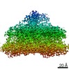 0977C  6lu1C 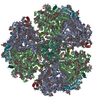 1jb0S S: Starting model for refinement C: citing same article ( |
|---|---|
| Similar structure data |


-Links
 PDBj
PDBj


- Assembly
| Deposited unit | 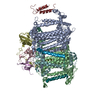
| ||||||||
|---|---|---|---|---|---|---|---|---|---|
| 1 |
| ||||||||
| Unit cell |
|
-Components
-Photosystem I ... , 12 types, 12 molecules ABCDEFIJKLMX
| #1: Protein | Mass: 83267.773 Da / Num. of mol.: 1 / Source method: isolated from a natural source Source: (natural) Thermosynechococcus elongatus BP-1 (bacteria)Strain: BP-1 / References: UniProt: P0A405, photosystem I |
|---|---|
| #2: Protein | Mass: 82992.453 Da / Num. of mol.: 1 / Source method: isolated from a natural source Source: (natural) Thermosynechococcus elongatus BP-1 (bacteria)Strain: BP-1 / References: UniProt: P0A407, photosystem I |
| #3: Protein | Mass: 8809.207 Da / Num. of mol.: 1 / Source method: isolated from a natural source Source: (natural) Thermosynechococcus elongatus (strain BP-1) (bacteria)Strain: BP-1 / References: UniProt: P0A415, photosystem I |
| #4: Protein | Mass: 15389.494 Da / Num. of mol.: 1 / Source method: isolated from a natural source Source: (natural) Thermosynechococcus elongatus (strain BP-1) (bacteria)Strain: BP-1 / References: UniProt: P0A420 |
| #5: Protein | Mass: 8399.485 Da / Num. of mol.: 1 / Source method: isolated from a natural source Source: (natural) Thermosynechococcus elongatus (strain BP-1) (bacteria)Strain: BP-1 / References: UniProt: P0A423 |
| #6: Protein | Mass: 17716.586 Da / Num. of mol.: 1 / Source method: isolated from a natural source Source: (natural) Thermosynechococcus elongatus BP-1 (bacteria)Strain: BP-1 / References: UniProt: P0A401 |
| #7: Protein/peptide | Mass: 4297.234 Da / Num. of mol.: 1 / Source method: isolated from a natural source Source: (natural) Thermosynechococcus elongatus BP-1 (bacteria)Strain: BP-1 / References: UniProt: P0A427 |
| #8: Protein/peptide | Mass: 4770.698 Da / Num. of mol.: 1 / Source method: isolated from a natural source Source: (natural) Thermosynechococcus elongatus BP-1 (bacteria)Strain: BP-1 / References: UniProt: P0A429 |
| #9: Protein | Mass: 8483.983 Da / Num. of mol.: 1 / Source method: isolated from a natural source Source: (natural) Thermosynechococcus elongatus BP-1 (bacteria)Strain: BP-1 / References: UniProt: P0A425 |
| #10: Protein | Mass: 16261.685 Da / Num. of mol.: 1 / Source method: isolated from a natural source Source: (natural) Thermosynechococcus elongatus (strain BP-1) (bacteria)Strain: BP-1 / References: UniProt: Q8DGB4 |
| #11: Protein/peptide | Mass: 3426.115 Da / Num. of mol.: 1 / Source method: isolated from a natural source Source: (natural) Thermosynechococcus elongatus (strain BP-1) (bacteria)Strain: BP-1 / References: UniProt: P0A403 |
| #12: Protein/peptide | Mass: 4424.317 Da / Num. of mol.: 1 / Source method: isolated from a natural source Source: (natural) Thermosynechococcus elongatus (strain BP-1) (bacteria)Strain: BP-1 / References: UniProt: Q8DKP6 |
-Details
| Has ligand of interest | N |
|---|---|
| Has protein modification | Y |
-Experimental details
-Experiment
| Experiment | Method: X-RAY DIFFRACTION / Number of used crystals: 1 |
|---|
- Sample preparation
| Crystal | Density Matthews: 4.57 Å3/Da / Density % sol: 73.09 % |
|---|---|
| Crystal grow | Temperature: 277.15 K / Method: vapor diffusion, hanging drop / pH: 6.8 / Details: no salt |
-Data collection
| Diffraction | Mean temperature: 100 K / Serial crystal experiment: N |
|---|---|
| Diffraction source | Source: SYNCHROTRON / Site: SPring-8 / Beamline: BL44XU / Wavelength: 0.9 Å |
| Detector | Type: RAYONIX MX300HE / Detector: CCD / Date: Jul 24, 2015 |
| Radiation | Protocol: SINGLE WAVELENGTH / Monochromatic (M) / Laue (L): M / Scattering type: x-ray |
| Radiation wavelength | Wavelength: 0.9 Å / Relative weight: 1 |
| Reflection | Resolution: 6.5→49.57 Å / Num. obs: 9572 / % possible obs: 98.5 % / Redundancy: 10.7 % / Biso Wilson estimate: 297.2 Å2 / Rmerge(I) obs: 0.05486 / Net I/σ(I): 29.58 |
| Reflection shell | Resolution: 6.5→6.73 Å / Redundancy: 11.3 % / Rmerge(I) obs: 0.3354 / Mean I/σ(I) obs: 7.39 / Num. unique obs: 949 / % possible all: 100 |
- Processing
| Software |
| ||||||||||||||||||||||||||||||||||||||||||||||||||||||||||||||||||||||||||||||||||||||||||||||||||||||||||||||||||||||||||||||||||||||||||||||||||||||||||||||||||||||||||||||||||||||
|---|---|---|---|---|---|---|---|---|---|---|---|---|---|---|---|---|---|---|---|---|---|---|---|---|---|---|---|---|---|---|---|---|---|---|---|---|---|---|---|---|---|---|---|---|---|---|---|---|---|---|---|---|---|---|---|---|---|---|---|---|---|---|---|---|---|---|---|---|---|---|---|---|---|---|---|---|---|---|---|---|---|---|---|---|---|---|---|---|---|---|---|---|---|---|---|---|---|---|---|---|---|---|---|---|---|---|---|---|---|---|---|---|---|---|---|---|---|---|---|---|---|---|---|---|---|---|---|---|---|---|---|---|---|---|---|---|---|---|---|---|---|---|---|---|---|---|---|---|---|---|---|---|---|---|---|---|---|---|---|---|---|---|---|---|---|---|---|---|---|---|---|---|---|---|---|---|---|---|---|---|---|---|---|
| Refinement | Method to determine structure: MOLECULAR REPLACEMENT Starting model: 1JB0 Resolution: 6.5→49.57 Å / Cor.coef. Fo:Fc: 0.705 / Cor.coef. Fo:Fc free: 0.599 / SU B: 469.165 / SU ML: 4.299 / Cross valid method: THROUGHOUT / ESU R Free: 4.447 / Details: HYDROGENS HAVE BEEN ADDED IN THE RIDING POSITIONS
| ||||||||||||||||||||||||||||||||||||||||||||||||||||||||||||||||||||||||||||||||||||||||||||||||||||||||||||||||||||||||||||||||||||||||||||||||||||||||||||||||||||||||||||||||||||||
| Solvent computation | Ion probe radii: 0.8 Å / Shrinkage radii: 0.8 Å / VDW probe radii: 1.2 Å | ||||||||||||||||||||||||||||||||||||||||||||||||||||||||||||||||||||||||||||||||||||||||||||||||||||||||||||||||||||||||||||||||||||||||||||||||||||||||||||||||||||||||||||||||||||||
| Displacement parameters | Biso mean: 179.9 Å2
| ||||||||||||||||||||||||||||||||||||||||||||||||||||||||||||||||||||||||||||||||||||||||||||||||||||||||||||||||||||||||||||||||||||||||||||||||||||||||||||||||||||||||||||||||||||||
| Refinement step | Cycle: LAST / Resolution: 6.5→49.57 Å
| ||||||||||||||||||||||||||||||||||||||||||||||||||||||||||||||||||||||||||||||||||||||||||||||||||||||||||||||||||||||||||||||||||||||||||||||||||||||||||||||||||||||||||||||||||||||
| Refine LS restraints |
|