Movie
Movie Controller

[English] 日本語
 Yorodumi
- PDB-6lu1: Cyanobacterial PSI Monomer from T. elongatus by Single Particle C... -
+ Open data

- Basic information
| Entry | Database: PDB / ID: 6lu1 | |||||||||
|---|---|---|---|---|---|---|---|---|---|---|
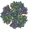
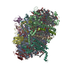
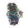
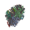
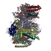
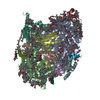
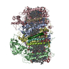
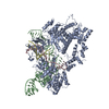
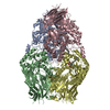
| Title | Cyanobacterial PSI Monomer from T. elongatus by Single Particle CRYO-EM at 3.2 A Resolution | |||||||||
 Components Components |
| |||||||||
 Keywords Keywords | MEMBRANE PROTEIN / monomer / complex / photosystem / photosynthesis | |||||||||
| Function / homology |  Function and homology information Function and homology informationphotosystem I reaction center / photosystem I / photosynthetic electron transport in photosystem I / photosystem I / plasma membrane-derived thylakoid membrane / chlorophyll binding / photosynthesis / 4 iron, 4 sulfur cluster binding / electron transfer activity / oxidoreductase activity ...photosystem I reaction center / photosystem I / photosynthetic electron transport in photosystem I / photosystem I / plasma membrane-derived thylakoid membrane / chlorophyll binding / photosynthesis / 4 iron, 4 sulfur cluster binding / electron transfer activity / oxidoreductase activity / magnesium ion binding / metal ion binding Similarity search - Function | |||||||||
| Biological species |   Thermosynechococcus elongatus BP-1 (bacteria)Thermosynechococcus elongatus (bacteria) Thermosynechococcus elongatus BP-1 (bacteria)Thermosynechococcus elongatus (bacteria) | |||||||||
| Method | ELECTRON MICROSCOPY / single particle reconstruction / cryo EM / Resolution: 3.2 Å | |||||||||
 Authors Authors | Kurisu, G. / Coruh, O. / Tanaka, H. / Gerle, C. / Kawamoto, A. / Kato, T. / Namba, K. / Nowaczyk, M.M. / Rogner, M. / Misumi, Y. ...Kurisu, G. / Coruh, O. / Tanaka, H. / Gerle, C. / Kawamoto, A. / Kato, T. / Namba, K. / Nowaczyk, M.M. / Rogner, M. / Misumi, Y. / Frank, A. / Eithar, E.M. | |||||||||
| Funding support |  Japan, 2items Japan, 2items
| |||||||||
 Citation Citation | Journal: Commun Biol / Year: 2021 Title: Cryo-EM structure of a functional monomeric Photosystem I from Thermosynechococcus elongatus reveals red chlorophyll cluster. Authors: Orkun Çoruh / Anna Frank / Hideaki Tanaka / Akihiro Kawamoto / Eithar El-Mohsnawy / Takayuki Kato / Keiichi Namba / Christoph Gerle / Marc M Nowaczyk / Genji Kurisu /   Abstract: A high-resolution structure of trimeric cyanobacterial Photosystem I (PSI) from Thermosynechococcus elongatus was reported as the first atomic model of PSI almost 20 years ago. However, the monomeric ...A high-resolution structure of trimeric cyanobacterial Photosystem I (PSI) from Thermosynechococcus elongatus was reported as the first atomic model of PSI almost 20 years ago. However, the monomeric PSI structure has not yet been reported despite long-standing interest in its structure and extensive spectroscopic characterization of the loss of red chlorophylls upon monomerization. Here, we describe the structure of monomeric PSI from Thermosynechococcus elongatus BP-1. Comparison with the trimer structure gave detailed insights into monomerization-induced changes in both the central trimerization domain and the peripheral regions of the complex. Monomerization-induced loss of red chlorophylls is assigned to a cluster of chlorophylls adjacent to PsaX. Based on our findings, we propose a role of PsaX in the stabilization of red chlorophylls and that lipids of the surrounding membrane present a major source of thermal energy for uphill excitation energy transfer from red chlorophylls to P700. | |||||||||
| History |
|
- Structure visualization
| Movie |
Movie viewer |
|---|---|
| Structure viewer | Molecule: MolmilJmol/JSmol |
- Downloads & links
-Download
| PDBx/mmCIF format | 6lu1.cif.gz | 493.2 KB | Display | PDBx/mmCIF format |
|---|---|---|---|---|
| PDB format | pdb6lu1.ent.gz | 417 KB | Display | PDB format |
| PDBx/mmJSON format | 6lu1.json.gz | Tree view | PDBx/mmJSON format | |
| Others |  Other downloads Other downloads |
-Validation report
| Arichive directory | https://data.pdbj.org/pub/pdb/validation_reports/lu/6lu1ftp://data.pdbj.org/pub/pdb/validation_reports/lu/6lu1 | HTTPS FTP |
|---|
-Related structure data
| Related structure data | 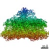 0977MC 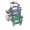 7bw2C M: map data used to model this data C: citing same article ( |
|---|---|
| Similar structure data | |
| EM raw data | EMPIAR-10352 (Title: 3.2 Å resolution structure of a functional monomeric Photosystem I from Thermosynechococcus elongatus BP-1 by single particle cryo-EM with a 200 kV CRYO ARM electron microscope Data size: 226.1 Data #1: Raw, non aligned micrograph movies in compressed TIFF format. [micrographs - multiframe]) |


-Links
 PDBj
PDBj


- Assembly
| Deposited unit | 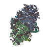
|
|---|---|
| 1 |
|
-Components
-Photosystem I P700 chlorophyll a apoprotein ... , 2 types, 2 molecules AB
| #1: Protein | Mass: 83267.773 Da / Num. of mol.: 1 / Source method: isolated from a natural source Source: (natural) Thermosynechococcus elongatus BP-1 (bacteria)Strain: BP-1 / References: UniProt: P0A405, photosystem I |
|---|---|
| #2: Protein | Mass: 83123.648 Da / Num. of mol.: 1 / Source method: isolated from a natural source Source: (natural) Thermosynechococcus elongatus (strain BP-1) (bacteria)Strain: BP-1 / References: UniProt: P0A407, photosystem I |
-Protein , 1 types, 1 molecules C
| #3: Protein | Mass: 8809.207 Da / Num. of mol.: 1 / Source method: isolated from a natural source Source: (natural) Thermosynechococcus elongatus (strain BP-1) (bacteria)Strain: BP-1 / References: UniProt: P0A415, photosystem I |
|---|
-Photosystem I reaction center subunit ... , 7 types, 7 molecules DEFIJLM
| #4: Protein | Mass: 15389.494 Da / Num. of mol.: 1 / Source method: isolated from a natural source Source: (natural) Thermosynechococcus elongatus (strain BP-1) (bacteria)Strain: BP-1 / References: UniProt: P0A420 |
|---|---|
| #5: Protein | Mass: 8399.485 Da / Num. of mol.: 1 / Source method: isolated from a natural source Source: (natural) Thermosynechococcus elongatus (strain BP-1) (bacteria)Strain: BP-1 / References: UniProt: P0A423 |
| #6: Protein | Mass: 17716.586 Da / Num. of mol.: 1 / Source method: isolated from a natural source Source: (natural) Thermosynechococcus elongatus BP-1 (bacteria)Strain: BP-1 / References: UniProt: P0A401 |
| #7: Protein/peptide | Mass: 4297.234 Da / Num. of mol.: 1 / Source method: isolated from a natural source Source: (natural) Thermosynechococcus elongatus BP-1 (bacteria)Strain: BP-1 / References: UniProt: P0A427 |
| #8: Protein/peptide | Mass: 4770.698 Da / Num. of mol.: 1 / Source method: isolated from a natural source Source: (natural) Thermosynechococcus elongatus BP-1 (bacteria)Strain: BP-1 / References: UniProt: P0A429 |
| #9: Protein | Mass: 16261.685 Da / Num. of mol.: 1 / Source method: isolated from a natural source Source: (natural) Thermosynechococcus elongatus (strain BP-1) (bacteria)Strain: BP-1 / References: UniProt: Q8DGB4 |
| #10: Protein/peptide | Mass: 3426.115 Da / Num. of mol.: 1 / Source method: isolated from a natural source Source: (natural) Thermosynechococcus elongatus (strain BP-1) (bacteria)Strain: BP-1 / References: UniProt: P0A403 |
-Non-polymers , 7 types, 121 molecules 


| #11: Chemical | ChemComp-CLA /  Mass: 893.489 Da / Num. of mol.: 82 / Source method: obtained synthetically / Formula: C55H72MgN4O5 Mass: 893.489 Da / Num. of mol.: 82 / Source method: obtained synthetically / Formula: C55H72MgN4O5#12: Chemical |  Mass: 450.696 Da / Num. of mol.: 2 / Source method: obtained synthetically / Formula: C31H46O2 Mass: 450.696 Da / Num. of mol.: 2 / Source method: obtained synthetically / Formula: C31H46O2#13: Chemical | ChemComp-BCR /  Mass: 536.873 Da / Num. of mol.: 26 / Source method: obtained synthetically / Formula: C40H56 Mass: 536.873 Da / Num. of mol.: 26 / Source method: obtained synthetically / Formula: C40H56#14: Chemical |  Mass: 722.970 Da / Num. of mol.: 2 / Source method: obtained synthetically / Formula: C38H75O10P / Comment: phospholipid*YM Mass: 722.970 Da / Num. of mol.: 2 / Source method: obtained synthetically / Formula: C38H75O10P / Comment: phospholipid*YM#15: Chemical |  Mass: 351.640 Da / Num. of mol.: 3 / Source method: obtained synthetically / Formula: Fe4S4 Mass: 351.640 Da / Num. of mol.: 3 / Source method: obtained synthetically / Formula: Fe4S4#16: Chemical | ChemComp-LMG / |  Mass: 787.158 Da / Num. of mol.: 1 / Source method: isolated from a natural source / Formula: C45H86O10 Mass: 787.158 Da / Num. of mol.: 1 / Source method: isolated from a natural source / Formula: C45H86O10#17: Water | ChemComp-HOH / | Mass: 18.015 Da / Num. of mol.: 5 / Source method: isolated from a natural source / Formula: H2O |
|---|
-Details
| Has ligand of interest | N |
|---|
-Experimental details
-Experiment
| Experiment | Method: ELECTRON MICROSCOPY |
|---|---|
| EM experiment | Aggregation state: PARTICLE / 3D reconstruction method: single particle reconstruction |
- Sample preparation
| Component | Name: Cyanobacterial PSI Monomer from T. elongatus / Type: COMPLEX Details: The monomers in this sample are generated by the monomerization of trimers of PSI during the purification process. Entity ID: #1-#10 / Source: NATURAL | ||||||||||||||||||||
|---|---|---|---|---|---|---|---|---|---|---|---|---|---|---|---|---|---|---|---|---|---|
| Molecular weight | Value: 350 kDa/nm / Experimental value: NO | ||||||||||||||||||||
| Source (natural) | Organism: Thermosynechococcus elongatus BP-1 (bacteria) | ||||||||||||||||||||
| Buffer solution | pH: 7.5 | ||||||||||||||||||||
| Buffer component |
| ||||||||||||||||||||
| Specimen | Conc.: 5 mg/ml / Embedding applied: NO / Shadowing applied: NO / Staining applied: NO / Vitrification applied: YES / Details: Sample was monodisperse. | ||||||||||||||||||||
| Specimen support | Grid material: COPPER / Grid mesh size: 300 divisions/in. / Grid type: Quantifoil R1.2/1.3 | ||||||||||||||||||||
| Vitrification | Instrument: FEI VITROBOT MARK IV / Cryogen name: ETHANE / Humidity: 95 % / Chamber temperature: 277.15 K |
- Electron microscopy imaging
| Microscopy | Model: JEOL CRYO ARM 200 |
|---|---|
| Electron gun | Electron source:  FIELD EMISSION GUN / Accelerating voltage: 200 kV / Illumination mode: FLOOD BEAM FIELD EMISSION GUN / Accelerating voltage: 200 kV / Illumination mode: FLOOD BEAM |
| Electron lens | Mode: BRIGHT FIELD / Nominal magnification: 60000 X / Calibrated magnification: 56497 X / Calibrated defocus min: 500 nm / Calibrated defocus max: 3500 nm / Cs: 1.4 mm / Alignment procedure: COMA FREE |
| Specimen holder | Cryogen: NITROGEN / Specimen holder model: JEOL / Temperature (max): 100.4 K / Temperature (min): 100.4 K |
| Image recording | Average exposure time: 12 sec. / Electron dose: 1.34 e/Å2 / Detector mode: COUNTING / Film or detector model: GATAN K2 SUMMIT (4k x 4k) / Num. of grids imaged: 1 |
| Image scans | Sampling size: 5 µm / Width: 3710 / Height: 3838 / Movie frames/image: 60 / Used frames/image: 1-60 |
- Processing
| Software | Name: PHENIX / Version: 1.14_3260: / Classification: refinement | ||||||||||||||||||||||||||||||||||||
|---|---|---|---|---|---|---|---|---|---|---|---|---|---|---|---|---|---|---|---|---|---|---|---|---|---|---|---|---|---|---|---|---|---|---|---|---|---|
| EM software |
| ||||||||||||||||||||||||||||||||||||
| CTF correction | Type: PHASE FLIPPING ONLY | ||||||||||||||||||||||||||||||||||||
| Particle selection | Num. of particles selected: 182018 | ||||||||||||||||||||||||||||||||||||
| Symmetry | Point symmetry: C1 (asymmetric) | ||||||||||||||||||||||||||||||||||||
| 3D reconstruction | Resolution: 3.2 Å / Resolution method: FSC 0.143 CUT-OFF / Num. of particles: 46105 / Algorithm: BACK PROJECTION / Num. of class averages: 1 / Symmetry type: POINT | ||||||||||||||||||||||||||||||||||||
| Atomic model building | B value: 45 / Protocol: OTHER / Space: REAL | ||||||||||||||||||||||||||||||||||||
| Atomic model building | PDB-ID: 1JB0 Accession code: 1JB0 / Source name: PDB / Type: experimental model | ||||||||||||||||||||||||||||||||||||
| Refinement | Highest resolution: 3.2 Å | ||||||||||||||||||||||||||||||||||||
| Refine LS restraints |
|