 Movie
Movie Controller

[English] 日本語
 Yorodumi
- PDB-5exe: Crystal structure of oxalate oxidoreductase from Moorella thermoa... -
+ Open data

- Basic information
| Entry | Database: PDB / ID: 5exe | ||||||
|---|---|---|---|---|---|---|---|
| Title | Crystal structure of oxalate oxidoreductase from Moorella thermoacetica bound with carboxy-TPP adduct | ||||||
 Components Components | (Oxalate oxidoreductase subunit ...) x 3 | ||||||
 Keywords Keywords | OXIDOREDUCTASE / oxalate / OFOR / thiamine | ||||||
| Function / homology |  Function and homology information Function and homology informationoxalate oxidoreductase / oxidoreductase activity, acting on the aldehyde or oxo group of donors, iron-sulfur protein as acceptor / oxalate catabolic process / thiamine pyrophosphate binding / 4 iron, 4 sulfur cluster binding / response to oxidative stress / metal ion binding Similarity search - Function | ||||||
| Biological species |  Moorella thermoacetica (bacteria) Moorella thermoacetica (bacteria) | ||||||
| Method |  X-RAY DIFFRACTION / SYNCHROTRON / MOLECULAR REPLACEMENT / Resolution: 1.88 Å X-RAY DIFFRACTION / SYNCHROTRON / MOLECULAR REPLACEMENT / Resolution: 1.88 Å | ||||||
 Authors Authors | Gibson, M.I. / Chen, P.Y.-T. / Drennan, C.L. | ||||||
 Citation Citation | Journal: Proc.Natl.Acad.Sci.USA / Year: 2016 Title: One-carbon chemistry of oxalate oxidoreductase captured by X-ray crystallography. Authors: Gibson, M.I. / Chen, P.Y. / Johnson, A.C. / Pierce, E. / Can, M. / Ragsdale, S.W. / Drennan, C.L. | ||||||
| History |
|
- Structure visualization
| Structure viewer | Molecule: MolmilJmol/JSmol |
|---|
- Downloads & links
-Download
| PDBx/mmCIF format | 5exe.cif.gz | 468.2 KB | Display | PDBx/mmCIF format |
|---|---|---|---|---|
| PDB format | pdb5exe.ent.gz | 371 KB | Display | PDB format |
| PDBx/mmJSON format | 5exe.json.gz | Tree view | PDBx/mmJSON format | |
| Others |  Other downloads Other downloads |
-Validation report
| Arichive directory | https://data.pdbj.org/pub/pdb/validation_reports/ex/5exeftp://data.pdbj.org/pub/pdb/validation_reports/ex/5exe | HTTPS FTP |
|---|
-Related structure data
| Related structure data |  5exdC  5c4iS C: citing same article ( S: Starting model for refinement |
|---|---|
| Similar structure data |
-Links
 PDBj
PDBj


- Assembly
| Deposited unit | 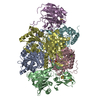
| ||||||||
|---|---|---|---|---|---|---|---|---|---|
| 1 |
| ||||||||
| Unit cell |
|
-Components
-Oxalate oxidoreductase subunit ... , 3 types, 6 molecules ADBECF
| #1: Protein | Mass: 43737.645 Da / Num. of mol.: 2 / Source method: isolated from a natural source Source: (natural)  Moorella thermoacetica (strain ATCC 39073) (bacteria) Moorella thermoacetica (strain ATCC 39073) (bacteria)Strain: ATCC 39073 / References: UniProt: Q2RI41, oxalate oxidoreductase #2: Protein | Mass: 33960.750 Da / Num. of mol.: 2 / Source method: isolated from a natural source Source: (natural) Moorella thermoacetica (strain ATCC 39073) (bacteria)Strain: ATCC 39073 / References: UniProt: Q2RI40, oxalate oxidoreductase #3: Protein | Mass: 34277.535 Da / Num. of mol.: 2 / Source method: isolated from a natural source Source: (natural) Moorella thermoacetica (strain ATCC 39073) (bacteria)Strain: ATCC 39073 / References: UniProt: Q2RI42, oxalate oxidoreductase |
|---|
-Non-polymers , 5 types, 2444 molecules 


| #4: Chemical | ChemComp-SF4 /  Mass: 351.640 Da / Num. of mol.: 6 / Source method: obtained synthetically / Formula: Fe4S4 Mass: 351.640 Da / Num. of mol.: 6 / Source method: obtained synthetically / Formula: Fe4S4#5: Chemical |  Mass: 468.316 Da / Num. of mol.: 2 / Source method: obtained synthetically / Formula: C13H18N4O9P2S Mass: 468.316 Da / Num. of mol.: 2 / Source method: obtained synthetically / Formula: C13H18N4O9P2S#6: Chemical |  Mass: 24.305 Da / Num. of mol.: 3 / Source method: obtained synthetically / Formula: Mg Mass: 24.305 Da / Num. of mol.: 3 / Source method: obtained synthetically / Formula: Mg#7: Chemical | ChemComp-NA / |  Mass: 22.990 Da / Num. of mol.: 1 / Source method: obtained synthetically / Formula: Na Mass: 22.990 Da / Num. of mol.: 1 / Source method: obtained synthetically / Formula: Na#8: Water | ChemComp-HOH / | Mass: 18.015 Da / Num. of mol.: 2432 / Source method: isolated from a natural source / Formula: H2O |
|---|
-Experimental details
-Experiment
| Experiment | Method: X-RAY DIFFRACTION |
|---|
- Sample preparation
| Crystal | Density Matthews: 2.96 Å3/Da / Density % sol: 58.41 % |
|---|---|
| Crystal grow | Temperature: 298 K / Method: vapor diffusion, hanging drop / pH: 7 Details: Crystals were grown in a Coy anaerobic chamber under an Ar/H2 gas mixture. OOR was mixed with the well solution containing PEG 3000 and Tacsimate (pH 7.0), in drops composed of 1 uL well ...Details: Crystals were grown in a Coy anaerobic chamber under an Ar/H2 gas mixture. OOR was mixed with the well solution containing PEG 3000 and Tacsimate (pH 7.0), in drops composed of 1 uL well solution and 1 uL OOR (30 mg/ml) with the addition of 0.2 uL of 100 mM oxalic acid (pH 8.0) to the crystallization drop |
-Data collection
| Diffraction | Mean temperature: 100 K |
|---|---|
| Diffraction source | Source: SYNCHROTRON / Site: APS  / Beamline: 24-ID-C / Wavelength: 0.9792 Å / Beamline: 24-ID-C / Wavelength: 0.9792 Å |
| Detector | Type: DECTRIS PILATUS 6M / Detector: PIXEL / Date: Jul 27, 2014 |
| Radiation | Protocol: SINGLE WAVELENGTH / Monochromatic (M) / Laue (L): M / Scattering type: x-ray |
| Radiation wavelength | Wavelength: 0.9792 Å / Relative weight: 1 |
| Reflection | Resolution: 1.88→50 Å / Num. obs: 200377 / % possible obs: 94.4 % / Redundancy: 5.9 % / Rsym value: 0.129 / Net I/σ(I): 7.7 |
| Reflection shell | Resolution: 1.88→1.91 Å / Redundancy: 4.1 % / Rmerge(I) obs: 0.882 / Mean I/σ(I) obs: 1.9 / % possible all: 75 |
- Processing
| Software |
| |||||||||||||||||||||||||||||||||||||||||||||||||||||||||||||||||||||||||||||||||||||||||||||||||||||||||||||||||||||||||||||||||||||||||||||||||||||||||||||||||||||||||||||||||||||||||||||||||||||||||||||||||||||||||
|---|---|---|---|---|---|---|---|---|---|---|---|---|---|---|---|---|---|---|---|---|---|---|---|---|---|---|---|---|---|---|---|---|---|---|---|---|---|---|---|---|---|---|---|---|---|---|---|---|---|---|---|---|---|---|---|---|---|---|---|---|---|---|---|---|---|---|---|---|---|---|---|---|---|---|---|---|---|---|---|---|---|---|---|---|---|---|---|---|---|---|---|---|---|---|---|---|---|---|---|---|---|---|---|---|---|---|---|---|---|---|---|---|---|---|---|---|---|---|---|---|---|---|---|---|---|---|---|---|---|---|---|---|---|---|---|---|---|---|---|---|---|---|---|---|---|---|---|---|---|---|---|---|---|---|---|---|---|---|---|---|---|---|---|---|---|---|---|---|---|---|---|---|---|---|---|---|---|---|---|---|---|---|---|---|---|---|---|---|---|---|---|---|---|---|---|---|---|---|---|---|---|---|---|---|---|---|---|---|---|---|---|---|---|---|---|---|---|---|
| Refinement | Method to determine structure: MOLECULAR REPLACEMENT Starting model: 5C4I Resolution: 1.88→48.704 Å / SU ML: 0.22 / Cross valid method: FREE R-VALUE / σ(F): 1.35 / Phase error: 22.64 / Stereochemistry target values: ML
| |||||||||||||||||||||||||||||||||||||||||||||||||||||||||||||||||||||||||||||||||||||||||||||||||||||||||||||||||||||||||||||||||||||||||||||||||||||||||||||||||||||||||||||||||||||||||||||||||||||||||||||||||||||||||
| Solvent computation | Shrinkage radii: 0.9 Å / VDW probe radii: 1.11 Å / Solvent model: FLAT BULK SOLVENT MODEL | |||||||||||||||||||||||||||||||||||||||||||||||||||||||||||||||||||||||||||||||||||||||||||||||||||||||||||||||||||||||||||||||||||||||||||||||||||||||||||||||||||||||||||||||||||||||||||||||||||||||||||||||||||||||||
| Refinement step | Cycle: LAST / Resolution: 1.88→48.704 Å
| |||||||||||||||||||||||||||||||||||||||||||||||||||||||||||||||||||||||||||||||||||||||||||||||||||||||||||||||||||||||||||||||||||||||||||||||||||||||||||||||||||||||||||||||||||||||||||||||||||||||||||||||||||||||||
| Refine LS restraints |
| |||||||||||||||||||||||||||||||||||||||||||||||||||||||||||||||||||||||||||||||||||||||||||||||||||||||||||||||||||||||||||||||||||||||||||||||||||||||||||||||||||||||||||||||||||||||||||||||||||||||||||||||||||||||||
| LS refinement shell |
|
