 Movie
Movie Controller

[English] 日本語
 Yorodumi
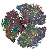
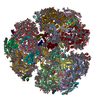
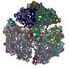
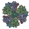
- PDB-7s3d: Structure of photosystem I with bound ferredoxin from Synechococc... -
+ Open data

- Basic information
| Entry | Database: PDB / ID: 7s3d | |||||||||
|---|---|---|---|---|---|---|---|---|---|---|
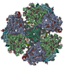
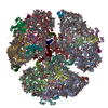
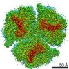
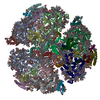
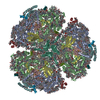
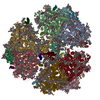
| Title | Structure of photosystem I with bound ferredoxin from Synechococcus sp. PCC 7335 acclimated to far-red light | |||||||||
 Components Components |
| |||||||||
 Keywords Keywords | PHOTOSYNTHESIS / Photosystem I / Far-red light photoacclimation / Chlorophyll f / Ferredoxin / PsaF / PsaJ | |||||||||
| Function / homology |  Function and homology information Function and homology informationphotosystem I reaction center / photosystem I / photosystem I / plasma membrane-derived thylakoid membrane / chlorophyll binding / photosynthesis / endomembrane system / electron transport chain / 2 iron, 2 sulfur cluster binding / 4 iron, 4 sulfur cluster binding ...photosystem I reaction center / photosystem I / photosystem I / plasma membrane-derived thylakoid membrane / chlorophyll binding / photosynthesis / endomembrane system / electron transport chain / 2 iron, 2 sulfur cluster binding / 4 iron, 4 sulfur cluster binding / electron transfer activity / oxidoreductase activity / magnesium ion binding / metal ion binding Similarity search - Function | |||||||||
| Biological species |  Synechococcus sp. PCC 7335 (bacteria) Synechococcus sp. PCC 7335 (bacteria) | |||||||||
| Method | ELECTRON MICROSCOPY / single particle reconstruction / cryo EM / Resolution: 2.91 Å | |||||||||
 Authors Authors | Gisriel, C.J. / Flesher, D.A. / Shen, G. / Wang, J. / Ho, M. / Brudvig, G.W. / Bryant, D.A. | |||||||||
| Funding support |  United States, 2items United States, 2items
| |||||||||
 Citation Citation | Journal: J Biol Chem / Year: 2022 Title: Structure of a photosystem I-ferredoxin complex from a marine cyanobacterium provides insights into far-red light photoacclimation. Authors: Christopher J Gisriel / David A Flesher / Gaozhong Shen / Jimin Wang / Ming-Yang Ho / Gary W Brudvig / Donald A Bryant /  Abstract: Far-red light photoacclimation exhibited by some cyanobacteria allows these organisms to use the far-red region of the solar spectrum (700-800 nm) for photosynthesis. Part of this process includes ...Far-red light photoacclimation exhibited by some cyanobacteria allows these organisms to use the far-red region of the solar spectrum (700-800 nm) for photosynthesis. Part of this process includes the replacement of six photosystem I (PSI) subunits with isoforms that confer the binding of chlorophyll (Chl) f molecules that absorb far-red light (FRL). However, the exact sites at which Chl f molecules are bound are still challenging to determine. To aid in the identification of Chl f-binding sites, we solved the cryo-EM structure of PSI from far-red light-acclimated cells of the cyanobacterium Synechococcus sp. PCC 7335. We identified six sites that bind Chl f with high specificity and three additional sites that are likely to bind Chl f at lower specificity. All of these binding sites are in the core-antenna regions of PSI, and Chl f was not observed among the electron transfer cofactors. This structural analysis also reveals both conserved and nonconserved Chl f-binding sites, the latter of which exemplify the diversity in FRL-PSI among species. We found that the FRL-PSI structure also contains a bound soluble ferredoxin, PetF1, at low occupancy, which suggests that ferredoxin binds less transiently than expected according to the canonical view of ferredoxin-binding to facilitate electron transfer. We suggest that this may result from structural changes in FRL-PSI that occur specifically during FRL photoacclimation. | |||||||||
| History |
|
- Structure visualization
| Movie |
Movie viewer |
|---|---|
| Structure viewer | Molecule: MolmilJmol/JSmol |
- Downloads & links
-Download
| PDBx/mmCIF format | 7s3d.cif.gz | 1.6 MB | Display | PDBx/mmCIF format |
|---|---|---|---|---|
| PDB format | pdb7s3d.ent.gz | 1.4 MB | Display | PDB format |
| PDBx/mmJSON format | 7s3d.json.gz | Tree view | PDBx/mmJSON format | |
| Others |  Other downloads Other downloads |
-Validation report
| Arichive directory | https://data.pdbj.org/pub/pdb/validation_reports/s3/7s3dftp://data.pdbj.org/pub/pdb/validation_reports/s3/7s3d | HTTPS FTP |
|---|
-Related structure data
| Related structure data | 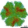 24821MC M: map data used to model this data C: citing same article ( |
|---|---|
| Similar structure data |
-Links
 PDBj
PDBj


- Assembly
| Deposited unit | 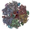
|
|---|---|
| 1 |
|
-Components
-Photosystem I P700 chlorophyll a apoprotein ... , 2 types, 6 molecules AGaBHb
| #1: Protein | Mass: 86411.227 Da / Num. of mol.: 3 / Source method: isolated from a natural source / Source: (natural) Synechococcus sp. PCC 7335 (bacteria) / Strain: ATCC 29403 / PCC 7335 / References: UniProt: B4WP20, photosystem I#2: Protein | Mass: 83207.648 Da / Num. of mol.: 3 / Source method: isolated from a natural source / Source: (natural) Synechococcus sp. PCC 7335 (bacteria) / Strain: ATCC 29403 / PCC 7335 / References: UniProt: B4WP21, photosystem I |
|---|
-Protein , 6 types, 18 molecules CNcDOdFQfIRiLUlXWx
| #3: Protein | Mass: 8809.169 Da / Num. of mol.: 3 / Source method: isolated from a natural source / Source: (natural) Synechococcus sp. PCC 7335 (bacteria)#4: Protein | Mass: 17051.336 Da / Num. of mol.: 3 / Source method: isolated from a natural source / Source: (natural) Synechococcus sp. PCC 7335 (bacteria) / Strain: ATCC 29403 / PCC 7335 / References: UniProt: B4WFP8#6: Protein | Mass: 18569.213 Da / Num. of mol.: 3 / Source method: isolated from a natural source / Source: (natural) Synechococcus sp. PCC 7335 (bacteria) / Strain: ATCC 29403 / PCC 7335 / References: UniProt: B4WP24#7: Protein | Mass: 7668.838 Da / Num. of mol.: 3 / Source method: isolated from a natural source / Source: (natural) Synechococcus sp. PCC 7335 (bacteria) / Strain: ATCC 29403 / PCC 7335 / References: UniProt: B4WP23#10: Protein | Mass: 18632.201 Da / Num. of mol.: 3 / Source method: isolated from a natural source / Source: (natural) Synechococcus sp. PCC 7335 (bacteria) / Strain: ATCC 29403 / PCC 7335 / References: UniProt: B4WP22#12: Protein | Mass: 10835.725 Da / Num. of mol.: 3 / Source method: isolated from a natural source / Source: (natural) Synechococcus sp. PCC 7335 (bacteria) / Strain: ATCC 29403 / PCC 7335 / References: UniProt: B4WFX2 |
|---|
-Photosystem I reaction center subunit ... , 3 types, 9 molecules EPeJSjKTk
| #5: Protein | Mass: 7955.112 Da / Num. of mol.: 3 / Source method: isolated from a natural source / Source: (natural) Synechococcus sp. PCC 7335 (bacteria) / Strain: ATCC 29403 / PCC 7335 / References: UniProt: B4WSJ5#8: Protein/peptide | Mass: 5170.094 Da / Num. of mol.: 3 / Source method: isolated from a natural source / Source: (natural) Synechococcus sp. PCC 7335 (bacteria) / Strain: ATCC 29403 / PCC 7335 / References: UniProt: B4WP25#9: Protein | Mass: 8195.834 Da / Num. of mol.: 3 / Source method: isolated from a natural source / Source: (natural) Synechococcus sp. PCC 7335 (bacteria) / Strain: ATCC 29403 / PCC 7335 / References: UniProt: B4WL17 |
|---|
-Protein/peptide / Sugars , 2 types, 42 molecules MVm
| #11: Protein/peptide | Mass: 3366.065 Da / Num. of mol.: 3 / Source method: isolated from a natural source / Source: (natural) Synechococcus sp. PCC 7335 (bacteria)#21: Sugar | ChemComp-LMT /  Type: D-saccharide / Mass: 510.615 Da / Num. of mol.: 39 / Source method: obtained synthetically / Formula: C24H46O11 / Comment: detergent*YM Type: D-saccharide / Mass: 510.615 Da / Num. of mol.: 39 / Source method: obtained synthetically / Formula: C24H46O11 / Comment: detergent*YM |
|---|
-Non-polymers , 12 types, 711 molecules 


| #13: Chemical |  Mass: 893.489 Da / Num. of mol.: 3 / Source method: isolated from a natural source / Formula: C55H72MgN4O5 Mass: 893.489 Da / Num. of mol.: 3 / Source method: isolated from a natural source / Formula: C55H72MgN4O5#14: Chemical | ChemComp-CLA /  Mass: 893.489 Da / Num. of mol.: 255 / Source method: isolated from a natural source / Formula: C55H72MgN4O5 Mass: 893.489 Da / Num. of mol.: 255 / Source method: isolated from a natural source / Formula: C55H72MgN4O5#15: Chemical | ChemComp-F6C /  Mass: 905.457 Da / Num. of mol.: 18 / Source method: isolated from a natural source / Formula: C55H68MgN4O6 / Feature type: SUBJECT OF INVESTIGATION Mass: 905.457 Da / Num. of mol.: 18 / Source method: isolated from a natural source / Formula: C55H68MgN4O6 / Feature type: SUBJECT OF INVESTIGATION#16: Chemical | ChemComp-PQN /  Mass: 450.696 Da / Num. of mol.: 6 / Source method: isolated from a natural source / Formula: C31H46O2 Mass: 450.696 Da / Num. of mol.: 6 / Source method: isolated from a natural source / Formula: C31H46O2#17: Chemical | ChemComp-SF4 /  Mass: 351.640 Da / Num. of mol.: 9 / Source method: isolated from a natural source / Formula: Fe4S4 Mass: 351.640 Da / Num. of mol.: 9 / Source method: isolated from a natural source / Formula: Fe4S4#18: Chemical | ChemComp-BCR /  Mass: 536.873 Da / Num. of mol.: 57 / Source method: isolated from a natural source / Formula: C40H56 Mass: 536.873 Da / Num. of mol.: 57 / Source method: isolated from a natural source / Formula: C40H56#19: Chemical | ChemComp-LHG /  Mass: 722.970 Da / Num. of mol.: 12 / Source method: obtained synthetically / Formula: C38H75O10P / Comment: phospholipid*YM Mass: 722.970 Da / Num. of mol.: 12 / Source method: obtained synthetically / Formula: C38H75O10P / Comment: phospholipid*YM#20: Chemical | ChemComp-LMG /  Mass: 787.158 Da / Num. of mol.: 9 / Source method: obtained synthetically / Formula: C45H86O10 Mass: 787.158 Da / Num. of mol.: 9 / Source method: obtained synthetically / Formula: C45H86O10#22: Chemical |  Mass: 35.453 Da / Num. of mol.: 3 / Source method: obtained synthetically / Formula: Cl Mass: 35.453 Da / Num. of mol.: 3 / Source method: obtained synthetically / Formula: Cl#23: Chemical |  Mass: 40.078 Da / Num. of mol.: 3 / Source method: obtained synthetically / Formula: Ca Mass: 40.078 Da / Num. of mol.: 3 / Source method: obtained synthetically / Formula: Ca#24: Chemical |  Mass: 175.820 Da / Num. of mol.: 3 / Source method: isolated from a natural source / Formula: Fe2S2 Mass: 175.820 Da / Num. of mol.: 3 / Source method: isolated from a natural source / Formula: Fe2S2#25: Water | ChemComp-HOH / | Mass: 18.015 Da / Num. of mol.: 333 / Source method: isolated from a natural source / Formula: H2O |
|---|
-Details
| Has ligand of interest | Y |
|---|
-Experimental details
-Experiment
| Experiment | Method: ELECTRON MICROSCOPY |
|---|---|
| EM experiment | Aggregation state: PARTICLE / 3D reconstruction method: single particle reconstruction |
- Sample preparation
| Component | Name: Far-red light-acclimated Photosystem I from Synechococcus sp. PCC 7335 Type: COMPLEX / Entity ID: #1-#12 / Source: NATURAL |
|---|---|
| Source (natural) | Organism: Synechococcus sp. PCC 7335 (bacteria) |
| Buffer solution | pH: 6.5 |
| Specimen | Embedding applied: NO / Shadowing applied: NO / Staining applied: NO / Vitrification applied: YES |
| Vitrification | Cryogen name: ETHANE |
- Electron microscopy imaging
| Experimental equipment |  Model: Titan Krios / Image courtesy: FEI Company |
|---|---|
| Microscopy | Model: FEI TITAN KRIOS |
| Electron gun | Electron source:  FIELD EMISSION GUN / Accelerating voltage: 300 kV / Illumination mode: FLOOD BEAM FIELD EMISSION GUN / Accelerating voltage: 300 kV / Illumination mode: FLOOD BEAM |
| Electron lens | Mode: BRIGHT FIELD |
| Image recording | Electron dose: 40.8 e/Å2 / Film or detector model: GATAN K3 (6k x 4k) |
- Processing
| CTF correction | Type: PHASE FLIPPING AND AMPLITUDE CORRECTION |
|---|---|
| 3D reconstruction | Resolution: 2.91 Å / Resolution method: FSC 0.143 CUT-OFF / Num. of particles: 286672 / Symmetry type: POINT |
