- EMDB-24821: Structure of photosystem I with bound ferredoxin from Synechococc... -
+
Open data
ID or keywords:
Loading...
-
Basic information
Entry
Database: EMDB / ID: EMD-24821
Title
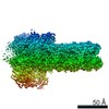
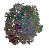
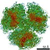
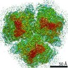
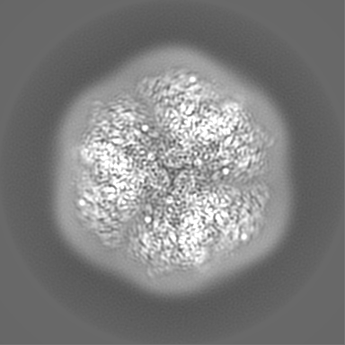
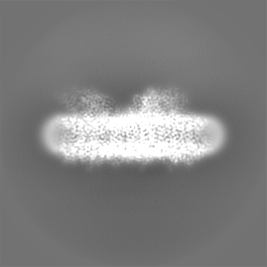
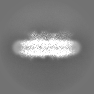
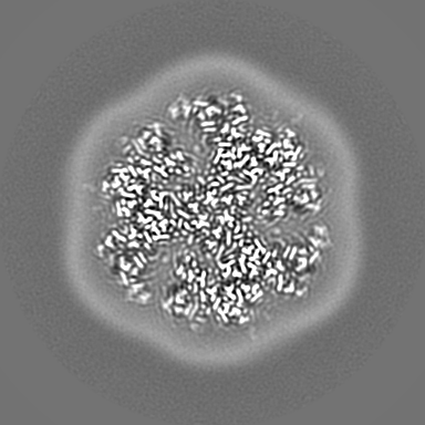
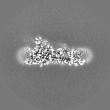
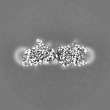
Structure of photosystem I with bound ferredoxin from Synechococcus sp. PCC 7335 acclimated to far-red light
Map data
Sharpened map for Far-red light-acclimated PSI from Synechococcus sp. PCC 7335
Sample
Complex: Far-red light-acclimated Photosystem I from Synechococcus sp. PCC 7335
Protein or peptide: x 12 types
Ligand: x 13 types
Keywords
Photosystem I / Far-red light photoacclimation / Chlorophyll f / Ferredoxin / PsaF / PsaJ / PHOTOSYNTHESIS
Function / homology
Function and homology information
photosystem I reaction center / photosystem I / photosystem I / plasma membrane-derived thylakoid membrane / chlorophyll binding / photosynthesis / endomembrane system / electron transport chain / 2 iron, 2 sulfur cluster binding / 4 iron, 4 sulfur cluster binding ...photosystem I reaction center / photosystem I / photosystem I / plasma membrane-derived thylakoid membrane / chlorophyll binding / photosynthesis / endomembrane system / electron transport chain / 2 iron, 2 sulfur cluster binding / 4 iron, 4 sulfur cluster binding / electron transfer activity / oxidoreductase activity / magnesium ion binding / metal ion binding Similarity search - Function
Photosystem I reaction center subunit PsaK / Ferredoxin [2Fe-2S], plant / Photosystem I reaction centre subunit PsaK / Photosystem I reaction centre subunit PsaK superfamily / Photosystem I reaction center subunit V/PsaK / Photosystem I psaG / psaK / Photosystem I PsaL, reaction centre subunit XI / Photosystem I, reaction centre subunit XI / Photosystem I PsaL, reaction centre subunit XI superfamily / Photosystem I reaction centre subunit XI ...Photosystem I reaction center subunit PsaK / Ferredoxin [2Fe-2S], plant / Photosystem I reaction centre subunit PsaK / Photosystem I reaction centre subunit PsaK superfamily / Photosystem I reaction center subunit V/PsaK / Photosystem I psaG / psaK / Photosystem I PsaL, reaction centre subunit XI / Photosystem I, reaction centre subunit XI / Photosystem I PsaL, reaction centre subunit XI superfamily / Photosystem I reaction centre subunit XI / Photosystem I reaction centre subunit VIII superfamily / Photosystem I PsaF, reaction centre subunit III / Photosystem I PsaF, reaction centre subunit III superfamily / Photosystem I reaction centre subunit III / Photosystem I PsaD / Photosystem I, reaction centre subunit PsaD superfamily / PsaD / Photosystem I PsaE, reaction centre subunit IV / Photosystem I reaction centre subunit IV / PsaE / Photosystem I PsaJ, reaction centre subunit IX superfamily / Photosystem I PsaJ, reaction centre subunit IX / Photosystem I reaction centre subunit IX / PsaJ / Photosystem I PsaA / Photosystem I PsaB / Photosystem I PsaA/PsaB, conserved site / Photosystem I psaA and psaB proteins signature. / Photosystem I PsaA/PsaB / Photosystem I PsaA/PsaB superfamily / Photosystem I psaA/psaB protein / 2Fe-2S ferredoxin, iron-sulphur binding site / 2Fe-2S ferredoxin-type iron-sulfur binding region signature. / Electron transport accessory-like domain superfamily / 2Fe-2S iron-sulfur cluster binding domain / Beta-grasp domain superfamily / 2Fe-2S ferredoxin-type iron-sulfur binding domain profile. / 2Fe-2S ferredoxin-type iron-sulfur binding domain / 2Fe-2S ferredoxin-like superfamily Similarity search - Domain/homology
Photosystem I reaction center subunit II / Ferredoxin-1 / Photosystem I reaction center subunit PsaK / Photosystem I P700 chlorophyll a apoprotein A1 / Photosystem I P700 chlorophyll a apoprotein A2 / Photosystem I reaction center subunit XI / Photosystem I reaction center subunit VIII / Photosystem I reaction center subunit III / Photosystem I reaction center subunit IX / Photosystem I reaction center subunit IV Similarity search - Component
Biological species
Synechococcus sp. PCC 7335 (bacteria)
Method
single particle reconstruction / cryo EM / Resolution: 2.91 Å
Journal: J Biol Chem / Year: 2022 Title: Structure of a photosystem I-ferredoxin complex from a marine cyanobacterium provides insights into far-red light photoacclimation. Authors: Christopher J Gisriel / David A Flesher / Gaozhong Shen / Jimin Wang / Ming-Yang Ho / Gary W Brudvig / Donald A Bryant / Abstract: Far-red light photoacclimation exhibited by some cyanobacteria allows these organisms to use the far-red region of the solar spectrum (700-800 nm) for photosynthesis. Part of this process includes ...Far-red light photoacclimation exhibited by some cyanobacteria allows these organisms to use the far-red region of the solar spectrum (700-800 nm) for photosynthesis. Part of this process includes the replacement of six photosystem I (PSI) subunits with isoforms that confer the binding of chlorophyll (Chl) f molecules that absorb far-red light (FRL). However, the exact sites at which Chl f molecules are bound are still challenging to determine. To aid in the identification of Chl f-binding sites, we solved the cryo-EM structure of PSI from far-red light-acclimated cells of the cyanobacterium Synechococcus sp. PCC 7335. We identified six sites that bind Chl f with high specificity and three additional sites that are likely to bind Chl f at lower specificity. All of these binding sites are in the core-antenna regions of PSI, and Chl f was not observed among the electron transfer cofactors. This structural analysis also reveals both conserved and nonconserved Chl f-binding sites, the latter of which exemplify the diversity in FRL-PSI among species. We found that the FRL-PSI structure also contains a bound soluble ferredoxin, PetF1, at low occupancy, which suggests that ferredoxin binds less transiently than expected according to the canonical view of ferredoxin-binding to facilitate electron transfer. We suggest that this may result from structural changes in FRL-PSI that occur specifically during FRL photoacclimation.
History
Deposition
Sep 5, 2021
-
Header (metadata) release
Nov 24, 2021
-
Map release
Nov 24, 2021
-
Update
Jun 5, 2024
-
Current status
Jun 5, 2024
Processing site: RCSB / Status: Released
-
Structure visualization
Movie
Surface view with section colored by density value
In the structure databanks used in Yorodumi, some data are registered as the other names, "COVID-19 virus" and "2019-nCoV". Here are the details of the virus and the list of structure data.
Jan 31, 2019. EMDB accession codes are about to change! (news from PDBe EMDB page)
EMDB accession codes are about to change! (news from PDBe EMDB page)
The allocation of 4 digits for EMDB accession codes will soon come to an end. Whilst these codes will remain in use, new EMDB accession codes will include an additional digit and will expand incrementally as the available range of codes is exhausted. The current 4-digit format prefixed with “EMD-” (i.e. EMD-XXXX) will advance to a 5-digit format (i.e. EMD-XXXXX), and so on. It is currently estimated that the 4-digit codes will be depleted around Spring 2019, at which point the 5-digit format will come into force.
The EM Navigator/Yorodumi systems omit the EMD- prefix.
Related info.:Q: What is EMD? / ID/Accession-code notation in Yorodumi/EM Navigator
Yorodumi is a browser for structure data from EMDB, PDB, SASBDB, etc.
This page is also the successor to EM Navigator detail page, and also detail information page/front-end page for Omokage search.
The word "yorodu" (or yorozu) is an old Japanese word meaning "ten thousand". "mi" (miru) is to see.
Related info.:EMDB / PDB / SASBDB / Comparison of 3 databanks / Yorodumi Search / Aug 31, 2016. New EM Navigator & Yorodumi / Yorodumi Papers / Jmol/JSmol / Function and homology information / Changes in new EM Navigator and Yorodumi
 Movie
Movie Controller

 Yorodumi
 Open data
 Basic information
 Map data
 Sample
 Keywords
 Function and homology information
 Synechococcus sp. PCC 7335 (bacteria)
 Authors
 United States, 2 items
  Citation
 Structure visualization
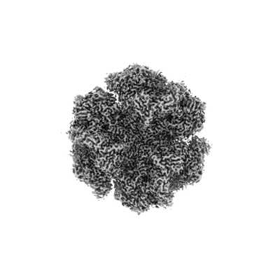
 Downloads & links
 emd_24821.png
 http://ftp.pdbj.org/pub/emdb/structures/EMD-24821


 Z (Sec.)
 Y (Row.)
 X (Col.)


 Sample components


 Processing
 Electron microscopy
 FIELD EMISSION GUN

