 Movie
Movie Controller

[English] 日本語
 Yorodumi
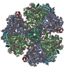
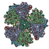
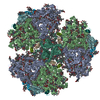
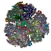
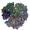
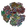
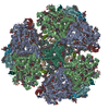
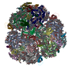
- PDB-6vpv: Trimeric Photosystem I from the High-Light Tolerant Cyanobacteria... -
+ Open data

- Basic information
| Entry | Database: PDB / ID: 6vpv | ||||||
|---|---|---|---|---|---|---|---|
| Title | Trimeric Photosystem I from the High-Light Tolerant Cyanobacteria Cyanobacterium Aponinum | ||||||
 Components Components |
| ||||||
 Keywords Keywords | PHOTOSYNTHESIS / Photosystem I / Membrane Protein / PSI | ||||||
| Function / homology |  Function and homology information Function and homology information: / photosystem I reaction center / photosystem I / photosynthetic electron transport in photosystem I / photosystem I / plasma membrane-derived thylakoid membrane / chlorophyll binding / membrane => GO:0016020 / photosynthesis / endomembrane system ...: / photosystem I reaction center / photosystem I / photosynthetic electron transport in photosystem I / photosystem I / plasma membrane-derived thylakoid membrane / chlorophyll binding / membrane => GO:0016020 / photosynthesis / endomembrane system / 4 iron, 4 sulfur cluster binding / electron transfer activity / oxidoreductase activity / magnesium ion binding / metal ion binding Similarity search - Function | ||||||
| Biological species |  Cyanobacterium aponinum 0216 (bacteria) Cyanobacterium aponinum 0216 (bacteria) | ||||||
| Method | ELECTRON MICROSCOPY / single particle reconstruction / cryo EM / Resolution: 2.7 Å | ||||||
 Authors Authors | Dobson, Z. / Toporik, H. / Vaughn, N. / Lin, S. / Williams, D. / Fromme, P. / Mazor, Y. | ||||||
 Citation Citation | Journal: Elife / Year: 2021 Title: The structure of photosystem I from a high-light-tolerant cyanobacteria. Authors: Zachary Dobson / Safa Ahad / Jackson Vanlandingham / Hila Toporik / Natalie Vaughn / Michael Vaughn / Dewight Williams / Michael Reppert / Petra Fromme / Yuval Mazor /  Abstract: Photosynthetic organisms have adapted to survive a myriad of extreme environments from the earth's deserts to its poles, yet the proteins that carry out the light reactions of photosynthesis are ...Photosynthetic organisms have adapted to survive a myriad of extreme environments from the earth's deserts to its poles, yet the proteins that carry out the light reactions of photosynthesis are highly conserved from the cyanobacteria to modern day crops. To investigate adaptations of the photosynthetic machinery in cyanobacteria to excessive light stress, we isolated a new strain of cyanobacteria, 0216, from the extreme light environment of the Sonoran Desert. Here we report the biochemical characterization and the 2.7 Å resolution structure of trimeric photosystem I from this high-light-tolerant cyanobacterium. The structure shows a new conformation of the PsaL C-terminus that supports trimer formation of cyanobacterial photosystem I. The spectroscopic analysis of this photosystem I revealed a decrease in far-red absorption, which is attributed to a decrease in the number of long- wavelength chlorophylls. Using these findings, we constructed two chimeric PSIs in sp. PCC 6803 demonstrating how unique structural features in photosynthetic complexes can change spectroscopic properties, allowing organisms to thrive under different environmental stresses. | ||||||
| History |
|
- Structure visualization
| Movie |
Movie viewer |
|---|---|
| Structure viewer | Molecule: MolmilJmol/JSmol |
- Downloads & links
-Download
| PDBx/mmCIF format | 6vpv.cif.gz | 1.6 MB | Display | PDBx/mmCIF format |
|---|---|---|---|---|
| PDB format | pdb6vpv.ent.gz | 1.4 MB | Display | PDB format |
| PDBx/mmJSON format | 6vpv.json.gz | Tree view | PDBx/mmJSON format | |
| Others |  Other downloads Other downloads |
-Validation report
| Arichive directory | https://data.pdbj.org/pub/pdb/validation_reports/vp/6vpvftp://data.pdbj.org/pub/pdb/validation_reports/vp/6vpv | HTTPS FTP |
|---|
-Related structure data
| Related structure data | 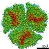 21320MC M: map data used to model this data C: citing same article ( |
|---|---|
| Similar structure data |

-Links
 PDBj
PDBj


- Assembly
| Deposited unit | 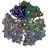
|
|---|---|
| 1 |
|
-Components
-Photosystem I P700 chlorophyll a apoprotein ... , 2 types, 6 molecules Aa1Bb2
| #1: Protein | Mass: 81754.039 Da / Num. of mol.: 3 / Source method: isolated from a natural source / Source: (natural) Cyanobacterium aponinum 0216 (bacteria) / Strain: 0216 / References: UniProt: A0A2G3P9X3, photosystem I#2: Protein | Mass: 81865.945 Da / Num. of mol.: 3 / Source method: isolated from a natural source / Source: (natural) Cyanobacterium aponinum 0216 (bacteria) / Strain: 0216 / References: UniProt: K9Z2J7, photosystem I |
|---|
-Protein , 2 types, 6 molecules Cc3Ff6
| #3: Protein | Mass: 8692.039 Da / Num. of mol.: 3 / Source method: isolated from a natural source / Source: (natural) Cyanobacterium aponinum 0216 (bacteria) / Strain: 0216 / References: UniProt: A0A2G3PD04, photosystem I#6: Protein | Mass: 15665.845 Da / Num. of mol.: 3 / Source method: isolated from a natural source / Source: (natural) Cyanobacterium aponinum 0216 (bacteria) / Strain: 0216 / References: UniProt: A0A2G3PEV5 |
|---|
-Photosystem I reaction center subunit ... , 7 types, 21 molecules Dd4Ee5Ii7Jj8Kk9Ll0Mmz
| #4: Protein | Mass: 15647.748 Da / Num. of mol.: 3 / Source method: isolated from a natural source / Source: (natural) Cyanobacterium aponinum 0216 (bacteria) / Strain: 0216 / References: UniProt: A0A2G3PAQ0#5: Protein | Mass: 7634.534 Da / Num. of mol.: 3 / Source method: isolated from a natural source / Source: (natural) Cyanobacterium aponinum 0216 (bacteria) / Strain: 0216 / References: UniProt: A0A2G3PEU8#7: Protein/peptide | Mass: 3913.726 Da / Num. of mol.: 3 / Source method: isolated from a natural source / Source: (natural) Cyanobacterium aponinum 0216 (bacteria) / Strain: 0216 / References: UniProt: K9Z9J3#8: Protein/peptide | Mass: 4354.158 Da / Num. of mol.: 3 / Source method: isolated from a natural source / Source: (natural) Cyanobacterium aponinum 0216 (bacteria) / Strain: 0216 / References: UniProt: A0A2G3PEV6#9: Protein | Mass: 7860.323 Da / Num. of mol.: 3 / Source method: isolated from a natural source / Source: (natural) Cyanobacterium aponinum 0216 (bacteria) / Strain: 0216 / References: UniProt: A0A2G3P6W6#10: Protein | Mass: 16779.295 Da / Num. of mol.: 3 / Source method: isolated from a natural source / Source: (natural) Cyanobacterium aponinum 0216 (bacteria) / Strain: 0216 / References: UniProt: A0A2G3P7D5#11: Protein/peptide | Mass: 3278.920 Da / Num. of mol.: 3 / Source method: isolated from a natural source / Source: (natural) Cyanobacterium aponinum 0216 (bacteria) / Strain: 0216 / References: UniProt: A0A2G3PA81 |
|---|
-Non-polymers , 8 types, 384 molecules 


| #12: Chemical |  Mass: 893.489 Da / Num. of mol.: 3 / Source method: obtained synthetically / Formula: C55H72MgN4O5 Mass: 893.489 Da / Num. of mol.: 3 / Source method: obtained synthetically / Formula: C55H72MgN4O5#13: Chemical | ChemComp-CLA /  Mass: 893.489 Da / Num. of mol.: 285 / Source method: obtained synthetically / Formula: C55H72MgN4O5 Mass: 893.489 Da / Num. of mol.: 285 / Source method: obtained synthetically / Formula: C55H72MgN4O5#14: Chemical | ChemComp-PQN /  Mass: 450.696 Da / Num. of mol.: 6 / Source method: obtained synthetically / Formula: C31H46O2 Mass: 450.696 Da / Num. of mol.: 6 / Source method: obtained synthetically / Formula: C31H46O2#15: Chemical | ChemComp-SF4 /  Mass: 351.640 Da / Num. of mol.: 9 / Source method: obtained synthetically / Formula: Fe4S4 Mass: 351.640 Da / Num. of mol.: 9 / Source method: obtained synthetically / Formula: Fe4S4#16: Chemical | ChemComp-BCR /  Mass: 536.873 Da / Num. of mol.: 69 / Source method: obtained synthetically / Formula: C40H56 Mass: 536.873 Da / Num. of mol.: 69 / Source method: obtained synthetically / Formula: C40H56#17: Chemical | ChemComp-LHG /  Mass: 722.970 Da / Num. of mol.: 6 / Source method: obtained synthetically / Formula: C38H75O10P / Comment: phospholipid*YM Mass: 722.970 Da / Num. of mol.: 6 / Source method: obtained synthetically / Formula: C38H75O10P / Comment: phospholipid*YM#18: Chemical |  Mass: 787.158 Da / Num. of mol.: 3 / Source method: obtained synthetically / Formula: C45H86O10 Mass: 787.158 Da / Num. of mol.: 3 / Source method: obtained synthetically / Formula: C45H86O10#19: Chemical |  Mass: 795.116 Da / Num. of mol.: 3 / Source method: obtained synthetically / Formula: C41H78O12S Mass: 795.116 Da / Num. of mol.: 3 / Source method: obtained synthetically / Formula: C41H78O12S |
|---|
-Details
| Has ligand of interest | N |
|---|---|
| Has protein modification | Y |
-Experimental details
-Experiment
| Experiment | Method: ELECTRON MICROSCOPY |
|---|---|
| EM experiment | Aggregation state: PARTICLE / 3D reconstruction method: single particle reconstruction |
- Sample preparation
| Component | Name: Trimeric PSI from a High Light tolerant cyanobacteria, Cyanobacterium aponinum Type: COMPLEX / Entity ID: #1-#11 / Source: NATURAL |
|---|---|
| Source (natural) | Organism: Cyanobacterium aponinum 0216 (bacteria) / Strain: 216 |
| Buffer solution | pH: 8 |
| Specimen | Embedding applied: NO / Shadowing applied: NO / Staining applied: NO / Vitrification applied: YES |
| Specimen support | Details: unspecified |
| Vitrification | Cryogen name: ETHANE |
- Electron microscopy imaging
| Experimental equipment |  Model: Titan Krios / Image courtesy: FEI Company |
|---|---|
| Microscopy | Model: FEI TITAN KRIOS |
| Electron gun | Electron source:  FIELD EMISSION GUN / Accelerating voltage: 300 kV / Illumination mode: FLOOD BEAM FIELD EMISSION GUN / Accelerating voltage: 300 kV / Illumination mode: FLOOD BEAM |
| Electron lens | Mode: BRIGHT FIELD |
| Image recording | Electron dose: 1.53 e/Å2 / Detector mode: SUPER-RESOLUTION / Film or detector model: GATAN K2 SUMMIT (4k x 4k) |
- Processing
| EM software |
| ||||||||||||||||||||||||
|---|---|---|---|---|---|---|---|---|---|---|---|---|---|---|---|---|---|---|---|---|---|---|---|---|---|
| CTF correction | Type: PHASE FLIPPING AND AMPLITUDE CORRECTION | ||||||||||||||||||||||||
| Symmetry | Point symmetry: C3 (3 fold cyclic) | ||||||||||||||||||||||||
| 3D reconstruction | Resolution: 2.7 Å / Resolution method: FSC 0.143 CUT-OFF / Num. of particles: 75290 / Symmetry type: POINT |
