Movie
Movie Controller

[English] 日本語
 Yorodumi
- PDB-7k7h: Density-fitted Model Structure of Antibody Variable Domains of Ty... -
+ Open data

- Basic information
| Entry | Database: PDB / ID: 7k7h | ||||||||||||
|---|---|---|---|---|---|---|---|---|---|---|---|---|---|
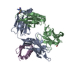
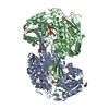
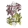
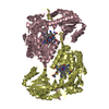
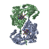
| Title | Density-fitted Model Structure of Antibody Variable Domains of TyTx1 in Complex with PltB pentamer of Typhoid Toxin | ||||||||||||
 Components Components |
| ||||||||||||
 Keywords Keywords | TOXIN / Typhoid Toxin / A2B5 / Antibody / Fab | ||||||||||||
| Function / homology | Bordetella pertussis toxin B, subunit 2/3, C-terminal / Pertussis toxin, subunit 2 and 3, C-terminal domain / Bordetella pertussis toxin A / Pertussis toxin, subunit 1 / Enterotoxin / NAD+ poly-ADP-ribosyltransferase activity / extracellular region / Pertussis like toxin subunit B / Pertussis toxin-like subunit ArtA Function and homology information Function and homology information | ||||||||||||
| Biological species |  Salmonella enterica subsp. enterica serovar Typhi str. CT18 (bacteria) Salmonella enterica subsp. enterica serovar Typhi str. CT18 (bacteria) | ||||||||||||
| Method | ELECTRON MICROSCOPY / single particle reconstruction / cryo EM / Resolution: 3 Å | ||||||||||||
 Authors Authors | Nguyen, T. / Feathers, J.R. / Fromme, J.C. / Song, J. | ||||||||||||
| Funding support |  United States, 3items United States, 3items
| ||||||||||||
 Citation Citation | Journal: Cell Rep / Year: 2021 Title: The structural basis of Salmonella AB toxin neutralization by antibodies targeting the glycan-receptor binding subunits. Authors: Tri Nguyen / Angelene F Richards / Durga P Neupane / J Ryan Feathers / Yi-An Yang / Ji Hyun Sim / Haewon Byun / Sohyoung Lee / Changhwan Ahn / Greta Van Slyke / J Christopher Fromme / ...Authors: Tri Nguyen / Angelene F Richards / Durga P Neupane / J Ryan Feathers / Yi-An Yang / Ji Hyun Sim / Haewon Byun / Sohyoung Lee / Changhwan Ahn / Greta Van Slyke / J Christopher Fromme / Nicholas J Mantis / Jeongmin Song / Abstract: Many bacterial pathogens secrete AB toxins comprising two functionally distinct yet complementary "A" and "B" subunits to benefit the pathogens during infection. The lectin-like pentameric B subunits ...Many bacterial pathogens secrete AB toxins comprising two functionally distinct yet complementary "A" and "B" subunits to benefit the pathogens during infection. The lectin-like pentameric B subunits recognize specific sets of host glycans to deliver the toxin into target host cells. Here, we offer the molecular mechanism by which neutralizing antibodies, which have the potential to bind to all glycan-receptor binding sites and thus completely inhibit toxin binding to host cells, are inhibited from exerting this action. Cryogenic electron microscopy (cryo-EM)-based analyses indicate that the skewed positioning of the toxin A subunit(s) toward one side of the toxin B pentamer inhibited neutralizing antibody binding to the laterally located epitopes, rendering some glycan-receptor binding sites that remained available for the toxin binding and endocytosis process, which is strikingly different from the counterpart antibodies recognizing the far side-located epitopes. These results highlight additional features of the toxin-antibody interactions and offer important insights into anti-toxin strategies. | ||||||||||||
| History |
|
- Structure visualization
| Movie |
Movie viewer |
|---|---|
| Structure viewer | Molecule: MolmilJmol/JSmol |
- Downloads & links
-Download
| PDBx/mmCIF format | 7k7h.cif.gz | 148.6 KB | Display | PDBx/mmCIF format |
|---|---|---|---|---|
| PDB format | pdb7k7h.ent.gz | 115.2 KB | Display | PDB format |
| PDBx/mmJSON format | 7k7h.json.gz | Tree view | PDBx/mmJSON format | |
| Others |  Other downloads Other downloads |
-Validation report
| Arichive directory | https://data.pdbj.org/pub/pdb/validation_reports/k7/7k7hftp://data.pdbj.org/pub/pdb/validation_reports/k7/7k7h | HTTPS FTP |
|---|
-Related structure data
| Related structure data | 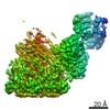 22699MC  7k7iC M: map data used to model this data C: citing same article ( |
|---|---|
| Similar structure data |


-Links
 PDBj
PDBj


- Assembly
| Deposited unit | 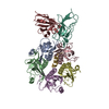
|
|---|---|
| 1 |
|
-Components
| #1: Protein | Mass: 12563.042 Da / Num. of mol.: 5 Source method: isolated from a genetically manipulated source Source: (gene. exp.) Salmonella enterica subsp. enterica serovar Typhi str. CT18 (bacteria)Gene: pltB, D4F19_07865, D4F39_13740, D4G09_02615, D4X81_03235, D5848_06295, D5891_16360, D5B50_14740, D5C10_10475, D6331_06440, D6K86_15515, D6P24_09080, D6Q71_07195, D7N07_01470, D8R98_02600, D8S38_ ...Gene: pltB, D4F19_07865, D4F39_13740, D4G09_02615, D4X81_03235, D5848_06295, D5891_16360, D5B50_14740, D5C10_10475, D6331_06440, D6K86_15515, D6P24_09080, D6Q71_07195, D7N07_01470, D8R98_02600, D8S38_09485, DL104_04040, DLF44_02615, DM364_15630, DMA85_08120, DMV05_17400, DN022_06665, DN116_07470, DN223_02675, DNJ32_13915, DNL67_05455, DNM39_02515, DNV82_15785, DNV95_15160, DOH59_08615, DP757_14390, DPC06_03915, DPJ15_14935, DPS97_10830, DQ802_09840, DQD72_07750, DQJ57_09285, DRE79_02610, DRW87_14355, DRX58_02590, DRX79_07315, DS260_02735, DS269_05775, DS339_07525, DS529_00005, DSM93_00460, DST18_05335, DTV88_06700, DU090_09370, DUQ83_10880, DUW14_00135, DVF55_05015, EDK96_01785, EIT32_03595, EIT43_00570, YL55_10165 Production host: #2: Antibody | | Mass: 12679.397 Da / Num. of mol.: 1 Source method: isolated from a genetically manipulated source Source: (gene. exp.) #3: Antibody | | Mass: 12802.131 Da / Num. of mol.: 1 Source method: isolated from a genetically manipulated source Source: (gene. exp.) #4: Protein/peptide | | Mass: 1665.970 Da / Num. of mol.: 1 Source method: isolated from a genetically manipulated source Source: (gene. exp.) Salmonella enterica subsp. enterica serovar Typhi str. CT18 (bacteria)Gene: artA, AFK99_24650, AN748_04695, AOB35_04685, AOC72_15785, AVA64_02290, AVS30_04695, AVS34_04420, AVS35_03980, AYK09_02505, BJP54_09640, BU463_03975, BW003_04370, BXS07_04190, C9E99_16570, C9F07_ ...Gene: artA, AFK99_24650, AN748_04695, AOB35_04685, AOC72_15785, AVA64_02290, AVS30_04695, AVS34_04420, AVS35_03980, AYK09_02505, BJP54_09640, BU463_03975, BW003_04370, BXS07_04190, C9E99_16570, C9F07_13170, C9F10_26685, CBK82_04610, CN028_04870, D6R15_03955, DFQ52_06390, DK703_09395, DLM27_02680, DN997_01215, DNM08_05965, DNM99_06525, DPT12_02550, DS310_07935, E0V17_06020, EDK86_05845, EJO30_06115 Production host: Has protein modification | Y | |
|---|
-Experimental details
-Experiment
| Experiment | Method: ELECTRON MICROSCOPY |
|---|---|
| EM experiment | Aggregation state: PARTICLE / 3D reconstruction method: single particle reconstruction |
- Sample preparation
| Component |
| ||||||||||||||||||||||||||||
|---|---|---|---|---|---|---|---|---|---|---|---|---|---|---|---|---|---|---|---|---|---|---|---|---|---|---|---|---|---|
| Molecular weight |
| ||||||||||||||||||||||||||||
| Source (natural) |
| ||||||||||||||||||||||||||||
| Source (recombinant) |
| ||||||||||||||||||||||||||||
| Buffer solution | pH: 7.5 | ||||||||||||||||||||||||||||
| Buffer component |
| ||||||||||||||||||||||||||||
| Specimen | Conc.: 0.2 mg/ml / Embedding applied: NO / Shadowing applied: NO / Staining applied: NO / Vitrification applied: YES Details: Freshly prepared size-exclusion-chromatography purified complex of TyTx1 Fab and Typhoid toxin catalytically inactivated toxin double mutant. | ||||||||||||||||||||||||||||
| Specimen support | Grid material: COPPER / Grid mesh size: 300 divisions/in. / Grid type: Quantifoil R1.2/1.3 | ||||||||||||||||||||||||||||
| Vitrification | Instrument: FEI VITROBOT MARK IV / Cryogen name: ETHANE / Humidity: 100 % / Chamber temperature: 277.15 K / Details: blot for 2.5 second before plunging |
- Electron microscopy imaging
| Experimental equipment |  Model: Talos Arctica / Image courtesy: FEI Company |
|---|---|
| Microscopy | Model: FEI TALOS ARCTICA |
| Electron gun | Electron source:  FIELD EMISSION GUN / Accelerating voltage: 200 kV / Illumination mode: FLOOD BEAM FIELD EMISSION GUN / Accelerating voltage: 200 kV / Illumination mode: FLOOD BEAM |
| Electron lens | Mode: BRIGHT FIELD / Nominal magnification: 49000 X / Nominal defocus max: 1500 nm / Nominal defocus min: 800 nm / Cs: 2.7 mm / C2 aperture diameter: 50 µm |
| Specimen holder | Cryogen: NITROGEN |
| Image recording | Electron dose: 53 e/Å2 / Film or detector model: GATAN K3 (6k x 4k) |
| EM imaging optics | Energyfilter name: GIF Bioquantum / Energyfilter slit width: 20 eV |
- Processing
| Software |
| ||||||||||||||||||||||||||||||||||||||||||||||||||||||||
|---|---|---|---|---|---|---|---|---|---|---|---|---|---|---|---|---|---|---|---|---|---|---|---|---|---|---|---|---|---|---|---|---|---|---|---|---|---|---|---|---|---|---|---|---|---|---|---|---|---|---|---|---|---|---|---|---|---|
| EM software |
| ||||||||||||||||||||||||||||||||||||||||||||||||||||||||
| CTF correction | Type: PHASE FLIPPING AND AMPLITUDE CORRECTION | ||||||||||||||||||||||||||||||||||||||||||||||||||||||||
| Symmetry | Point symmetry: C1 (asymmetric) | ||||||||||||||||||||||||||||||||||||||||||||||||||||||||
| 3D reconstruction | Resolution: 3 Å / Resolution method: FSC 0.143 CUT-OFF / Num. of particles: 326766 / Symmetry type: POINT | ||||||||||||||||||||||||||||||||||||||||||||||||||||||||
| Atomic model building | Details: Initial local fitting was done using Chimera and then Coot was used for rebuilding Fab variable domains into correct sequences. Refinement was performed using Real Space Refine in PHENIX and ...Details: Initial local fitting was done using Chimera and then Coot was used for rebuilding Fab variable domains into correct sequences. Refinement was performed using Real Space Refine in PHENIX and was iterated with manual building in Coot. | ||||||||||||||||||||||||||||||||||||||||||||||||||||||||
| Atomic model building | 3D fitting-ID: 1
| ||||||||||||||||||||||||||||||||||||||||||||||||||||||||
| Refinement | Cross valid method: NONE | ||||||||||||||||||||||||||||||||||||||||||||||||||||||||
| Refine LS restraints |
|