| Entry | Database: PDB / ID: 4lem
|
|---|
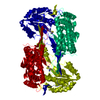
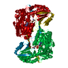
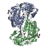
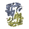
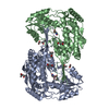
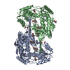
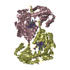
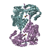
| Title | Crystal structure of the Delta-pyrroline-5-carboxylate dehydrogenase from Mycobacterium tuberculosis |
|---|
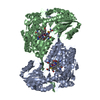
 Components Components | 1-pyrroline-5-carboxylate dehydrogenase |
|---|
 Keywords Keywords | OXIDOREDUCTASE / Rossmann fold / dehydrogenase |
|---|
| Function / homology |  Function and homology information Function and homology information
proline dehydrogenase activity / L-glutamate gamma-semialdehyde dehydrogenase / L-glutamate gamma-semialdehyde dehydrogenase activity / : / cytoplasmic side of plasma membrane / nucleotide binding / metal ion bindingSimilarity search - Function Delta-1-pyrroline-5-carboxylate dehydrogenase / : / Aldehyde Dehydrogenase; Chain A, domain 2 / Aldehyde Dehydrogenase; Chain A, domain 2 / Aldehyde Dehydrogenase; Chain A, domain 1 / Aldehyde Dehydrogenase; Chain A, domain 1 / Aldehyde dehydrogenase, cysteine active site / Aldehyde dehydrogenases cysteine active site. / Aldehyde dehydrogenase domain / Aldehyde dehydrogenase family ...Delta-1-pyrroline-5-carboxylate dehydrogenase / : / Aldehyde Dehydrogenase; Chain A, domain 2 / Aldehyde Dehydrogenase; Chain A, domain 2 / Aldehyde Dehydrogenase; Chain A, domain 1 / Aldehyde Dehydrogenase; Chain A, domain 1 / Aldehyde dehydrogenase, cysteine active site / Aldehyde dehydrogenases cysteine active site. / Aldehyde dehydrogenase domain / Aldehyde dehydrogenase family / Aldehyde dehydrogenase, C-terminal / Aldehyde dehydrogenase, N-terminal / Aldehyde/histidinol dehydrogenase / 3-Layer(aba) Sandwich / Alpha BetaSimilarity search - Domain/homology |
|---|
| Biological species |   Mycobacterium tuberculosis (bacteria) Mycobacterium tuberculosis (bacteria) |
|---|
| Method |  X-RAY DIFFRACTION / SYNCHROTRON / MOLECULAR REPLACEMENT / Resolution: 2.27 Å X-RAY DIFFRACTION / SYNCHROTRON / MOLECULAR REPLACEMENT / Resolution: 2.27 Å |
|---|
 Authors Authors | Lagautriere, T. / Bashiri, G. / Baker, E.N. |
|---|
 Citation Citation | Journal: J.Struct.Biol. / Year: 2015
Title: Use of a "silver bullet" to resolve crystal lattice dislocation disorder: A cobalamin complex of Delta (1)-pyrroline-5-carboxylate dehydrogenase from Mycobacterium tuberculosis.
Authors: Lagautriere, T. / Bashiri, G. / Baker, E.N. |
|---|
| History | | Deposition | Jun 26, 2013 | Deposition site: RCSB / Processing site: RCSB |
|---|
| Revision 1.0 | May 14, 2014 | Provider: repository / Type: Initial release |
|---|
| Revision 1.1 | May 21, 2014 | Group: Database references |
|---|
| Revision 1.2 | Nov 12, 2014 | Group: Structure summary |
|---|
| Revision 1.3 | Mar 4, 2015 | Group: Database references |
|---|
| Revision 1.4 | Nov 6, 2024 | Group: Data collection / Database references ...Data collection / Database references / Derived calculations / Structure summary
Category: chem_comp_atom / chem_comp_bond ...chem_comp_atom / chem_comp_bond / database_2 / pdbx_entry_details / pdbx_modification_feature / pdbx_struct_conn_angle / struct_conn / struct_ref_seq_dif / struct_site
Item: _database_2.pdbx_DOI / _database_2.pdbx_database_accession ..._database_2.pdbx_DOI / _database_2.pdbx_database_accession / _pdbx_struct_conn_angle.ptnr1_auth_asym_id / _pdbx_struct_conn_angle.ptnr1_auth_comp_id / _pdbx_struct_conn_angle.ptnr1_auth_seq_id / _pdbx_struct_conn_angle.ptnr1_label_asym_id / _pdbx_struct_conn_angle.ptnr1_label_comp_id / _pdbx_struct_conn_angle.ptnr1_label_seq_id / _pdbx_struct_conn_angle.ptnr2_auth_asym_id / _pdbx_struct_conn_angle.ptnr2_auth_seq_id / _pdbx_struct_conn_angle.ptnr2_label_asym_id / _pdbx_struct_conn_angle.ptnr3_auth_asym_id / _pdbx_struct_conn_angle.ptnr3_auth_comp_id / _pdbx_struct_conn_angle.ptnr3_auth_seq_id / _pdbx_struct_conn_angle.ptnr3_label_asym_id / _pdbx_struct_conn_angle.ptnr3_label_comp_id / _pdbx_struct_conn_angle.ptnr3_label_seq_id / _pdbx_struct_conn_angle.value / _struct_conn.pdbx_dist_value / _struct_conn.pdbx_leaving_atom_flag / _struct_conn.ptnr1_auth_asym_id / _struct_conn.ptnr1_auth_comp_id / _struct_conn.ptnr1_auth_seq_id / _struct_conn.ptnr1_label_asym_id / _struct_conn.ptnr1_label_atom_id / _struct_conn.ptnr1_label_comp_id / _struct_conn.ptnr1_label_seq_id / _struct_conn.ptnr2_auth_asym_id / _struct_conn.ptnr2_auth_comp_id / _struct_conn.ptnr2_auth_seq_id / _struct_conn.ptnr2_label_asym_id / _struct_conn.ptnr2_label_atom_id / _struct_conn.ptnr2_label_comp_id / _struct_ref_seq_dif.details / _struct_site.pdbx_auth_asym_id / _struct_site.pdbx_auth_comp_id / _struct_site.pdbx_auth_seq_id |
|---|
|
|---|
 Movie
Movie Controller

 Yorodumi
 Open data
 Basic information
 Structure visualization
 Downloads & links
 Other downloads


 PDBj
PDBj

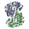
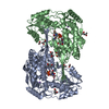
 Assembly
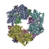

 Mass: 1330.356 Da / Num. of mol.: 4 / Source method: obtained synthetically / Formula: C62H89CoN13O14P
 Mass: 24.305 Da / Num. of mol.: 3 / Source method: obtained synthetically / Formula: Mg
 Mass: 18.015 Da / Num. of mol.: 2514 / Source method: isolated from a natural source / Formula: H2O
 Sample preparation
 / Beamline: MX2 / Wavelength: 0.953691 Å
 Processing