 Movie
Movie Controller

[English] 日本語
 Yorodumi
- PDB-4ns3: Crystal structure of the Delta-pyrroline-5-carboxylate dehydrogen... -
+ Open data

- Basic information
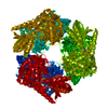
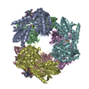
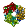
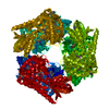
| Entry | Database: PDB / ID: 4ns3 | ||||||
|---|---|---|---|---|---|---|---|
| Title | Crystal structure of the Delta-pyrroline-5-carboxylate dehydrogenase from Mycobacterium tuberculosis bound with NAD and cobalamin | ||||||
 Components Components | Delta-1-pyrroline-5-carboxylate dehydrogenase | ||||||
 Keywords Keywords | OXIDOREDUCTASE / Rossmann Fold / dehydrogenase / dehydrogenation | ||||||
| Function / homology |  Function and homology information Function and homology informationproline dehydrogenase activity / L-glutamate gamma-semialdehyde dehydrogenase / L-glutamate gamma-semialdehyde dehydrogenase activity / : / cytoplasmic side of plasma membrane / nucleotide binding / metal ion binding Similarity search - Function | ||||||
| Biological species |   Mycobacterium tuberculosis (bacteria) Mycobacterium tuberculosis (bacteria) | ||||||
| Method |  X-RAY DIFFRACTION / SYNCHROTRON / MOLECULAR REPLACEMENT / Resolution: 2.38 Å X-RAY DIFFRACTION / SYNCHROTRON / MOLECULAR REPLACEMENT / Resolution: 2.38 Å | ||||||
 Authors Authors | Lagautriere, T. / Bashiri, G. / Baker, E.N. | ||||||
 Citation Citation | Journal: J.Struct.Biol. / Year: 2015 Title: Use of a "silver bullet" to resolve crystal lattice dislocation disorder: A cobalamin complex of Delta (1)-pyrroline-5-carboxylate dehydrogenase from Mycobacterium tuberculosis. Authors: Lagautriere, T. / Bashiri, G. / Baker, E.N. | ||||||
| History |
|
- Structure visualization
| Structure viewer | Molecule: MolmilJmol/JSmol |
|---|
- Downloads & links
-Download
| PDBx/mmCIF format | 4ns3.cif.gz | 679.5 KB | Display | PDBx/mmCIF format |
|---|---|---|---|---|
| PDB format | pdb4ns3.ent.gz | 554.9 KB | Display | PDB format |
| PDBx/mmJSON format | 4ns3.json.gz | Tree view | PDBx/mmJSON format | |
| Others |  Other downloads Other downloads |
-Validation report
| Arichive directory | https://data.pdbj.org/pub/pdb/validation_reports/ns/4ns3ftp://data.pdbj.org/pub/pdb/validation_reports/ns/4ns3 | HTTPS FTP |
|---|
-Related structure data


-Links
 PDBj
PDBj

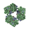
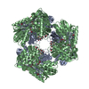
- Assembly
| Deposited unit | 
| |||||||||||||||||||||||||||||||||||||||||||||||||||||||||||||||||||||||||||||||||||||||||||||||||||||||||||||||||||||||||||||||||||||||||||||||||||||||||||||||||||||||||||||||||||||||||||||||||||||||||||||||||||||||||||||||||||||||
|---|---|---|---|---|---|---|---|---|---|---|---|---|---|---|---|---|---|---|---|---|---|---|---|---|---|---|---|---|---|---|---|---|---|---|---|---|---|---|---|---|---|---|---|---|---|---|---|---|---|---|---|---|---|---|---|---|---|---|---|---|---|---|---|---|---|---|---|---|---|---|---|---|---|---|---|---|---|---|---|---|---|---|---|---|---|---|---|---|---|---|---|---|---|---|---|---|---|---|---|---|---|---|---|---|---|---|---|---|---|---|---|---|---|---|---|---|---|---|---|---|---|---|---|---|---|---|---|---|---|---|---|---|---|---|---|---|---|---|---|---|---|---|---|---|---|---|---|---|---|---|---|---|---|---|---|---|---|---|---|---|---|---|---|---|---|---|---|---|---|---|---|---|---|---|---|---|---|---|---|---|---|---|---|---|---|---|---|---|---|---|---|---|---|---|---|---|---|---|---|---|---|---|---|---|---|---|---|---|---|---|---|---|---|---|---|---|---|---|---|---|---|---|---|---|---|---|---|---|---|---|---|---|
| 1 |
| |||||||||||||||||||||||||||||||||||||||||||||||||||||||||||||||||||||||||||||||||||||||||||||||||||||||||||||||||||||||||||||||||||||||||||||||||||||||||||||||||||||||||||||||||||||||||||||||||||||||||||||||||||||||||||||||||||||||
| Unit cell |
| |||||||||||||||||||||||||||||||||||||||||||||||||||||||||||||||||||||||||||||||||||||||||||||||||||||||||||||||||||||||||||||||||||||||||||||||||||||||||||||||||||||||||||||||||||||||||||||||||||||||||||||||||||||||||||||||||||||||
| Noncrystallographic symmetry (NCS) | NCS domain:
NCS domain segments: Component-ID: _ / Refine code: _
|