Protocol: SINGLE WAVELENGTH / Monochromatic (M) / Laue (L): M / Scattering type: x-ray
Radiation wavelength
Wavelength: 0.953688 Å / Relative weight: 1
Reflection
Resolution: 1.6→29.8 Å / Num. obs: 93414 / % possible obs: 99.8 %
Reflection shell
Resolution (Å)
Diffraction-ID
% possible all
1.6-1.69
1
98.7
5.07-29.84
1
99.3
-
Processing
Software
Name
Version
Classification
Blu-Ice
ice
datacollection
MOLREP
phasing
REFMAC
5.6.0117
refinement
XDS
datareduction
SCALA
datascaling
Refinement
Method to determine structure: MOLECULAR REPLACEMENT / Resolution: 1.6→29.8 Å / Cor.coef. Fo:Fc: 0.972 / Cor.coef. Fo:Fc free: 0.957 / SU B: 1.434 / SU ML: 0.049 / Cross valid method: THROUGHOUT / ESU R: 0.069 / ESU R Free: 0.073 / Stereochemistry target values: MAXIMUM LIKELIHOOD / Details: HYDROGENS HAVE BEEN USED IF PRESENT IN THE INPUT
Rfactor
Num. reflection
% reflection
Selection details
Rfree
0.20203
4908
5 %
RANDOM
Rwork
0.17025
-
-
-
obs
0.17183
93414
99.72 %
-
Solvent computation
Ion probe radii: 0.8 Å / Shrinkage radii: 0.8 Å / VDW probe radii: 1.2 Å / Solvent model: MASK
Displacement parameters
Biso mean: 26.008 Å2
Baniso -1
Baniso -2
Baniso -3
1-
0.14 Å2
0.07 Å2
0 Å2
2-
-
0.14 Å2
0 Å2
3-
-
-
-0.21 Å2
Refinement step
Cycle: LAST / Resolution: 1.6→29.8 Å
Protein
Nucleic acid
Ligand
Solvent
Total
Num. atoms
4108
0
27
389
4524
Refine LS restraints
Refine-ID
Type
Dev ideal
Dev ideal target
Number
X-RAY DIFFRACTION
r_bond_refined_d
0.028
0.019
4242
X-RAY DIFFRACTION
r_angle_refined_deg
2.644
1.954
5791
X-RAY DIFFRACTION
r_dihedral_angle_1_deg
6.642
5
536
X-RAY DIFFRACTION
r_dihedral_angle_2_deg
35.725
22.969
192
X-RAY DIFFRACTION
r_dihedral_angle_3_deg
13.203
15
622
X-RAY DIFFRACTION
r_dihedral_angle_4_deg
23.558
15
36
X-RAY DIFFRACTION
r_chiral_restr
0.207
0.2
642
X-RAY DIFFRACTION
r_gen_planes_refined
0.017
0.021
3298
LS refinement shell
Resolution: 1.602→1.644 Å / Total num. of bins used: 20
Rfactor
Num. reflection
% reflection
Rfree
0.343
334
-
Rwork
0.316
6205
-
obs
-
-
97.09 %
+
About Yorodumi
-
News
-
Feb 9, 2022. New format data for meta-information of EMDB entries
New format data for meta-information of EMDB entries
Version 3 of the EMDB header file is now the official format.
The previous official version 1.9 will be removed from the archive.
In the structure databanks used in Yorodumi, some data are registered as the other names, "COVID-19 virus" and "2019-nCoV". Here are the details of the virus and the list of structure data.
Jan 31, 2019. EMDB accession codes are about to change! (news from PDBe EMDB page)
EMDB accession codes are about to change! (news from PDBe EMDB page)
The allocation of 4 digits for EMDB accession codes will soon come to an end. Whilst these codes will remain in use, new EMDB accession codes will include an additional digit and will expand incrementally as the available range of codes is exhausted. The current 4-digit format prefixed with “EMD-” (i.e. EMD-XXXX) will advance to a 5-digit format (i.e. EMD-XXXXX), and so on. It is currently estimated that the 4-digit codes will be depleted around Spring 2019, at which point the 5-digit format will come into force.
The EM Navigator/Yorodumi systems omit the EMD- prefix.
Related info.:Q: What is EMD? / ID/Accession-code notation in Yorodumi/EM Navigator
Yorodumi is a browser for structure data from EMDB, PDB, SASBDB, etc.
This page is also the successor to EM Navigator detail page, and also detail information page/front-end page for Omokage search.
The word "yorodu" (or yorozu) is an old Japanese word meaning "ten thousand". "mi" (miru) is to see.
Related info.:EMDB / PDB / SASBDB / Comparison of 3 databanks / Yorodumi Search / Aug 31, 2016. New EM Navigator & Yorodumi / Yorodumi Papers / Jmol/JSmol / Function and homology information / Changes in new EM Navigator and Yorodumi
 Movie
Movie Controller

 Yorodumi
 Open data
 Basic information
 Components
 Keywords
 Function and homology information
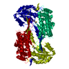
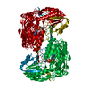
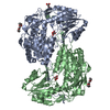
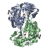
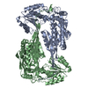
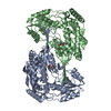
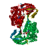
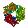
 Mycobacterium tuberculosis (bacteria)
 X-RAY DIFFRACTION /
  Authors
 Citation
 Structure visualization
 Downloads & links
 Other downloads


 PDBj
PDBj

 Assembly

 Mass: 663.425 Da / Num. of mol.: 1 / Source method: obtained synthetically / Formula: C21H27N7O14P2 / Comment: NAD*YM
 Mass: 18.015 Da / Num. of mol.: 389 / Source method: isolated from a natural source / Formula: H2O
 Sample preparation
 / Beamline: MX2 / Wavelength: 0.953688 Å
 Processing