 Movie
Movie Controller

[English] 日本語
 Yorodumi
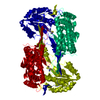
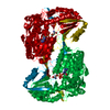
- PDB-4ihi: Crystal structure of the Delta-pyrroline-5-carboxylate dehydrogen... -
+ Open data

- Basic information
| Entry | Database: PDB / ID: 4ihi | ||||||
|---|---|---|---|---|---|---|---|
| Title | Crystal structure of the Delta-pyrroline-5-carboxylate dehydrogenase from Mycobacterium tuberculosis bound with NAD | ||||||
 Components Components | Delta-1-pyrroline-5-carboxylate dehydrogenase | ||||||
 Keywords Keywords | OXIDOREDUCTASE / Rossmann fold / pyrroline-5-carboxylate dehtdrogenase / pyrroline-5-carboxylic acid / dehydrogenation | ||||||
| Function / homology |  Function and homology information Function and homology informationproline dehydrogenase activity / L-glutamate gamma-semialdehyde dehydrogenase / L-glutamate gamma-semialdehyde dehydrogenase activity / : / cytoplasmic side of plasma membrane / nucleotide binding / metal ion binding Similarity search - Function | ||||||
| Biological species |   Mycobacterium tuberculosis (bacteria) Mycobacterium tuberculosis (bacteria) | ||||||
| Method |  X-RAY DIFFRACTION / SYNCHROTRON / MOLECULAR REPLACEMENT / Resolution: 2.25 Å X-RAY DIFFRACTION / SYNCHROTRON / MOLECULAR REPLACEMENT / Resolution: 2.25 Å | ||||||
 Authors Authors | Lagautriere, T. / Bashiri, G. / Baker, E.N. | ||||||
 Citation Citation | Journal: Acta Crystallogr.,Sect.D / Year: 2014 Title: Characterization of the proline-utilization pathway in Mycobacterium tuberculosis through structural and functional studies. Authors: Lagautriere, T. / Bashiri, G. / Paterson, N.G. / Berney, M. / Cook, G.M. / Baker, E.N. | ||||||
| History |
|
- Structure visualization
| Structure viewer | Molecule: MolmilJmol/JSmol |
|---|
- Downloads & links
-Download
| PDBx/mmCIF format | 4ihi.cif.gz | 129.2 KB | Display | PDBx/mmCIF format |
|---|---|---|---|---|
| PDB format | pdb4ihi.ent.gz | 98.6 KB | Display | PDB format |
| PDBx/mmJSON format | 4ihi.json.gz | Tree view | PDBx/mmJSON format | |
| Others |  Other downloads Other downloads |
-Validation report
| Arichive directory | https://data.pdbj.org/pub/pdb/validation_reports/ih/4ihiftp://data.pdbj.org/pub/pdb/validation_reports/ih/4ihi | HTTPS FTP |
|---|
-Related structure data


-Links
 PDBj
PDBj

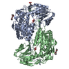
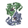
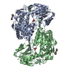
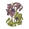
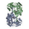
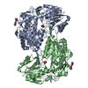
- Assembly
| Deposited unit | 
| ||||||||
|---|---|---|---|---|---|---|---|---|---|
| 1 | 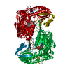
| ||||||||
| 2 | x 6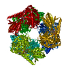
| ||||||||
| 3 | 
| ||||||||
| Unit cell |
|
-Components
| #1: Protein | Mass: 61134.566 Da / Num. of mol.: 1 / Mutation: G505D Source method: isolated from a genetically manipulated source Source: (gene. exp.) Mycobacterium tuberculosis (bacteria) / Strain: H37Rv / Gene: MT1224, pruA, rocA, Rv1187 / Production host: Mycobacterium smegmatis (bacteria) / Strain (production host): Mc2 4517 / References: UniProt: O50443, EC: 1.5.1.12 |
|---|---|
| #2: Chemical | ChemComp-NAD /   Mass: 663.425 Da / Num. of mol.: 1 / Source method: obtained synthetically / Formula: C21H27N7O14P2 / Comment: NAD*YM Mass: 663.425 Da / Num. of mol.: 1 / Source method: obtained synthetically / Formula: C21H27N7O14P2 / Comment: NAD*YM |
| #3: Water | ChemComp-HOH /  Mass: 18.015 Da / Num. of mol.: 450 / Source method: isolated from a natural source / Formula: H2O Mass: 18.015 Da / Num. of mol.: 450 / Source method: isolated from a natural source / Formula: H2O |
-Experimental details
-Experiment
| Experiment | Method: X-RAY DIFFRACTION / Number of used crystals: 1 |
|---|
- Sample preparation
| Crystal | Density Matthews: 6.25 Å3/Da / Density % sol: 80.31 % |
|---|---|
| Crystal grow | Temperature: 291 K / Method: vapor diffusion, hanging drop / pH: 8.1 Details: 0.1 M Bicine/Tris pH 8.1, 12% polyethylene glycol (PEG) 1000, 12% PEG 3350, 12% 2-methyl-2,4-pentandiol (MPD), 0.03 M sodium nitrate, 0.03 M disodium hydrogen phosphate and 0.03 M ammonium ...Details: 0.1 M Bicine/Tris pH 8.1, 12% polyethylene glycol (PEG) 1000, 12% PEG 3350, 12% 2-methyl-2,4-pentandiol (MPD), 0.03 M sodium nitrate, 0.03 M disodium hydrogen phosphate and 0.03 M ammonium sulfate, VAPOR DIFFUSION, HANGING DROP, temperature 291K |
-Data collection
| Diffraction | Mean temperature: 100 K | |||||||||
|---|---|---|---|---|---|---|---|---|---|---|
| Diffraction source | Source: SYNCHROTRON / Site: Australian Synchrotron  / Beamline: MX2 / Wavelength: 0.953688 Å / Beamline: MX2 / Wavelength: 0.953688 Å | |||||||||
| Detector | Type: ADSC QUANTUM 315r / Detector: CCD / Date: Jul 22, 2011 | |||||||||
| Radiation | Protocol: SINGLE WAVELENGTH / Monochromatic (M) / Laue (L): M / Scattering type: x-ray | |||||||||
| Radiation wavelength | Wavelength: 0.953688 Å / Relative weight: 1 | |||||||||
| Reflection | Resolution: 1.91→29.65 Å / Num. obs: 73091 / % possible obs: 99.8 % | |||||||||
| Reflection shell |
|
- Processing
| Software |
| ||||||||||||||||||||||||||||||||||||||||||||||||||||||||||||||||||||||||||||||||||||||||||||||||||||||||||||||||||||||||||||||||||||||||||||||||||||||||||||||||||||||||||
|---|---|---|---|---|---|---|---|---|---|---|---|---|---|---|---|---|---|---|---|---|---|---|---|---|---|---|---|---|---|---|---|---|---|---|---|---|---|---|---|---|---|---|---|---|---|---|---|---|---|---|---|---|---|---|---|---|---|---|---|---|---|---|---|---|---|---|---|---|---|---|---|---|---|---|---|---|---|---|---|---|---|---|---|---|---|---|---|---|---|---|---|---|---|---|---|---|---|---|---|---|---|---|---|---|---|---|---|---|---|---|---|---|---|---|---|---|---|---|---|---|---|---|---|---|---|---|---|---|---|---|---|---|---|---|---|---|---|---|---|---|---|---|---|---|---|---|---|---|---|---|---|---|---|---|---|---|---|---|---|---|---|---|---|---|---|---|---|---|---|---|---|
| Refinement | Method to determine structure: MOLECULAR REPLACEMENT / Resolution: 2.25→29.51 Å / Cor.coef. Fo:Fc: 0.832 / Cor.coef. Fo:Fc free: 0.796 / SU B: 4.619 / SU ML: 0.12 / Cross valid method: THROUGHOUT / ESU R: 0.192 / ESU R Free: 0.184 / Stereochemistry target values: MAXIMUM LIKELIHOOD Details: HYDROGENS HAVE BEEN ADDED IN THE RIDING POSITIONS. STRUCTURE CONSISTS IN AN ARRANGEMENT OF ORDERED AND DISORDERED LAYERS RESULTING IN GAPS BETWEEN ORDERED MOLECULES. ALTHOUGH NO MOLECULES ...Details: HYDROGENS HAVE BEEN ADDED IN THE RIDING POSITIONS. STRUCTURE CONSISTS IN AN ARRANGEMENT OF ORDERED AND DISORDERED LAYERS RESULTING IN GAPS BETWEEN ORDERED MOLECULES. ALTHOUGH NO MOLECULES ARE MODELLED WITHIN THIS EMPTY LAYER, MAD DATA FROM SEMET-PRUA CRYSTALS INDICATES THAT PROTEIN ELECTRON DENSITY EXISTS WITHING THESE GAPS AND SUGGESTS A MOTION OF THE ENTIRE LAYER.
| ||||||||||||||||||||||||||||||||||||||||||||||||||||||||||||||||||||||||||||||||||||||||||||||||||||||||||||||||||||||||||||||||||||||||||||||||||||||||||||||||||||||||||
| Solvent computation | Ion probe radii: 0.8 Å / Shrinkage radii: 0.8 Å / VDW probe radii: 1.2 Å / Solvent model: MASK | ||||||||||||||||||||||||||||||||||||||||||||||||||||||||||||||||||||||||||||||||||||||||||||||||||||||||||||||||||||||||||||||||||||||||||||||||||||||||||||||||||||||||||
| Displacement parameters | Biso mean: 15.087 Å2
| ||||||||||||||||||||||||||||||||||||||||||||||||||||||||||||||||||||||||||||||||||||||||||||||||||||||||||||||||||||||||||||||||||||||||||||||||||||||||||||||||||||||||||
| Refinement step | Cycle: LAST / Resolution: 2.25→29.51 Å
| ||||||||||||||||||||||||||||||||||||||||||||||||||||||||||||||||||||||||||||||||||||||||||||||||||||||||||||||||||||||||||||||||||||||||||||||||||||||||||||||||||||||||||
| Refine LS restraints |
| ||||||||||||||||||||||||||||||||||||||||||||||||||||||||||||||||||||||||||||||||||||||||||||||||||||||||||||||||||||||||||||||||||||||||||||||||||||||||||||||||||||||||||
| LS refinement shell | Resolution: 2.25→2.308 Å / Total num. of bins used: 20
|
