 Movie
Movie Controller


+ Open data

- Basic information
| Entry | Database: PDB / ID: 7cy7 | ||||||||||||
|---|---|---|---|---|---|---|---|---|---|---|---|---|---|
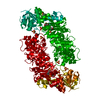
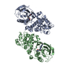
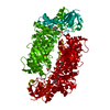
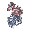
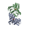
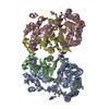
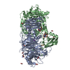
| Title | Crystal Structure of CMD1 in complex with DNA | ||||||||||||
 Components Components |
| ||||||||||||
 Keywords Keywords | TRANSFERASE / TET / Vitamin C / dioxygenase / 5gmC / DNA modification / 2-oxoglutarate | ||||||||||||
| Function / homology |  Function and homology information Function and homology informationmethylcytosine to 5-glyceryl-methylcytosine dioxygenase activity / regulation of photosynthesis / Oxidoreductases; Acting on paired donors, with incorporation or reduction of molecular oxygen; Miscellaneous / chromosomal 5-methylcytosine DNA demethylation, oxidation pathway / dioxygenase activity / detection of maltose stimulus / maltose transport complex / carbohydrate transport / carbohydrate transmembrane transporter activity / maltose binding ...methylcytosine to 5-glyceryl-methylcytosine dioxygenase activity / regulation of photosynthesis / Oxidoreductases; Acting on paired donors, with incorporation or reduction of molecular oxygen; Miscellaneous / chromosomal 5-methylcytosine DNA demethylation, oxidation pathway / dioxygenase activity / detection of maltose stimulus / maltose transport complex / carbohydrate transport / carbohydrate transmembrane transporter activity / maltose binding / maltose transport / maltodextrin transmembrane transport / ATP-binding cassette (ABC) transporter complex, substrate-binding subunit-containing / ATP-binding cassette (ABC) transporter complex / cell chemotaxis / outer membrane-bounded periplasmic space / periplasmic space / iron ion binding / DNA damage response / membrane / nucleus Similarity search - Function | ||||||||||||
| Biological species |    Chlamydomonas reinhardtii (plant) Chlamydomonas reinhardtii (plant)synthetic construct (others) | ||||||||||||
| Method |  X-RAY DIFFRACTION / SYNCHROTRON / MOLECULAR REPLACEMENT / Resolution: 2.15 Å X-RAY DIFFRACTION / SYNCHROTRON / MOLECULAR REPLACEMENT / Resolution: 2.15 Å | ||||||||||||
 Authors Authors | Li, W. / Zhang, T. / Sun, M. / Ding, J. | ||||||||||||
| Funding support |  China, 3items China, 3items
| ||||||||||||
 Citation Citation | Journal: Nat Commun / Year: 2021 Title: Molecular mechanism for vitamin C-derived C 5 -glyceryl-methylcytosine DNA modification catalyzed by algal TET homologue CMD1. Authors: Li, W. / Zhang, T. / Sun, M. / Shi, Y. / Zhang, X.J. / Xu, G.L. / Ding, J. | ||||||||||||
| History |
|
- Structure visualization
| Structure viewer | Molecule: MolmilJmol/JSmol |
|---|
- Downloads & links
-Download
| PDBx/mmCIF format | 7cy7.cif.gz | 388.9 KB | Display | PDBx/mmCIF format |
|---|---|---|---|---|
| PDB format | pdb7cy7.ent.gz | 308 KB | Display | PDB format |
| PDBx/mmJSON format | 7cy7.json.gz | Tree view | PDBx/mmJSON format | |
| Others |  Other downloads Other downloads |
-Validation report
| Arichive directory | https://data.pdbj.org/pub/pdb/validation_reports/cy/7cy7ftp://data.pdbj.org/pub/pdb/validation_reports/cy/7cy7 | HTTPS FTP |
|---|
-Related structure data
| Related structure data | 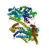 7cy4SC 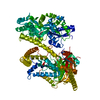 7cy5C 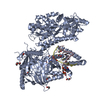 7cy6C 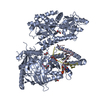 7cy8C S: Starting model for refinement C: citing same article ( |
|---|---|
| Similar structure data |


-Links
 PDBj
PDBj


- Assembly
| Deposited unit | 
| ||||||||
|---|---|---|---|---|---|---|---|---|---|
| 1 |
| ||||||||
| Unit cell |
|
-Components
-Protein / DNA chain , 2 types, 3 molecules ACD
| #1: Protein | Mass: 98913.453 Da / Num. of mol.: 1 / Mutation: D108A, K109A, E198A, N199A, E385A, K388A, D389A Source method: isolated from a genetically manipulated source Details: The fusion protein of Maltodextrin-binding protein UNP RESIDUES 27-392), linker, 5-methylcytosine-modifying enzyme 1 (UNP RESIDUES 1-532) and tags Source: (gene. exp.) Chlamydomonas reinhardtii (plant)Gene: CMD1, CHLRE_12g553400v5 / Production host: References: UniProt: A0A376KDN7, UniProt: A0A2K3D5Z7, UniProt: P0AEX9*PLUS, Oxidoreductases; Acting on paired donors, with incorporation or reduction of molecular oxygen; Miscellaneous |
|---|---|
| #2: DNA chain | Mass: 4297.778 Da / Num. of mol.: 2 / Source method: obtained synthetically / Source: (synth.) synthetic construct (others) |
-Non-polymers , 4 types, 412 molecules 


| #3: Chemical | ChemComp-EDO /  Mass: 62.068 Da / Num. of mol.: 8 / Source method: obtained synthetically / Formula: C2H6O2 Mass: 62.068 Da / Num. of mol.: 8 / Source method: obtained synthetically / Formula: C2H6O2#4: Chemical | ChemComp-IPA / |  Mass: 60.095 Da / Num. of mol.: 1 / Source method: obtained synthetically / Formula: C3H8O / Feature type: SUBJECT OF INVESTIGATION Mass: 60.095 Da / Num. of mol.: 1 / Source method: obtained synthetically / Formula: C3H8O / Feature type: SUBJECT OF INVESTIGATION#5: Chemical | ChemComp-FE2 / |  Mass: 55.845 Da / Num. of mol.: 1 / Source method: obtained synthetically / Formula: Fe / Feature type: SUBJECT OF INVESTIGATION Mass: 55.845 Da / Num. of mol.: 1 / Source method: obtained synthetically / Formula: Fe / Feature type: SUBJECT OF INVESTIGATION#6: Water | ChemComp-HOH / | Mass: 18.015 Da / Num. of mol.: 402 / Source method: isolated from a natural source / Formula: H2O |
|---|
-Details
| Has ligand of interest | Y |
|---|
-Experimental details
-Experiment
| Experiment | Method: X-RAY DIFFRACTION / Number of used crystals: 1 |
|---|
- Sample preparation
| Crystal | Density Matthews: 2.8 Å3/Da / Density % sol: 56.15 % |
|---|---|
| Crystal grow | Temperature: 290 K / Method: vapor diffusion, hanging drop / pH: 5 Details: 15% (v/v) 2-propanol, 0.1 M sodium citrate, pH 5.0, and 10% (w/v) PEG 10000 |
-Data collection
| Diffraction | Mean temperature: 100 K / Serial crystal experiment: N |
|---|---|
| Diffraction source | Source: SYNCHROTRON / Site: NFPSS / Beamline: BL19U1 / Wavelength: 0.9785 Å |
| Detector | Type: DECTRIS PILATUS3 S 6M / Detector: PIXEL / Date: May 30, 2020 |
| Radiation | Protocol: SINGLE WAVELENGTH / Monochromatic (M) / Laue (L): M / Scattering type: x-ray |
| Radiation wavelength | Wavelength: 0.9785 Å / Relative weight: 1 |
| Reflection | Resolution: 2.15→50.01 Å / Num. obs: 60566 / % possible obs: 96.7 % / Redundancy: 6.6 % / CC1/2: 1 / Rmerge(I) obs: 0.067 / Net I/σ(I): 15.8 |
| Reflection shell | Resolution: 2.15→2.21 Å / Rmerge(I) obs: 0.52 / Mean I/σ(I) obs: 2 / Num. unique obs: 3028 / CC1/2: 0.82 |
- Processing
| Software |
| ||||||||||||||||||||||||||||||||||||||||||||||||||||||||||||||||||||||||||||||||||||||||||||||||||||
|---|---|---|---|---|---|---|---|---|---|---|---|---|---|---|---|---|---|---|---|---|---|---|---|---|---|---|---|---|---|---|---|---|---|---|---|---|---|---|---|---|---|---|---|---|---|---|---|---|---|---|---|---|---|---|---|---|---|---|---|---|---|---|---|---|---|---|---|---|---|---|---|---|---|---|---|---|---|---|---|---|---|---|---|---|---|---|---|---|---|---|---|---|---|---|---|---|---|---|---|---|---|
| Refinement | Method to determine structure: MOLECULAR REPLACEMENT Starting model: 7CY4 Resolution: 2.15→50.01 Å / Cor.coef. Fo:Fc: 0.95 / Cor.coef. Fo:Fc free: 0.932 / SU B: 11.678 / SU ML: 0.131 / Cross valid method: THROUGHOUT / σ(F): 0 / ESU R Free: 0.177 / Stereochemistry target values: MAXIMUM LIKELIHOOD Details: HYDROGENS HAVE BEEN ADDED IN THE RIDING POSITIONS U VALUES : WITH TLS ADDED
| ||||||||||||||||||||||||||||||||||||||||||||||||||||||||||||||||||||||||||||||||||||||||||||||||||||
| Solvent computation | Ion probe radii: 0.8 Å / Shrinkage radii: 0.8 Å / VDW probe radii: 1.2 Å / Solvent model: MASK | ||||||||||||||||||||||||||||||||||||||||||||||||||||||||||||||||||||||||||||||||||||||||||||||||||||
| Displacement parameters | Biso max: 189.11 Å2 / Biso mean: 40.4 Å2 / Biso min: 12.18 Å2
| ||||||||||||||||||||||||||||||||||||||||||||||||||||||||||||||||||||||||||||||||||||||||||||||||||||
| Refinement step | Cycle: final / Resolution: 2.15→50.01 Å
| ||||||||||||||||||||||||||||||||||||||||||||||||||||||||||||||||||||||||||||||||||||||||||||||||||||
| Refine LS restraints |
| ||||||||||||||||||||||||||||||||||||||||||||||||||||||||||||||||||||||||||||||||||||||||||||||||||||
| LS refinement shell | Resolution: 2.15→2.206 Å / Rfactor Rfree error: 0 / Total num. of bins used: 20
| ||||||||||||||||||||||||||||||||||||||||||||||||||||||||||||||||||||||||||||||||||||||||||||||||||||
| Refinement TLS params. | Method: refined / Refine-ID: X-RAY DIFFRACTION
| ||||||||||||||||||||||||||||||||||||||||||||||||||||||||||||||||||||||||||||||||||||||||||||||||||||
| Refinement TLS group |
|
