 Movie
Movie Controller


+ Open data

- Basic information
| Entry | Database: PDB / ID: 7cec | |||||||||
|---|---|---|---|---|---|---|---|---|---|---|
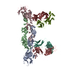
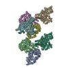
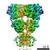
| Title | Structure of alpha6beta1 integrin in complex with laminin-511 | |||||||||
 Components Components |
| |||||||||
 Keywords Keywords | CELL ADHESION/IMMUNE SYSTEM / Integrin / Laminin / Fv-clasp / CELL ADHESION / CELL ADHESION-IMMUNE SYSTEM complex | |||||||||
| Function / homology |  Function and homology information Function and homology informationintegrin alpha6-beta4 complex / laminin-522 trimer / laminin-523 trimer / laminin-332 trimer / neuronal-glial interaction involved in cerebral cortex radial glia guided migration / laminin-411 trimer / laminin-521 trimer / laminin-211 trimer / Developmental Lineage of Mammary Gland Alveolar Cells / Developmental Lineage of Mammary Gland Luminal Epithelial Cells ...integrin alpha6-beta4 complex / laminin-522 trimer / laminin-523 trimer / laminin-332 trimer / neuronal-glial interaction involved in cerebral cortex radial glia guided migration / laminin-411 trimer / laminin-521 trimer / laminin-211 trimer / Developmental Lineage of Mammary Gland Alveolar Cells / Developmental Lineage of Mammary Gland Luminal Epithelial Cells / laminin-111 trimer / laminin-511 trimer / regulation of basement membrane organization / integrin alpha8-beta1 complex / neuregulin binding / myoblast fate specification / extracellular matrix of synaptic cleft / L1CAM interactions / integrin alpha3-beta1 complex / integrin alpha6-beta1 complex / integrin alpha7-beta1 complex / integrin alpha10-beta1 complex / integrin alpha11-beta1 complex / positive regulation of glutamate uptake involved in transmission of nerve impulse / Type I hemidesmosome assembly / cardiac cell fate specification / integrin alpha5-beta1 complex / integrin alpha9-beta1 complex / ectodermal cell differentiation / regulation of collagen catabolic process / nail development / integrin alpha1-beta1 complex / integrin binding involved in cell-matrix adhesion / hemidesmosome assembly / integrin alpha4-beta1 complex / cell adhesion receptor activity / collagen binding involved in cell-matrix adhesion / integrin alpha2-beta1 complex / Localization of the PINCH-ILK-PARVIN complex to focal adhesions / formation of radial glial scaffolds / cell-cell adhesion mediated by integrin / glycosphingolipid binding / Other semaphorin interactions / positive regulation of integrin-mediated signaling pathway / Formation of the ureteric bud / cerebellar climbing fiber to Purkinje cell synapse / postsynapse organization / CD40 signaling pathway / calcium-independent cell-matrix adhesion / positive regulation of fibroblast growth factor receptor signaling pathway / reactive gliosis / myelin sheath abaxonal region / integrin alphav-beta1 complex / basement membrane organization / skin morphogenesis / CHL1 interactions / cardiac muscle cell myoblast differentiation / regulation of synapse pruning / Fibronectin matrix formation / Attachment of bacteria to epithelial cells / MET interacts with TNS proteins / protein complex involved in cell-matrix adhesion / Laminin interactions / Developmental Lineage of Mammary Stem Cells / primordial germ cell migration / EGR2 and SOX10-mediated initiation of Schwann cell myelination / Platelet Adhesion to exposed collagen / endoderm development / leukocyte tethering or rolling / Formation of the dystrophin-glycoprotein complex (DGC) / insulin-like growth factor I binding / negative regulation of cell adhesion / myoblast fusion / positive regulation of vascular endothelial growth factor signaling pathway / cell migration involved in sprouting angiogenesis / axon extension / Elastic fibre formation / cardiac muscle cell differentiation / cell-substrate junction assembly / myoblast differentiation / mesodermal cell differentiation / central nervous system neuron differentiation / Differentiation of Keratinocytes in Interfollicular Epidermis in Mammalian Skin / cell projection organization / wound healing, spreading of epidermal cells / positive regulation of fibroblast migration / Assembly of collagen fibrils and other multimeric structures / negative regulation of Rho protein signal transduction / odontogenesis / integrin complex / regulation of spontaneous synaptic transmission / skeletal system morphogenesis / cell adhesion mediated by integrin / Molecules associated with elastic fibres / MET activates PTK2 signaling / heterotypic cell-cell adhesion / leukocyte migration / Basigin interactions / lamellipodium assembly / negative regulation of vasoconstriction Similarity search - Function | |||||||||
| Biological species |  Homo sapiens (human) Homo sapiens (human) | |||||||||
| Method | ELECTRON MICROSCOPY / single particle reconstruction / cryo EM / Resolution: 3.9 Å | |||||||||
 Authors Authors | Arimori, T. / Miyazaki, N. / Takagi, J. | |||||||||
| Funding support |  Japan, 2items Japan, 2items
| |||||||||
 Citation Citation | Journal: Nat Commun / Year: 2021 Title: Structural mechanism of laminin recognition by integrin. Authors: Takao Arimori / Naoyuki Miyazaki / Emiko Mihara / Mamoru Takizawa / Yukimasa Taniguchi / Carlos Cabañas / Kiyotoshi Sekiguchi / Junichi Takagi /  Abstract: Recognition of laminin by integrin receptors is central to the epithelial cell adhesion to basement membrane, but the structural background of this molecular interaction remained elusive. Here, we ...Recognition of laminin by integrin receptors is central to the epithelial cell adhesion to basement membrane, but the structural background of this molecular interaction remained elusive. Here, we report the structures of the prototypic laminin receptor α6β1 integrin alone and in complex with three-chain laminin-511 fragment determined via crystallography and cryo-electron microscopy, respectively. The laminin-integrin interface is made up of several binding sites located on all five subunits, with the laminin γ1 chain C-terminal portion providing focal interaction using two carboxylate anchor points to bridge metal-ion dependent adhesion site of integrin β1 subunit and Asn189 of integrin α6 subunit. Laminin α5 chain also contributes to the affinity and specificity by making electrostatic interactions with large surface on the β-propeller domain of α6, part of which comprises an alternatively spliced X1 region. The propeller sheet corresponding to this region shows unusually high mobility, suggesting its unique role in ligand capture. | |||||||||
| History |
|
- Structure visualization
| Movie |
Movie viewer |
|---|---|
| Structure viewer | Molecule: MolmilJmol/JSmol |
- Downloads & links
-Download
| PDBx/mmCIF format | 7cec.cif.gz | 426.3 KB | Display | PDBx/mmCIF format |
|---|---|---|---|---|
| PDB format | pdb7cec.ent.gz | 325.8 KB | Display | PDB format |
| PDBx/mmJSON format | 7cec.json.gz | Tree view | PDBx/mmJSON format | |
| Others |  Other downloads Other downloads |
-Validation report
| Arichive directory | https://data.pdbj.org/pub/pdb/validation_reports/ce/7cecftp://data.pdbj.org/pub/pdb/validation_reports/ce/7cec | HTTPS FTP |
|---|
-Related structure data
| Related structure data | 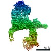 30342MC 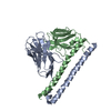 7ceaC  7cebC M: map data used to model this data C: citing same article ( |
|---|---|
| Similar structure data |


-Links
 PDBj
PDBj


- Assembly
| Deposited unit | 
|
|---|---|
| 1 |
|
-Components
-Protein , 2 types, 2 molecules AB
| #1: Protein | Mass: 69776.023 Da / Num. of mol.: 1 Source method: isolated from a genetically manipulated source Source: (gene. exp.) Homo sapiens (human) / Gene: ITGA6 / Production host: Homo sapiens (human) / References: UniProt: P23229 |
|---|---|
| #2: Protein | Mass: 50510.930 Da / Num. of mol.: 1 Source method: isolated from a genetically manipulated source Source: (gene. exp.) Homo sapiens (human) / Gene: ITGB1, FNRB, MDF2, MSK12 / Production host: Homo sapiens (human) / References: UniProt: P05556 |
-Laminin subunit ... , 3 types, 3 molecules CDE
| #3: Protein | Mass: 73414.203 Da / Num. of mol.: 1 / Fragment: E8 fragment / Mutation: I2723C Source method: isolated from a genetically manipulated source Source: (gene. exp.) Homo sapiens (human) / Gene: LAMA5, KIAA0533, KIAA1907 / Production host: Homo sapiens (human) / References: UniProt: O15230 |
|---|---|
| #4: Protein | Mass: 8552.753 Da / Num. of mol.: 1 / Fragment: E8 fragment Source method: isolated from a genetically manipulated source Source: (gene. exp.) Homo sapiens (human) / Gene: LAMB1 / Production host: Homo sapiens (human) / References: UniProt: P07942 |
| #5: Protein | Mass: 9483.724 Da / Num. of mol.: 1 / Fragment: E8 fragment / Mutation: D1585C Source method: isolated from a genetically manipulated source Source: (gene. exp.) Homo sapiens (human) / Gene: LAMC1, LAMB2 / Production host: Homo sapiens (human) / References: UniProt: P11047 |
-Antibody , 4 types, 4 molecules FGHI
| #6: Antibody | Mass: 19463.992 Da / Num. of mol.: 1 Source method: isolated from a genetically manipulated source Details: chimera of TS2/16 VH(S112C)-SARAH Source: (gene. exp.) Homo sapiens (human)Production host:  |
|---|---|
| #7: Antibody | Mass: 18270.742 Da / Num. of mol.: 1 Source method: isolated from a genetically manipulated source Details: chimera of TS2/16 VL-SARAH(S37C) Source: (gene. exp.) Homo sapiens (human)Production host: |
| #8: Antibody | Mass: 19760.207 Da / Num. of mol.: 1 Source method: isolated from a genetically manipulated source Details: chimera of HUTS-4 VH(S112C)-SARAH Source: (gene. exp.) Homo sapiens (human)Production host: |
| #9: Antibody | Mass: 17701.023 Da / Num. of mol.: 1 Source method: isolated from a genetically manipulated source Details: chimera of HUTS-4 VL(C87Y)-SARAH(S37C) Source: (gene. exp.) Homo sapiens (human)Production host: |
-Sugars , 2 types, 7 molecules 
| #10: Polysaccharide | 2-acetamido-2-deoxy-beta-D-glucopyranose-(1-4)-2-acetamido-2-deoxy-beta-D-glucopyranose Source method: isolated from a genetically manipulated source |
|---|---|
| #12: Sugar | ChemComp-NAG /  Type: D-saccharide, beta linking / Mass: 221.208 Da / Num. of mol.: 6 Type: D-saccharide, beta linking / Mass: 221.208 Da / Num. of mol.: 6Source method: isolated from a genetically manipulated source Formula: C8H15NO6 |
-Non-polymers , 2 types, 7 molecules 

| #11: Chemical | ChemComp-CA /  Mass: 40.078 Da / Num. of mol.: 4 / Source method: obtained synthetically / Formula: Ca Mass: 40.078 Da / Num. of mol.: 4 / Source method: obtained synthetically / Formula: Ca#13: Chemical |  Mass: 54.938 Da / Num. of mol.: 3 / Source method: obtained synthetically / Formula: Mn Mass: 54.938 Da / Num. of mol.: 3 / Source method: obtained synthetically / Formula: Mn |
|---|
-Details
| Has ligand of interest | N |
|---|---|
| Has protein modification | Y |
-Experimental details
-Experiment
| Experiment | Method: ELECTRON MICROSCOPY |
|---|---|
| EM experiment | Aggregation state: PARTICLE / 3D reconstruction method: single particle reconstruction |
- Sample preparation
| Component | Name: Quaternary complex of alpha6beta1 integrin, laminin-511, TS2/16 Fv-clasp, and HUTS-4 Fv-clasp Type: COMPLEX / Entity ID: #1-#9 / Source: RECOMBINANT | |||||||||||||||||||||||||
|---|---|---|---|---|---|---|---|---|---|---|---|---|---|---|---|---|---|---|---|---|---|---|---|---|---|---|
| Molecular weight | Value: 0.286 MDa / Experimental value: NO | |||||||||||||||||||||||||
| Source (natural) | Organism: Homo sapiens (human) | |||||||||||||||||||||||||
| Source (recombinant) | Organism: | |||||||||||||||||||||||||
| Buffer solution | pH: 7.5 | |||||||||||||||||||||||||
| Buffer component |
| |||||||||||||||||||||||||
| Specimen | Conc.: 0.07 mg/ml / Embedding applied: NO / Shadowing applied: NO / Staining applied: NO / Vitrification applied: YES | |||||||||||||||||||||||||
| Specimen support | Grid material: MOLYBDENUM / Grid mesh size: 300 divisions/in. / Grid type: Quantifoil R2/1 | |||||||||||||||||||||||||
| Vitrification | Instrument: FEI VITROBOT MARK IV / Cryogen name: ETHANE / Humidity: 100 % / Chamber temperature: 277 K |
- Electron microscopy imaging
| Experimental equipment |  Model: Titan Krios / Image courtesy: FEI Company |
|---|---|
| Microscopy | Model: FEI TITAN KRIOS |
| Electron gun | Electron source:  FIELD EMISSION GUN / Accelerating voltage: 300 kV / Illumination mode: FLOOD BEAM FIELD EMISSION GUN / Accelerating voltage: 300 kV / Illumination mode: FLOOD BEAM |
| Electron lens | Mode: BRIGHT FIELD / Nominal magnification: 59000 X / Nominal defocus max: 800 nm / Nominal defocus min: 600 nm / Alignment procedure: ZEMLIN TABLEAU |
| Specimen holder | Cryogen: NITROGEN / Specimen holder model: FEI TITAN KRIOS AUTOGRID HOLDER |
| Image recording | Electron dose: 40 e/Å2 / Detector mode: INTEGRATING / Film or detector model: FEI FALCON III (4k x 4k) / Num. of grids imaged: 1 / Num. of real images: 7768 |
| EM imaging optics | Phase plate: VOLTA PHASE PLATE |
| Image scans | Width: 4096 / Height: 4096 |
- Processing
| Software |
| ||||||||||||||||||||||||||||
|---|---|---|---|---|---|---|---|---|---|---|---|---|---|---|---|---|---|---|---|---|---|---|---|---|---|---|---|---|---|
| EM software |
| ||||||||||||||||||||||||||||
| CTF correction | Type: PHASE FLIPPING AND AMPLITUDE CORRECTION | ||||||||||||||||||||||||||||
| Particle selection | Num. of particles selected: 2660283 | ||||||||||||||||||||||||||||
| Symmetry | Point symmetry: C1 (asymmetric) | ||||||||||||||||||||||||||||
| 3D reconstruction | Resolution: 3.9 Å / Resolution method: FSC 0.143 CUT-OFF / Num. of particles: 429521 / Symmetry type: POINT | ||||||||||||||||||||||||||||
| Refinement | Stereochemistry target values: CDL v1.2 | ||||||||||||||||||||||||||||
| Refine LS restraints |
|
