 Movie
Movie Controller

[English] 日本語
 Yorodumi
- PDB-3hk9: Crystal structure of uronate isomerase from Bacillus halodurans c... -
+ Open data

- Basic information
| Entry | Database: PDB / ID: 3hk9 | ||||||
|---|---|---|---|---|---|---|---|
| Title | Crystal structure of uronate isomerase from Bacillus halodurans complexed with zinc and D-Glucuronate | ||||||
 Components Components | Uronate isomerase | ||||||
 Keywords Keywords | ISOMERASE / uronate isomerase / D-Glucuronate / Mechanism of the reaction | ||||||
| Function / homology | uronate isomerase, domain 2, chain A / uronate isomerase, domain 2, chain A / Metal-dependent hydrolase / Orthogonal Bundle / Mainly Alpha / metal ion binding / CARBONATE ION / D-glucuronic acid / BH0493 protein Function and homology information Function and homology information | ||||||
| Biological species |  Bacillus halodurans C-125 (bacteria) Bacillus halodurans C-125 (bacteria) | ||||||
| Method |  X-RAY DIFFRACTION / SYNCHROTRON / MOLECULAR REPLACEMENT / Resolution: 2.1 Å X-RAY DIFFRACTION / SYNCHROTRON / MOLECULAR REPLACEMENT / Resolution: 2.1 Å | ||||||
 Authors Authors | Fedorov, A.A. / Fedorov, E.V. / Nguyen, T.T. / Raushel, F.M. / Almo, S.C. | ||||||
 Citation Citation | Journal: Biochemistry / Year: 2009 Title: The mechanism of the reaction catalyzed by uronate isomerase illustrates how an isomerase may have evolved from a hydrolase within the amidohydrolase superfamily. Authors: Nguyen, T.T. / Fedorov, A.A. / Williams, L. / Fedorov, E.V. / Li, Y. / Xu, C. / Almo, S.C. / Raushel, F.M. | ||||||
| History |
|
- Structure visualization
| Structure viewer | Molecule: MolmilJmol/JSmol |
|---|
- Downloads & links
-Download
| PDBx/mmCIF format | 3hk9.cif.gz | 1019.4 KB | Display | PDBx/mmCIF format |
|---|---|---|---|---|
| PDB format | pdb3hk9.ent.gz | 847.7 KB | Display | PDB format |
| PDBx/mmJSON format | 3hk9.json.gz | Tree view | PDBx/mmJSON format | |
| Others |  Other downloads Other downloads |
-Validation report
| Arichive directory | https://data.pdbj.org/pub/pdb/validation_reports/hk/3hk9ftp://data.pdbj.org/pub/pdb/validation_reports/hk/3hk9 | HTTPS FTP |
|---|
-Related structure data
| Related structure data |  3hk5C  3hk7C  3hk8C  3hkaC  2q08S S: Starting model for refinement C: citing same article ( |
|---|---|
| Similar structure data |

-Links
 PDBj
PDBj


- Assembly
| Deposited unit | 
| ||||||||
|---|---|---|---|---|---|---|---|---|---|
| 1 | 
| ||||||||
| 2 | 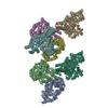
| ||||||||
| 3 | 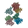
| ||||||||
| 4 | 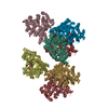
| ||||||||
| Unit cell |
|
-Components
-Protein / Sugars , 2 types, 24 molecules ABCDEFGHIJKL
| #1: Protein | Mass: 50006.852 Da / Num. of mol.: 12 Source method: isolated from a genetically manipulated source Source: (gene. exp.) Bacillus halodurans C-125 (bacteria) / Strain: C-125 / DSM 18197 / FERM 7344 / JCM 9153 / Gene: BH0493 / Production host: #2: Sugar | ChemComp-REL /  Type: D-saccharide / Mass: 194.139 Da / Num. of mol.: 12 Type: D-saccharide / Mass: 194.139 Da / Num. of mol.: 12Source method: isolated from a genetically manipulated source Formula: C6H10O7 |
|---|
-Non-polymers , 4 types, 1852 molecules 


| #3: Chemical | ChemComp-CO3 /  Mass: 60.009 Da / Num. of mol.: 12 / Source method: obtained synthetically / Formula: CO3 Mass: 60.009 Da / Num. of mol.: 12 / Source method: obtained synthetically / Formula: CO3#4: Chemical | ChemComp-ZN /  Mass: 65.409 Da / Num. of mol.: 12 / Source method: obtained synthetically / Formula: Zn Mass: 65.409 Da / Num. of mol.: 12 / Source method: obtained synthetically / Formula: Zn#5: Chemical | ChemComp-CL /  Mass: 35.453 Da / Num. of mol.: 4 / Source method: obtained synthetically / Formula: Cl Mass: 35.453 Da / Num. of mol.: 4 / Source method: obtained synthetically / Formula: Cl#6: Water | ChemComp-HOH / | Mass: 18.015 Da / Num. of mol.: 1824 / Source method: isolated from a natural source / Formula: H2O |
|---|
-Experimental details
-Experiment
| Experiment | Method: X-RAY DIFFRACTION / Number of used crystals: 1 |
|---|
- Sample preparation
| Crystal | Density Matthews: 2.99 Å3/Da / Density % sol: 58.83 % |
|---|---|
| Crystal grow | Temperature: 293 K / Method: vapor diffusion, hanging drop / pH: 7 Details: 20% PEG 3350, 0.2M Ammonium citrate, pH 7.0, VAPOR DIFFUSION, HANGING DROP, temperature 293.0K |
-Data collection
| Diffraction | Mean temperature: 100 K |
|---|---|
| Diffraction source | Source: SYNCHROTRON / Site: NSLS  / Beamline: X4A / Wavelength: 0.97915 Å / Beamline: X4A / Wavelength: 0.97915 Å |
| Detector | Type: ADSC QUANTUM 4 / Detector: CCD / Date: May 2, 2007 / Details: mirrors |
| Radiation | Monochromator: Si(111) CHANNEL / Protocol: SINGLE WAVELENGTH / Monochromatic (M) / Laue (L): M / Scattering type: x-ray |
| Radiation wavelength | Wavelength: 0.97915 Å / Relative weight: 1 |
| Reflection | Resolution: 2.1→25 Å / Num. all: 404122 / Num. obs: 404122 / % possible obs: 98.7 % / Observed criterion σ(F): 0 / Observed criterion σ(I): 0 / Biso Wilson estimate: 19.4 Å2 / Rmerge(I) obs: 0.074 |
- Processing
| Software |
| ||||||||||||||||||||||||||||||||||||
|---|---|---|---|---|---|---|---|---|---|---|---|---|---|---|---|---|---|---|---|---|---|---|---|---|---|---|---|---|---|---|---|---|---|---|---|---|---|
| Refinement | Method to determine structure: MOLECULAR REPLACEMENT Starting model: PDB entry 2Q08 Resolution: 2.1→24.95 Å / Rfactor Rfree error: 0.002 / Data cutoff high absF: 1778514.91 / Data cutoff low absF: 0 / Isotropic thermal model: RESTRAINED / Cross valid method: THROUGHOUT / σ(F): 0 / σ(I): 0 / Stereochemistry target values: Engh & Huber
| ||||||||||||||||||||||||||||||||||||
| Solvent computation | Solvent model: FLAT MODEL / Bsol: 47.3742 Å2 / ksol: 0.361085 e/Å3 | ||||||||||||||||||||||||||||||||||||
| Displacement parameters | Biso mean: 29.8 Å2
| ||||||||||||||||||||||||||||||||||||
| Refine analyze |
| ||||||||||||||||||||||||||||||||||||
| Refinement step | Cycle: LAST / Resolution: 2.1→24.95 Å
| ||||||||||||||||||||||||||||||||||||
| Refine LS restraints |
| ||||||||||||||||||||||||||||||||||||
| LS refinement shell | Resolution: 2.1→2.18 Å / Rfactor Rfree error: 0.007 / Total num. of bins used: 10
| ||||||||||||||||||||||||||||||||||||
| Xplor file |
|
