 Movie
Movie Controller


+ Open data

- Basic information
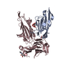
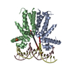
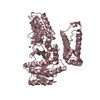
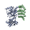
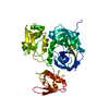
| Entry | Database: PDB / ID: 7cea | |||||||||
|---|---|---|---|---|---|---|---|---|---|---|
| Title | Crystal structure of HUTS-4 Fv-clasp fragment | |||||||||
 Components Components |
| |||||||||
 Keywords Keywords | IMMUNE SYSTEM / Antibody fragment / Fv-clasp / Integrin | |||||||||
| Function / homology | Immunoglobulins / Immunoglobulin-like / Sandwich / Mainly Beta Function and homology information Function and homology information | |||||||||
| Biological species |   Homo sapiens (human) Homo sapiens (human) | |||||||||
| Method |  X-RAY DIFFRACTION / SYNCHROTRON / MOLECULAR REPLACEMENT / Resolution: 2.55 Å X-RAY DIFFRACTION / SYNCHROTRON / MOLECULAR REPLACEMENT / Resolution: 2.55 Å | |||||||||
 Authors Authors | Arimori, T. / Takagi, J. | |||||||||
| Funding support |  Japan, 2items Japan, 2items
| |||||||||
 Citation Citation | Journal: Nat Commun / Year: 2021 Title: Structural mechanism of laminin recognition by integrin. Authors: Takao Arimori / Naoyuki Miyazaki / Emiko Mihara / Mamoru Takizawa / Yukimasa Taniguchi / Carlos Cabañas / Kiyotoshi Sekiguchi / Junichi Takagi /  Abstract: Recognition of laminin by integrin receptors is central to the epithelial cell adhesion to basement membrane, but the structural background of this molecular interaction remained elusive. Here, we ...Recognition of laminin by integrin receptors is central to the epithelial cell adhesion to basement membrane, but the structural background of this molecular interaction remained elusive. Here, we report the structures of the prototypic laminin receptor α6β1 integrin alone and in complex with three-chain laminin-511 fragment determined via crystallography and cryo-electron microscopy, respectively. The laminin-integrin interface is made up of several binding sites located on all five subunits, with the laminin γ1 chain C-terminal portion providing focal interaction using two carboxylate anchor points to bridge metal-ion dependent adhesion site of integrin β1 subunit and Asn189 of integrin α6 subunit. Laminin α5 chain also contributes to the affinity and specificity by making electrostatic interactions with large surface on the β-propeller domain of α6, part of which comprises an alternatively spliced X1 region. The propeller sheet corresponding to this region shows unusually high mobility, suggesting its unique role in ligand capture. | |||||||||
| History |
|
- Structure visualization
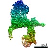
| Structure viewer | Molecule: MolmilJmol/JSmol |
|---|
- Downloads & links
-Download
| PDBx/mmCIF format | 7cea.cif.gz | 166.5 KB | Display | PDBx/mmCIF format |
|---|---|---|---|---|
| PDB format | pdb7cea.ent.gz | 112.9 KB | Display | PDB format |
| PDBx/mmJSON format | 7cea.json.gz | Tree view | PDBx/mmJSON format | |
| Others |  Other downloads Other downloads |
-Validation report
| Arichive directory | https://data.pdbj.org/pub/pdb/validation_reports/ce/7ceaftp://data.pdbj.org/pub/pdb/validation_reports/ce/7cea | HTTPS FTP |
|---|
-Related structure data
| Related structure data |  7cebC  7cecC  3qq9S  4kaqS 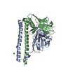 5xctS S: Starting model for refinement C: citing same article ( |
|---|---|
| Similar structure data |


-Links
 PDBj
PDBj


- Assembly
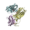
| Deposited unit | 
| ||||||||||
|---|---|---|---|---|---|---|---|---|---|---|---|
| 1 |
| ||||||||||
| Unit cell |
|
-Components
| #1: Antibody | Mass: 19760.207 Da / Num. of mol.: 1 Source method: isolated from a genetically manipulated source Details: chimera of HUTS-4 VH(S112C)-SARAH Source: (gene. exp.) Homo sapiens (human)Production host:  |
|---|---|
| #2: Antibody | Mass: 17701.023 Da / Num. of mol.: 1 Source method: isolated from a genetically manipulated source Details: chimera of HUTS-4 VL(C87Y)-SARAH(S37C) Source: (gene. exp.) Homo sapiens (human)Production host: |
| Has protein modification | Y |
-Experimental details
-Experiment
| Experiment | Method: X-RAY DIFFRACTION / Number of used crystals: 1 |
|---|
- Sample preparation
| Crystal | Density Matthews: 2.35 Å3/Da / Density % sol: 47.67 % |
|---|---|
| Crystal grow | Temperature: 293 K / Method: vapor diffusion, sitting drop / Details: 1.26 M ammonium sulfate, 0.1 M MES, pH 6.0 |
-Data collection
| Diffraction | Mean temperature: 100 K / Serial crystal experiment: N |
|---|---|
| Diffraction source | Source: SYNCHROTRON / Site: Photon Factory / Beamline: BL-17A / Wavelength: 0.98 Å |
| Detector | Type: DECTRIS EIGER X 16M / Detector: PIXEL / Date: Nov 20, 2018 |
| Radiation | Protocol: SINGLE WAVELENGTH / Monochromatic (M) / Laue (L): M / Scattering type: x-ray |
| Radiation wavelength | Wavelength: 0.98 Å / Relative weight: 1 |
| Reflection | Resolution: 2.55→44.183 Å / Num. obs: 11865 / % possible obs: 99.8 % / Redundancy: 13.5 % / Biso Wilson estimate: 69.53 Å2 / CC1/2: 0.999 / Rsym value: 0.104 / Net I/σ(I): 17.77 |
| Reflection shell | Resolution: 2.55→2.7 Å / Num. unique obs: 1860 / CC1/2: 0.899 |
- Processing
| Software |
| |||||||||||||||||||||||||||||||||||||||||||||||||||||||||||||||||||||||||||||||||||||||||||||||||||||||||||||||||||||||||||||
|---|---|---|---|---|---|---|---|---|---|---|---|---|---|---|---|---|---|---|---|---|---|---|---|---|---|---|---|---|---|---|---|---|---|---|---|---|---|---|---|---|---|---|---|---|---|---|---|---|---|---|---|---|---|---|---|---|---|---|---|---|---|---|---|---|---|---|---|---|---|---|---|---|---|---|---|---|---|---|---|---|---|---|---|---|---|---|---|---|---|---|---|---|---|---|---|---|---|---|---|---|---|---|---|---|---|---|---|---|---|---|---|---|---|---|---|---|---|---|---|---|---|---|---|---|---|---|
| Refinement | Method to determine structure: MOLECULAR REPLACEMENT Starting model: 3qq9, 4kaq, 5xct Resolution: 2.55→34.79 Å / SU ML: 0.3834 / Cross valid method: FREE R-VALUE / σ(F): 1.34 / Phase error: 38.7576 / Stereochemistry target values: CDL v1.2
| |||||||||||||||||||||||||||||||||||||||||||||||||||||||||||||||||||||||||||||||||||||||||||||||||||||||||||||||||||||||||||||
| Solvent computation | Shrinkage radii: 0.9 Å / VDW probe radii: 1.11 Å / Solvent model: FLAT BULK SOLVENT MODEL | |||||||||||||||||||||||||||||||||||||||||||||||||||||||||||||||||||||||||||||||||||||||||||||||||||||||||||||||||||||||||||||
| Displacement parameters | Biso mean: 92.34 Å2 | |||||||||||||||||||||||||||||||||||||||||||||||||||||||||||||||||||||||||||||||||||||||||||||||||||||||||||||||||||||||||||||
| Refinement step | Cycle: LAST / Resolution: 2.55→34.79 Å
| |||||||||||||||||||||||||||||||||||||||||||||||||||||||||||||||||||||||||||||||||||||||||||||||||||||||||||||||||||||||||||||
| Refine LS restraints |
| |||||||||||||||||||||||||||||||||||||||||||||||||||||||||||||||||||||||||||||||||||||||||||||||||||||||||||||||||||||||||||||
| LS refinement shell |
| |||||||||||||||||||||||||||||||||||||||||||||||||||||||||||||||||||||||||||||||||||||||||||||||||||||||||||||||||||||||||||||
| Refinement TLS params. | Method: refined / Refine-ID: X-RAY DIFFRACTION
| |||||||||||||||||||||||||||||||||||||||||||||||||||||||||||||||||||||||||||||||||||||||||||||||||||||||||||||||||||||||||||||
| Refinement TLS group |
|
