 Movie
Movie Controller

[English] 日本語
 Yorodumi
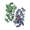
- PDB-5ua1: Mycobacterium tuberculosis KstR in complex with a 18-bp DNA operator -
+ Open data

- Basic information
| Entry | Database: PDB / ID: 5ua1 | ||||||
|---|---|---|---|---|---|---|---|
| Title | Mycobacterium tuberculosis KstR in complex with a 18-bp DNA operator | ||||||
 Components Components |
| ||||||
 Keywords Keywords | Transcription/DNA / KstR / DNA / complex / transcriptional regulator / TetR family transcriptional repressor / Structural Genomics TB Structural Genomics Consortium / TBSGC / Transcription-DNA complex | ||||||
| Function / homology |  Function and homology information Function and homology informationtranscription cis-regulatory region binding / DNA-binding transcription factor activity / regulation of DNA-templated transcription / protein homodimerization activity / DNA binding Similarity search - Function | ||||||
| Biological species |   Mycobacterium tuberculosis (bacteria)Mycobacterium tuberculosis H37Rv (bacteria) Mycobacterium tuberculosis (bacteria)Mycobacterium tuberculosis H37Rv (bacteria) | ||||||
| Method |  X-RAY DIFFRACTION / SYNCHROTRON / MOLECULAR REPLACEMENT / Resolution: 2.9 Å X-RAY DIFFRACTION / SYNCHROTRON / MOLECULAR REPLACEMENT / Resolution: 2.9 Å | ||||||
 Authors Authors | Ho, N.A.T. / Dawes, S.S. / Baker, E.N. / Lott, J.S. / TB Structural Genomics Consortium (TBSGC) | ||||||
| Funding support |  New Zealand, 1items New Zealand, 1items
| ||||||
 Citation Citation | Journal: To Be Published Title: Crystal structure of KstR in complex with cognate DNA operator Authors: Ho, N.A.T. / Dawes, S.S. / Baker, E.N. / Lott, J.S. | ||||||
| History |
|
- Structure visualization
| Structure viewer | Molecule: MolmilJmol/JSmol |
|---|
- Downloads & links
-Download
| PDBx/mmCIF format | 5ua1.cif.gz | 119.5 KB | Display | PDBx/mmCIF format |
|---|---|---|---|---|
| PDB format | pdb5ua1.ent.gz | 85.8 KB | Display | PDB format |
| PDBx/mmJSON format | 5ua1.json.gz | Tree view | PDBx/mmJSON format | |
| Others |  Other downloads Other downloads |
-Validation report
| Arichive directory | https://data.pdbj.org/pub/pdb/validation_reports/ua/5ua1ftp://data.pdbj.org/pub/pdb/validation_reports/ua/5ua1 | HTTPS FTP |
|---|
-Related structure data
| Related structure data | 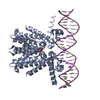 5ua2C 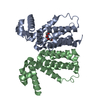 5cxgS S: Starting model for refinement C: citing same article ( |
|---|---|
| Similar structure data |


-Links
 PDBj
PDBj


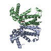
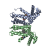
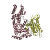
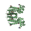
- Assembly
| Deposited unit | 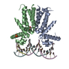
| ||||||||
|---|---|---|---|---|---|---|---|---|---|
| 1 | 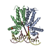
| ||||||||
| 2 | 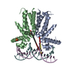
| ||||||||
| Unit cell |
|
-Components
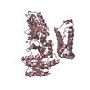
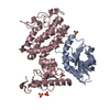
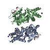
-Protein , 1 types, 2 molecules BA
| #1: Protein | Mass: 22352.510 Da / Num. of mol.: 2 Source method: isolated from a genetically manipulated source Source: (gene. exp.) Mycobacterium tuberculosis (strain ATCC 25618 / H37Rv) (bacteria)Strain: ATCC 25618 / H37Rv / Gene: kstR, Rv3574 / Production host: |
|---|
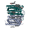
-DNA chain , 2 types, 4 molecules CEDF
| #2: DNA chain | Mass: 5514.603 Da / Num. of mol.: 2 / Source method: obtained synthetically Source: (synth.) Mycobacterium tuberculosis H37Rv (bacteria)#3: DNA chain | Mass: 5514.603 Da / Num. of mol.: 2 / Source method: obtained synthetically Source: (synth.) Mycobacterium tuberculosis H37Rv (bacteria) |
|---|
-Non-polymers , 3 types, 8 molecules 


| #4: Chemical |  Mass: 96.063 Da / Num. of mol.: 2 / Source method: obtained synthetically / Formula: SO4 Mass: 96.063 Da / Num. of mol.: 2 / Source method: obtained synthetically / Formula: SO4#5: Chemical |  Mass: 150.173 Da / Num. of mol.: 2 / Source method: obtained synthetically / Formula: C6H14O4 Mass: 150.173 Da / Num. of mol.: 2 / Source method: obtained synthetically / Formula: C6H14O4#6: Water | ChemComp-HOH / | Mass: 18.015 Da / Num. of mol.: 4 / Source method: isolated from a natural source / Formula: H2O |
|---|
-Experimental details
-Experiment
| Experiment | Method: X-RAY DIFFRACTION / Number of used crystals: 1 |
|---|
- Sample preparation
| Crystal | Density Matthews: 2.12 Å3/Da / Density % sol: 41.93 % |
|---|---|
| Crystal grow | Temperature: 291 K / Method: vapor diffusion / pH: 6.5 Details: 15% PEG 3350, 9% MPD, 0.2 M Li2SO4, 0.1 M imidazole-HCl |
-Data collection
| Diffraction | Mean temperature: 100 K |
|---|---|
| Diffraction source | Source: SYNCHROTRON / Site: Australian Synchrotron  / Beamline: MX2 / Wavelength: 0.9537 Å / Beamline: MX2 / Wavelength: 0.9537 Å |
| Detector | Type: ADSC QUANTUM 315r / Detector: CCD / Date: Nov 20, 2015 |
| Radiation | Protocol: SINGLE WAVELENGTH / Monochromatic (M) / Laue (L): M / Scattering type: x-ray |
| Radiation wavelength | Wavelength: 0.9537 Å / Relative weight: 1 |
| Reflection | Resolution: 2.9→48.56 Å / Num. obs: 24284 / % possible obs: 100 % / Redundancy: 3.9 % / CC1/2: 0.993 / Rmerge(I) obs: 0.155 / Net I/σ(I): 9.3 |
| Reflection shell | Resolution: 2.9→3 Å / Redundancy: 3.9 % / Rmerge(I) obs: 1.045 / Mean I/σ(I) obs: 1.9 / CC1/2: 0.47 / % possible all: 100 |
- Processing
| Software |
| ||||||||||||||||||||||||||||||||||||||||||||||||||||||||||||||||||||||
|---|---|---|---|---|---|---|---|---|---|---|---|---|---|---|---|---|---|---|---|---|---|---|---|---|---|---|---|---|---|---|---|---|---|---|---|---|---|---|---|---|---|---|---|---|---|---|---|---|---|---|---|---|---|---|---|---|---|---|---|---|---|---|---|---|---|---|---|---|---|---|---|
| Refinement | Method to determine structure: MOLECULAR REPLACEMENT Starting model: 5CXG Resolution: 2.9→46.853 Å / SU ML: 0.6 / Cross valid method: FREE R-VALUE / σ(F): 1.97 / Phase error: 30.51 / Stereochemistry target values: ML
| ||||||||||||||||||||||||||||||||||||||||||||||||||||||||||||||||||||||
| Solvent computation | Shrinkage radii: 0.9 Å / VDW probe radii: 1.11 Å / Solvent model: FLAT BULK SOLVENT MODEL | ||||||||||||||||||||||||||||||||||||||||||||||||||||||||||||||||||||||
| Refinement step | Cycle: LAST / Resolution: 2.9→46.853 Å
| ||||||||||||||||||||||||||||||||||||||||||||||||||||||||||||||||||||||
| Refine LS restraints |
| ||||||||||||||||||||||||||||||||||||||||||||||||||||||||||||||||||||||
| LS refinement shell |
|
