 Movie
Movie Controller

[English] 日本語
 Yorodumi
- PDB-4hyx: Crystal Structure Analysis of the Bacteriorhodopsin in Facial Amp... -
+ Open data

- Basic information
| Entry | Database: PDB / ID: 4hyx | ||||||
|---|---|---|---|---|---|---|---|
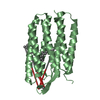
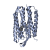
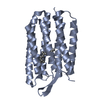
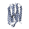
| Title | Crystal Structure Analysis of the Bacteriorhodopsin in Facial Amphiphile-4 DMPC Bicelle | ||||||
 Components Components | Bacteriorhodopsin | ||||||
 Keywords Keywords | PROTON TRANSPORT / Facial Amphiphile-4 DMPC Bicelle / Alpha helical 7 Transmembrane Protein / Photoreceptor / Proton Pump / Retinal Binding / Transmembrane | ||||||
| Function / homology |  Function and homology information Function and homology informationlight-driven active monoatomic ion transmembrane transporter activity / photoreceptor activity / phototransduction / monoatomic ion channel activity / proton transmembrane transport / plasma membrane Similarity search - Function | ||||||
| Biological species |  Halobacterium salinarum (Halophile) Halobacterium salinarum (Halophile) | ||||||
| Method |  X-RAY DIFFRACTION / SYNCHROTRON / MOLECULAR REPLACEMENT / Resolution: 1.99 Å X-RAY DIFFRACTION / SYNCHROTRON / MOLECULAR REPLACEMENT / Resolution: 1.99 Å | ||||||
 Authors Authors | Lee, S. / Stout, C.D. / Zhang, Q. | ||||||
 Citation Citation | Journal: Proc.Natl.Acad.Sci.USA / Year: 2013 Title: Steroid-based facial amphiphiles for stabilization and crystallization of membrane proteins. Authors: Lee, S.C. / Bennett, B.C. / Hong, W.X. / Fu, Y. / Baker, K.A. / Marcoux, J. / Robinson, C.V. / Ward, A.B. / Halpert, J.R. / Stevens, R.C. / Stout, C.D. / Yeager, M.J. / Zhang, Q. | ||||||
| History |
|
- Structure visualization
| Structure viewer | Molecule: MolmilJmol/JSmol |
|---|
- Downloads & links
-Download
| PDBx/mmCIF format | 4hyx.cif.gz | 105.9 KB | Display | PDBx/mmCIF format |
|---|---|---|---|---|
| PDB format | pdb4hyx.ent.gz | 80.6 KB | Display | PDB format |
| PDBx/mmJSON format | 4hyx.json.gz | Tree view | PDBx/mmJSON format | |
| Others |  Other downloads Other downloads |
-Validation report
| Arichive directory | https://data.pdbj.org/pub/pdb/validation_reports/hy/4hyxftp://data.pdbj.org/pub/pdb/validation_reports/hy/4hyx | HTTPS FTP |
|---|
-Related structure data


-Links
 PDBj
PDBj


- Assembly
| Deposited unit | 
| ||||||||||||||||||
|---|---|---|---|---|---|---|---|---|---|---|---|---|---|---|---|---|---|---|---|
| 1 | 
| ||||||||||||||||||
| 2 | 
| ||||||||||||||||||
| Unit cell |
| ||||||||||||||||||
| Noncrystallographic symmetry (NCS) | NCS domain:
NCS domain segments: Component-ID: _ / Ens-ID: 1 / Beg auth comp-ID: GLY / Beg label comp-ID: GLY / End auth comp-ID: PHE / End label comp-ID: PHE / Refine code: _ / Auth seq-ID: 6 - 230 / Label seq-ID: 19 - 243
|
-Components
-Protein , 1 types, 2 molecules AB
| #1: Protein | Mass: 28270.084 Da / Num. of mol.: 2 / Source method: isolated from a natural source / Source: (natural) Halobacterium salinarum (Halophile) / Strain: S9 / References: UniProt: P02945 |
|---|
-Non-polymers , 7 types, 56 molecules 


| #2: Chemical |  Mass: 284.436 Da / Num. of mol.: 2 / Source method: obtained synthetically / Formula: C20H28O Mass: 284.436 Da / Num. of mol.: 2 / Source method: obtained synthetically / Formula: C20H28O#3: Chemical | ChemComp-OCT /  Mass: 114.229 Da / Num. of mol.: 4 / Source method: obtained synthetically / Formula: C8H18 Mass: 114.229 Da / Num. of mol.: 4 / Source method: obtained synthetically / Formula: C8H18#4: Chemical | ChemComp-D10 /  Mass: 142.282 Da / Num. of mol.: 8 / Source method: obtained synthetically / Formula: C10H22 Mass: 142.282 Da / Num. of mol.: 8 / Source method: obtained synthetically / Formula: C10H22#5: Chemical | ChemComp-HEX / |  Mass: 86.175 Da / Num. of mol.: 1 / Source method: obtained synthetically / Formula: C6H14 Mass: 86.175 Da / Num. of mol.: 1 / Source method: obtained synthetically / Formula: C6H14#6: Chemical | ChemComp-C14 / |  Mass: 198.388 Da / Num. of mol.: 1 / Source method: obtained synthetically / Formula: C14H30 Mass: 198.388 Da / Num. of mol.: 1 / Source method: obtained synthetically / Formula: C14H30#7: Chemical | ChemComp-GOL / |  Mass: 92.094 Da / Num. of mol.: 1 / Source method: obtained synthetically / Formula: C3H8O3 Mass: 92.094 Da / Num. of mol.: 1 / Source method: obtained synthetically / Formula: C3H8O3#8: Water | ChemComp-HOH / | Mass: 18.015 Da / Num. of mol.: 39 / Source method: isolated from a natural source / Formula: H2O |
|---|
-Details
| Has protein modification | Y |
|---|
-Experimental details
-Experiment
| Experiment | Method: X-RAY DIFFRACTION / Number of used crystals: 1 |
|---|
- Sample preparation
| Crystal | Density Matthews: 2.36 Å3/Da / Density % sol: 47.87 % |
|---|---|
| Crystal grow | Temperature: 300.15 K / Method: vapor diffusion, sitting drop / pH: 3.4 Details: 3.6M Sodium Phosphate (pH 3.4), 3.5% Triethyleneglycol, 180mM Hexanediol, VAPOR DIFFUSION, SITTING DROP, temperature 300.15K |
-Data collection
| Diffraction | Mean temperature: 100 K |
|---|---|
| Diffraction source | Source: SYNCHROTRON / Site: SSRL  / Beamline: BL11-1 / Wavelength: 0.97945 Å / Beamline: BL11-1 / Wavelength: 0.97945 Å |
| Detector | Type: ADSC QUANTUM 315 / Detector: CCD / Date: Apr 15, 2011 |
| Radiation | Monochromator: Slide scattering bent cube-root I-beam single crystal; asymmetric cut 4.965 degs Protocol: SINGLE WAVELENGTH / Monochromatic (M) / Laue (L): M / Scattering type: x-ray |
| Radiation wavelength | Wavelength: 0.97945 Å / Relative weight: 1 |
| Reflection | Resolution: 2→56.46 Å / Num. all: 35212 / Num. obs: 32607 / % possible obs: 92.6 % / Observed criterion σ(F): 0 / Observed criterion σ(I): 0 / Redundancy: 2.1 % / Rmerge(I) obs: 0.074 / Net I/σ(I): 6.1 |
| Reflection shell | Resolution: 2→2.11 Å / Redundancy: 2.1 % / Rmerge(I) obs: 0.399 / Mean I/σ(I) obs: 2 / % possible all: 86 |
- Processing
| Software |
| ||||||||||||||||||||||||||||||||||||||||||||||||||||||||||||||||||||||||||||||||||||||||||||||||||||||||||||||||||||||||||||||||||||||||||||||||||||||||||||||||||||||||||
|---|---|---|---|---|---|---|---|---|---|---|---|---|---|---|---|---|---|---|---|---|---|---|---|---|---|---|---|---|---|---|---|---|---|---|---|---|---|---|---|---|---|---|---|---|---|---|---|---|---|---|---|---|---|---|---|---|---|---|---|---|---|---|---|---|---|---|---|---|---|---|---|---|---|---|---|---|---|---|---|---|---|---|---|---|---|---|---|---|---|---|---|---|---|---|---|---|---|---|---|---|---|---|---|---|---|---|---|---|---|---|---|---|---|---|---|---|---|---|---|---|---|---|---|---|---|---|---|---|---|---|---|---|---|---|---|---|---|---|---|---|---|---|---|---|---|---|---|---|---|---|---|---|---|---|---|---|---|---|---|---|---|---|---|---|---|---|---|---|---|---|---|
| Refinement | Method to determine structure: MOLECULAR REPLACEMENT / Resolution: 1.99→46.85 Å / Cor.coef. Fo:Fc: 0.948 / Cor.coef. Fo:Fc free: 0.933 / SU B: 2.009 / SU ML: 0.061 / Cross valid method: THROUGHOUT / ESU R: 0.171 / ESU R Free: 0.15 / Stereochemistry target values: MAXIMUM LIKELIHOOD / Details: HYDROGENS HAVE BEEN ADDED IN THE RIDING POSITIONS
| ||||||||||||||||||||||||||||||||||||||||||||||||||||||||||||||||||||||||||||||||||||||||||||||||||||||||||||||||||||||||||||||||||||||||||||||||||||||||||||||||||||||||||
| Solvent computation | Ion probe radii: 0.8 Å / Shrinkage radii: 0.8 Å / VDW probe radii: 1.2 Å / Solvent model: MASK | ||||||||||||||||||||||||||||||||||||||||||||||||||||||||||||||||||||||||||||||||||||||||||||||||||||||||||||||||||||||||||||||||||||||||||||||||||||||||||||||||||||||||||
| Displacement parameters | Biso mean: 33.358 Å2
| ||||||||||||||||||||||||||||||||||||||||||||||||||||||||||||||||||||||||||||||||||||||||||||||||||||||||||||||||||||||||||||||||||||||||||||||||||||||||||||||||||||||||||
| Refinement step | Cycle: LAST / Resolution: 1.99→46.85 Å
| ||||||||||||||||||||||||||||||||||||||||||||||||||||||||||||||||||||||||||||||||||||||||||||||||||||||||||||||||||||||||||||||||||||||||||||||||||||||||||||||||||||||||||
| Refine LS restraints |
| ||||||||||||||||||||||||||||||||||||||||||||||||||||||||||||||||||||||||||||||||||||||||||||||||||||||||||||||||||||||||||||||||||||||||||||||||||||||||||||||||||||||||||
| Refine LS restraints NCS | Ens-ID: 1 / Number: 14105 / Refine-ID: X-RAY DIFFRACTION / Type: interatomic distance / Rms dev position: 0.06 Å / Weight position: 0.05
| ||||||||||||||||||||||||||||||||||||||||||||||||||||||||||||||||||||||||||||||||||||||||||||||||||||||||||||||||||||||||||||||||||||||||||||||||||||||||||||||||||||||||||
| LS refinement shell | Resolution: 1.994→2.046 Å / Total num. of bins used: 20
|
