 Movie
Movie Controller


+ Open data

- Basic information
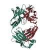
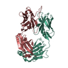
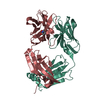
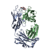
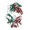
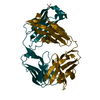
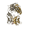
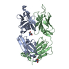
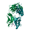
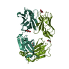
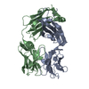
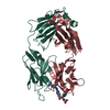
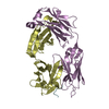
| Entry | Database: PDB / ID: 4kaq | |||||||||
|---|---|---|---|---|---|---|---|---|---|---|
| Title | Structure of rituximab Fab | |||||||||
 Components Components |
| |||||||||
 Keywords Keywords | IMMUNE SYSTEM / monoclonal antibody | |||||||||
| Function / homology | Immunoglobulins / Immunoglobulin-like / Sandwich / Mainly Beta Function and homology information Function and homology information | |||||||||
| Biological species |  Homo sapiens (human) Homo sapiens (human) | |||||||||
| Method |  X-RAY DIFFRACTION / MOLECULAR REPLACEMENT / Resolution: 2.48 Å X-RAY DIFFRACTION / MOLECULAR REPLACEMENT / Resolution: 2.48 Å | |||||||||
 Authors Authors | Bzymek, K.P. / Williams, J.C. | |||||||||
 Citation Citation | Journal: To be Published Title: Crystal structure of rituximab Authors: Bzymek, K.P. / Williams, J.C. | |||||||||
| History |
|
- Structure visualization
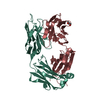
| Structure viewer | Molecule: MolmilJmol/JSmol |
|---|
- Downloads & links
-Download
| PDBx/mmCIF format | 4kaq.cif.gz | 100 KB | Display | PDBx/mmCIF format |
|---|---|---|---|---|
| PDB format | pdb4kaq.ent.gz | 75.9 KB | Display | PDB format |
| PDBx/mmJSON format | 4kaq.json.gz | Tree view | PDBx/mmJSON format | |
| Others |  Other downloads Other downloads |
-Validation report
| Arichive directory | https://data.pdbj.org/pub/pdb/validation_reports/ka/4kaqftp://data.pdbj.org/pub/pdb/validation_reports/ka/4kaq | HTTPS FTP |
|---|
-Related structure data
| Related structure data |  2oslS S: Starting model for refinement |
|---|---|
| Similar structure data |


-Links
 PDBj
PDBj

- Assembly
| Deposited unit | 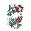
| ||||||||||||
|---|---|---|---|---|---|---|---|---|---|---|---|---|---|
| 1 |
| ||||||||||||
| Unit cell |
| ||||||||||||
| Components on special symmetry positions |
|
-Components
| #1: Antibody | Mass: 23078.623 Da / Num. of mol.: 1 Source method: isolated from a genetically manipulated source Details: Commercially available as Rituxin / Source: (gene. exp.) Homo sapiens, Mus musculus |
|---|---|
| #2: Antibody | Mass: 23716.512 Da / Num. of mol.: 1 Source method: isolated from a genetically manipulated source Details: Commercially available as Rituxin / Source: (gene. exp.) Homo sapiens, Mus musculus |
| #3: Water | ChemComp-HOH /  Mass: 18.015 Da / Num. of mol.: 252 / Source method: isolated from a natural source / Formula: H2O Mass: 18.015 Da / Num. of mol.: 252 / Source method: isolated from a natural source / Formula: H2O |
| Has protein modification | Y |
-Experimental details
-Experiment
| Experiment | Method: X-RAY DIFFRACTION / Number of used crystals: 1 |
|---|
- Sample preparation
| Crystal | Density Matthews: 2.2 Å3/Da / Density % sol: 44.02 % |
|---|---|
| Crystal grow | Temperature: 293 K / Method: vapor diffusion, hanging drop / pH: 7 Details: 22% PEG 3350, pH 7, VAPOR DIFFUSION, HANGING DROP, temperature 293K |
-Data collection
| Diffraction | Mean temperature: 100 K |
|---|---|
| Diffraction source | Source: ROTATING ANODE / Type: RIGAKU MICROMAX-007 HF / Wavelength: 1.5418 Å |
| Detector | Type: RIGAKU RAXIS IV++ / Detector: IMAGE PLATE / Date: Apr 8, 2013 |
| Radiation | Monochromator: Varimax / Protocol: SINGLE WAVELENGTH / Monochromatic (M) / Laue (L): M / Scattering type: x-ray |
| Radiation wavelength | Wavelength: 1.5418 Å / Relative weight: 1 |
| Reflection | Resolution: 2.48→30.32 Å / Num. all: 15014 / Num. obs: 14891 / % possible obs: 99.2 % / Observed criterion σ(F): 2 / Observed criterion σ(I): 2 / Redundancy: 4.6 % / Rmerge(I) obs: 0.058 / Net I/σ(I): 22.4 |
| Reflection shell | Resolution: 2.48→2.54 Å / Redundancy: 3.3 % / Rmerge(I) obs: 0.199 / Mean I/σ(I) obs: 6.3 / % possible all: 92.4 |
- Processing
| Software |
| ||||||||||||||||||||||||||||||||||||||||||
|---|---|---|---|---|---|---|---|---|---|---|---|---|---|---|---|---|---|---|---|---|---|---|---|---|---|---|---|---|---|---|---|---|---|---|---|---|---|---|---|---|---|---|---|
| Refinement | Method to determine structure: MOLECULAR REPLACEMENT Starting model: 2OSL Resolution: 2.48→30.32 Å / SU ML: 0.3 / σ(F): 2 / σ(I): 2 / Phase error: 20.64 / Stereochemistry target values: ML
| ||||||||||||||||||||||||||||||||||||||||||
| Solvent computation | Shrinkage radii: 0.9 Å / VDW probe radii: 1.11 Å / Solvent model: FLAT BULK SOLVENT MODEL | ||||||||||||||||||||||||||||||||||||||||||
| Refinement step | Cycle: LAST / Resolution: 2.48→30.32 Å
| ||||||||||||||||||||||||||||||||||||||||||
| Refine LS restraints |
| ||||||||||||||||||||||||||||||||||||||||||
| LS refinement shell |
|
