 Movie
Movie Controller

[English] 日本語
 Yorodumi
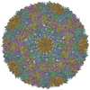
- PDB-3iyw: West Nile virus in complex with Fab fragments of MAb CR4354 (fitt... -
+ Open data

- Basic information
| Entry | Database: PDB / ID: 3iyw | |||||||||||||||
|---|---|---|---|---|---|---|---|---|---|---|---|---|---|---|---|---|
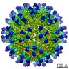
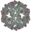
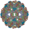
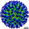
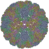
| Title | West Nile virus in complex with Fab fragments of MAb CR4354 (fitted coordinates of envelope proteins and Fab fragments of one icosahedral ASU) | |||||||||||||||
 Components Components |
| |||||||||||||||
 Keywords Keywords | VIRUS / virus-antibody complex / neutralizing Fab fragment / flavivirus / West Nile Virus / envelope protein / icosahedral virus | |||||||||||||||
| Function / homology |  Function and homology information Function and homology informationflavivirin / symbiont-mediated suppression of host JAK-STAT cascade via inhibition of STAT2 activity / symbiont-mediated suppression of host JAK-STAT cascade via inhibition of STAT1 activity / viral capsid / nucleoside-triphosphate phosphatase / double-stranded RNA binding / mRNA (guanine-N7)-methyltransferase / methyltransferase cap1 / methyltransferase cap1 activity / mRNA 5'-cap (guanine-N7-)-methyltransferase activity ...flavivirin / symbiont-mediated suppression of host JAK-STAT cascade via inhibition of STAT2 activity / symbiont-mediated suppression of host JAK-STAT cascade via inhibition of STAT1 activity / viral capsid / nucleoside-triphosphate phosphatase / double-stranded RNA binding / mRNA (guanine-N7)-methyltransferase / methyltransferase cap1 / methyltransferase cap1 activity / mRNA 5'-cap (guanine-N7-)-methyltransferase activity / RNA helicase activity / protein dimerization activity / symbiont-mediated suppression of host innate immune response / host cell perinuclear region of cytoplasm / host cell endoplasmic reticulum membrane / RNA helicase / symbiont-mediated suppression of host type I interferon-mediated signaling pathway / serine-type endopeptidase activity / symbiont-mediated activation of host autophagy / RNA-directed RNA polymerase / viral RNA genome replication / RNA-directed RNA polymerase activity / fusion of virus membrane with host endosome membrane / viral envelope / symbiont entry into host cell / virion attachment to host cell / host cell nucleus / virion membrane / structural molecule activity / ATP hydrolysis activity / proteolysis / extracellular region / ATP binding / metal ion binding Similarity search - Function | |||||||||||||||
| Biological species |  West Nile virus West Nile virus Homo sapiens (human) Homo sapiens (human) | |||||||||||||||
| Method | ELECTRON MICROSCOPY / single particle reconstruction / cryo EM / Resolution: 13.7 Å | |||||||||||||||
 Authors Authors | Rossmann, M.G. / Kaufmann, B. | |||||||||||||||
 Citation Citation | Journal: Proc Natl Acad Sci U S A / Year: 2010 Title: Neutralization of West Nile virus by cross-linking of its surface proteins with Fab fragments of the human monoclonal antibody CR4354. Authors: Bärbel Kaufmann / Matthew R Vogt / Jaap Goudsmit / Heather A Holdaway / Anastasia A Aksyuk / Paul R Chipman / Richard J Kuhn / Michael S Diamond / Michael G Rossmann /  Abstract: Many flaviviruses are significant human pathogens, with the humoral immune response playing an essential role in restricting infection and disease. CR4354, a human monoclonal antibody isolated from a ...Many flaviviruses are significant human pathogens, with the humoral immune response playing an essential role in restricting infection and disease. CR4354, a human monoclonal antibody isolated from a patient, neutralizes West Nile virus (WNV) infection at a postattachment stage in the viral life-cycle. Here, we determined the structure of WNV complexed with Fab fragments of CR4354 using cryoelectron microscopy. The outer glycoprotein shell of a mature WNV particle is formed by 30 rafts of three homodimers of the viral surface protein E. CR4354 binds to a discontinuous epitope formed by protein segments from two neighboring E molecules, but does not cause any detectable structural disturbance on the viral surface. The epitope occurs at two independent positions within an icosahedral asymmetric unit, resulting in 120 binding sites on the viral surface. The cross-linking of the six E monomers within one raft by four CR4354 Fab fragments suggests that the antibody neutralizes WNV by blocking the pH-induced rearrangement of the E protein required for virus fusion with the endosomal membrane. | |||||||||||||||
| History |
|
- Structure visualization
| Movie |
Movie viewer |
|---|---|
| Structure viewer | Molecule: MolmilJmol/JSmol |
- Downloads & links
-Download
| PDBx/mmCIF format | 3iyw.cif.gz | 410.1 KB | Display | PDBx/mmCIF format |
|---|---|---|---|---|
| PDB format | pdb3iyw.ent.gz | 332.8 KB | Display | PDB format |
| PDBx/mmJSON format | 3iyw.json.gz | Tree view | PDBx/mmJSON format | |
| Others |  Other downloads Other downloads |
-Validation report
| Arichive directory | https://data.pdbj.org/pub/pdb/validation_reports/iy/3iywftp://data.pdbj.org/pub/pdb/validation_reports/iy/3iyw | HTTPS FTP |
|---|
-Related structure data
| Related structure data |  5190MC 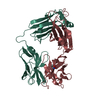 3n9gC M: map data used to model this data C: citing same article ( |
|---|---|
| Similar structure data |

-Links
 PDBj
PDBj


- Assembly
| Deposited unit | 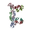
|
|---|---|
| 1 | x 60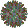
|
| 2 |
|
| 3 | x 5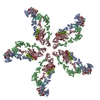
|
| 4 | x 6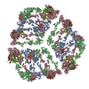
|
| 5 | 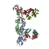
|
| Symmetry | Point symmetry: (Schoenflies symbol: I (icosahedral)) |
-Components
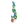
| #1: Protein | Mass: 43272.098 Da / Num. of mol.: 3 / Fragment: ectodomain of viral surface protein Source method: isolated from a genetically manipulated source Source: (gene. exp.) West Nile virus / Production host: Homo sapiens (human) / References: UniProt: Q91R02, UniProt: Q9Q6P4*PLUS#2: Antibody | Mass: 24745.699 Da / Num. of mol.: 2 / Fragment: Fab fragment, heavy chain Source method: isolated from a genetically manipulated source Source: (gene. exp.) Homo sapiens (human) / Cell (production host): B cells / Production host: Homo sapiens (human) / Tissue (production host): peripheral blood#3: Antibody | Mass: 22908.160 Da / Num. of mol.: 2 / Fragment: Fab fragment, light chain Source method: isolated from a genetically manipulated source Source: (gene. exp.) Homo sapiens (human) / Production host: Homo sapiens (human)#4: Polysaccharide | Source method: isolated from a genetically manipulated source Has protein modification | Y | |
|---|
-Experimental details
-Experiment
| Experiment | Method: ELECTRON MICROSCOPY |
|---|---|
| EM experiment | Aggregation state: PARTICLE / 3D reconstruction method: single particle reconstruction |
- Sample preparation
| Component | Name: West Nile virus in complex with Fab fragments of MAb CR4354 Type: VIRUS Details: THE ENTRY PRESENTED HERE DOES NOT CONTAIN THE COMPLETE BIOLOGICAL ASSEMBLY. COORDINATES FOR A COMPLETE MULTIMER REPRESENTING THE KNOWN BIOLOGICALLY SIGNIFICANT OLIGOMERIZATION STATE OF THE ...Details: THE ENTRY PRESENTED HERE DOES NOT CONTAIN THE COMPLETE BIOLOGICAL ASSEMBLY. COORDINATES FOR A COMPLETE MULTIMER REPRESENTING THE KNOWN BIOLOGICALLY SIGNIFICANT OLIGOMERIZATION STATE OF THE VIRUS-FAB COMPLEX CAN BE GENERATED BY APPLYING BIOMT TRANSFORMATIONS GIVEN. (entry represents one icoshedral ASU containing 3 envelope E protein molecules and 2 CR4354 Fab fragments) |
|---|---|
| Details of virus | Empty: NO / Enveloped: YES / Host category: VERTEBRATES / Isolate: STRAIN / Type: VIRION |
| Natural host | Organism: Homo sapiens |
| Buffer solution | pH: 8 / Details: 12mM Tris-HCl, 120mM NaCl, 1mM EDTA |
| Specimen | Embedding applied: NO / Shadowing applied: NO / Staining applied: NO / Vitrification applied: YES / Details: 12mM Tris-HCl, 120mM NaCl, 1mM EDTA |
| Vitrification | Instrument: HOMEMADE PLUNGER / Cryogen name: ETHANE Method: A small vial of ethane is placed inside a larger liquid nitrogen reservoir. The grid holding a few microliters of the sample is held in place at the bottom of a plunger by the means of fine ...Method: A small vial of ethane is placed inside a larger liquid nitrogen reservoir. The grid holding a few microliters of the sample is held in place at the bottom of a plunger by the means of fine tweezers. Once the ethane in the vial is completely frozen, it needs to be slightly melted. When the liquid ethane is ready, a piece of filter paper is then pressed against the sample to blot of excess buffer, sufficient to leave a thin layer on the grid. After a predetermined time, the filter paper is removed, and the plunger is allowed to drop into the liquid ethane. Once the grid enters the liquid ethane, the sample is rapidly frozen, and the grid is transferred under liquid nitrogen to a storage box immersed liquid nitrogen for later use in the microscope. |
- Electron microscopy imaging
| Microscopy | Model: FEI/PHILIPS CM300FEG/T / Date: Sep 9, 2009 / Details: low dose imaging |
|---|---|
| Electron gun | Electron source:  FIELD EMISSION GUN / Accelerating voltage: 300 kV / Illumination mode: FLOOD BEAM FIELD EMISSION GUN / Accelerating voltage: 300 kV / Illumination mode: FLOOD BEAM |
| Electron lens | Mode: BRIGHT FIELD / Nominal magnification: 45000 X / Calibrated magnification: 47244 X / Nominal defocus max: 3530 nm / Nominal defocus min: 1450 nm / Astigmatism: live FFT at 200K magnification / Camera length: 0 mm |
| Specimen holder | Specimen holder model: GATAN LIQUID NITROGEN / Specimen holder type: Eucentric / Temperature: 98 K / Tilt angle max: -9999 ° / Tilt angle min: -9999 ° |
| Image recording | Electron dose: 22 e/Å2 / Film or detector model: KODAK SO-163 FILM |
| Image scans | Sampling size: 6.35 µm / Details: scanned images binned 2x2 / Num. digital images: 69 / Od range: 1 / Scanner model: NIKON SUPER COOLSCAN 9000 |
| Radiation | Protocol: SINGLE WAVELENGTH / Monochromatic (M) / Laue (L): M |
| Radiation wavelength | Relative weight: 1 |
- Processing
| EM software |
| |||||||||||||||||||||
|---|---|---|---|---|---|---|---|---|---|---|---|---|---|---|---|---|---|---|---|---|---|---|
| CTF correction | Details: Each particle | |||||||||||||||||||||
| Symmetry | Point symmetry: I (icosahedral) | |||||||||||||||||||||
| 3D reconstruction | Method: common lines, Fourier method / Resolution: 13.7 Å / Resolution method: FSC 0.5 CUT-OFF / Num. of particles: 5006 Details: final map includes data to 13.0 Ang resolution (FCS at about 0.2 cut-off) ( Details about the particle: The particles were selected interactively at the computer terminal. ) Symmetry type: POINT | |||||||||||||||||||||
| Atomic model building |
| |||||||||||||||||||||
| Atomic model building |
| |||||||||||||||||||||
| Refinement step | Cycle: LAST
|
