[English] 日本語
 Yorodumi
- PDB-6zhx: Cryo-EM structure of the regulatory linker of ALC1 bound to the n... -
+ Open data
- Basic information
| Entry | Database: PDB / ID: 6zhx | |||||||||
|---|---|---|---|---|---|---|---|---|---|---|
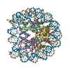
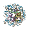
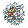
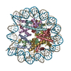
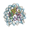
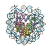
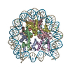
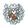
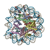
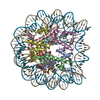
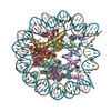
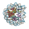
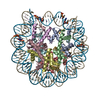
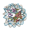
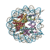
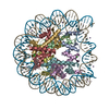
| Title | Cryo-EM structure of the regulatory linker of ALC1 bound to the nucleosome's acidic patch: nucleosome class. | |||||||||
 Components Components |
| |||||||||
 Keywords Keywords | DNA BINDING PROTEIN / ALC1 / CHD1L / chromatin remodeler / DNA damage response / nucleosome / NUCLEAR PROTEIN / GENE REGULATION | |||||||||
| Function / homology |  Function and homology information Function and homology informationpoly-ADP-D-ribose modification-dependent protein binding / ATP-dependent chromatin remodeler activity / histone reader activity / site of DNA damage / nucleosome binding / DNA helicase activity / Hydrolases; Acting on acid anhydrides; Acting on acid anhydrides to facilitate cellular and subcellular movement / Dual Incision in GG-NER / Formation of Incision Complex in GG-NER / structural constituent of chromatin ...poly-ADP-D-ribose modification-dependent protein binding / ATP-dependent chromatin remodeler activity / histone reader activity / site of DNA damage / nucleosome binding / DNA helicase activity / Hydrolases; Acting on acid anhydrides; Acting on acid anhydrides to facilitate cellular and subcellular movement / Dual Incision in GG-NER / Formation of Incision Complex in GG-NER / structural constituent of chromatin / heterochromatin formation / nucleosome / nucleosome assembly / site of double-strand break / chromatin remodeling / protein heterodimerization activity / DNA repair / nucleotide binding / DNA damage response / ATP hydrolysis activity / DNA binding / nucleoplasm / ATP binding / nucleus Similarity search - Function | |||||||||
| Biological species | synthetic construct (others)  Homo sapiens (human) Homo sapiens (human) | |||||||||
| Method | ELECTRON MICROSCOPY / single particle reconstruction / cryo EM / Resolution: 2.5 Å | |||||||||
 Authors Authors | Bacic, L. / Gaullier, G. / Croll, T.I. / Deindl, S. | |||||||||
| Funding support |  Sweden, 1items Sweden, 1items
| |||||||||
 Citation Citation |  Journal: Cell Rep / Year: 2020 Journal: Cell Rep / Year: 2020Title: Mechanistic Insights into Regulation of the ALC1 Remodeler by the Nucleosome Acidic Patch. Authors: Laura C Lehmann / Luka Bacic / Graeme Hewitt / Klaus Brackmann / Anton Sabantsev / Guillaume Gaullier / Sofia Pytharopoulou / Gianluca Degliesposti / Hanneke Okkenhaug / Song Tan / ...Authors: Laura C Lehmann / Luka Bacic / Graeme Hewitt / Klaus Brackmann / Anton Sabantsev / Guillaume Gaullier / Sofia Pytharopoulou / Gianluca Degliesposti / Hanneke Okkenhaug / Song Tan / Alessandro Costa / J Mark Skehel / Simon J Boulton / Sebastian Deindl /    Abstract: Upon DNA damage, the ALC1/CHD1L nucleosome remodeling enzyme (remodeler) is activated by binding to poly(ADP-ribose). How activated ALC1 recognizes the nucleosome, as well as how this recognition is ...Upon DNA damage, the ALC1/CHD1L nucleosome remodeling enzyme (remodeler) is activated by binding to poly(ADP-ribose). How activated ALC1 recognizes the nucleosome, as well as how this recognition is coupled to remodeling, is unknown. Here, we show that remodeling by ALC1 requires a wild-type acidic patch on the entry side of the nucleosome. The cryo-electron microscopy structure of a nucleosome-ALC1 linker complex reveals a regulatory linker segment that binds to the acidic patch. Mutations within this interface alter the dynamics of ALC1 recruitment to DNA damage and impede the ATPase and remodeling activities of ALC1. Full activation requires acidic patch-linker segment interactions that tether the remodeler to the nucleosome and couple ATP hydrolysis to nucleosome mobilization. Upon DNA damage, such a requirement may be used to modulate ALC1 activity via changes in the nucleosome acidic patches. #1: Journal: Protein Sci / Year: 2018 Title: UCSF ChimeraX: Meeting modern challenges in visualization and analysis. Authors: Thomas D Goddard / Conrad C Huang / Elaine C Meng / Eric F Pettersen / Gregory S Couch / John H Morris / Thomas E Ferrin /  Abstract: UCSF ChimeraX is next-generation software for the visualization and analysis of molecular structures, density maps, 3D microscopy, and associated data. It addresses challenges in the size, scope, and ...UCSF ChimeraX is next-generation software for the visualization and analysis of molecular structures, density maps, 3D microscopy, and associated data. It addresses challenges in the size, scope, and disparate types of data attendant with cutting-edge experimental methods, while providing advanced options for high-quality rendering (interactive ambient occlusion, reliable molecular surface calculations, etc.) and professional approaches to software design and distribution. This article highlights some specific advances in the areas of visualization and usability, performance, and extensibility. ChimeraX is free for noncommercial use and is available from http://www.rbvi.ucsf.edu/chimerax/ for Windows, Mac, and Linux. #2:  Journal: Acta Crystallogr D Struct Biol / Year: 2018 Journal: Acta Crystallogr D Struct Biol / Year: 2018Title: ISOLDE: a physically realistic environment for model building into low-resolution electron-density maps. Authors: Croll, T.I. #3:  Journal: IUCrJ / Year: 2020 Journal: IUCrJ / Year: 2020Title: Estimation of high-order aberrations and anisotropic magnification from cryo-EM data sets in Authors: Zivanov, J. / Nakane, T. / Scheres, S.H.W. #4:  Journal: J. Struct. Biol. / Year: 2012 Journal: J. Struct. Biol. / Year: 2012Title: RELION: implementation of a Bayesian approach to cryo-EM structure determination. Authors: Scheres, S.H. | |||||||||
| History |
|
- Structure visualization
| Movie |
 Movie viewer Movie viewer |
|---|---|
| Structure viewer | Molecule:  Molmil Molmil Jmol/JSmol Jmol/JSmol |
- Downloads & links
- Download
| PDBx/mmCIF format |  6zhx.cif.gz 6zhx.cif.gz | 296.5 KB | Display |  PDBx/mmCIF format PDBx/mmCIF format |
|---|---|---|---|---|
| PDB format |  pdb6zhx.ent.gz pdb6zhx.ent.gz | 219.8 KB | Display |  PDB format PDB format |
| PDBx/mmJSON format |  6zhx.json.gz 6zhx.json.gz | Tree view |  PDBx/mmJSON format PDBx/mmJSON format | |
| Others |  Other downloads Other downloads |
-Validation report
| Arichive directory |  https://data.pdbj.org/pub/pdb/validation_reports/zh/6zhx https://data.pdbj.org/pub/pdb/validation_reports/zh/6zhx ftp://data.pdbj.org/pub/pdb/validation_reports/zh/6zhx ftp://data.pdbj.org/pub/pdb/validation_reports/zh/6zhx | HTTPS FTP |
|---|
-Related structure data
| Related structure data |  11220MC  6zhyC M: map data used to model this data C: citing same article ( |
|---|---|
| Similar structure data | |
| EM raw data |  EMPIAR-10465 (Title: Single-particle cryo-EM dataset of the complex between a nucleosome and the regulatory linker of ALC1 EMPIAR-10465 (Title: Single-particle cryo-EM dataset of the complex between a nucleosome and the regulatory linker of ALC1Data size: 6.8 TB Data #1: Unaligned movies of the nucleosome - ALC1-linker complex - session_1_disk_1 [micrographs - multiframe] Data #2: Unaligned movies of the nucleosome - ALC1-linker complex - session_1_disk_2 [micrographs - multiframe] Data #3: Unaligned movies of the nucleosome - ALC1-linker complex - session_1_disk_3 [micrographs - multiframe] Data #4: Unaligned movies of the nucleosome - ALC1-linker complex - session_2_grid_1 [micrographs - multiframe] Data #5: Unaligned movies of the nucleosome - ALC1-linker complex - session_2_grid_2 [micrographs - multiframe] Data #6: Particles of the high-resolution nucleosome class [picked particles - multiframe - processed] Data #7: Particles of the hexasome class [picked particles - multiframe - processed]) |
| Experimental dataset #1 | Data reference:  10.6019/EMPIAR-10465 / Data set type: EMPIAR 10.6019/EMPIAR-10465 / Data set type: EMPIAR |
- Links
Links
- Assembly
| Deposited unit | 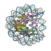
|
|---|---|
| 1 |
|
- Components
-Protein , 4 types, 8 molecules AEBFCGDH
| #1: Protein | Mass: 15403.062 Da / Num. of mol.: 2 / Mutation: C110A Source method: isolated from a genetically manipulated source Source: (gene. exp.)  #2: Protein | Mass: 11394.426 Da / Num. of mol.: 2 Source method: isolated from a genetically manipulated source Source: (gene. exp.)  #3: Protein | Mass: 14109.436 Da / Num. of mol.: 2 Source method: isolated from a genetically manipulated source Source: (gene. exp.)  #4: Protein | Mass: 13655.948 Da / Num. of mol.: 2 Source method: isolated from a genetically manipulated source Source: (gene. exp.)  |
|---|
-DNA (145-MER) Widom 601 ... , 2 types, 2 molecules IJ
| #5: DNA chain | Mass: 44520.383 Da / Num. of mol.: 1 Source method: isolated from a genetically manipulated source Source: (gene. exp.) synthetic construct (others) / Production host:  |
|---|---|
| #6: DNA chain | Mass: 44991.660 Da / Num. of mol.: 1 Source method: isolated from a genetically manipulated source Source: (gene. exp.) synthetic construct (others) / Production host:  |
-Protein/peptide / Non-polymers , 2 types, 148 molecules KL

| #7: Protein/peptide | Mass: 4073.678 Da / Num. of mol.: 2 / Source method: obtained synthetically / Details: Synthetic peptide biotinylated at its C-terminus. / Source: (synth.)  Homo sapiens (human) / References: UniProt: Q86WJ1, DNA helicase Homo sapiens (human) / References: UniProt: Q86WJ1, DNA helicase#8: Water | ChemComp-HOH / | |
|---|
-Experimental details
-Experiment
| Experiment | Method: ELECTRON MICROSCOPY |
|---|---|
| EM experiment | Aggregation state: PARTICLE / 3D reconstruction method: single particle reconstruction |
- Sample preparation
| Component |
| ||||||||||||||||||||||||||||||
|---|---|---|---|---|---|---|---|---|---|---|---|---|---|---|---|---|---|---|---|---|---|---|---|---|---|---|---|---|---|---|---|
| Molecular weight | Value: 0.206653 MDa / Experimental value: NO | ||||||||||||||||||||||||||||||
| Source (natural) |
| ||||||||||||||||||||||||||||||
| Source (recombinant) |
| ||||||||||||||||||||||||||||||
| Buffer solution | pH: 7.5 | ||||||||||||||||||||||||||||||
| Buffer component |
| ||||||||||||||||||||||||||||||
| Specimen | Conc.: 1.32 mg/ml / Embedding applied: NO / Shadowing applied: NO / Staining applied: NO / Vitrification applied: YES | ||||||||||||||||||||||||||||||
| Specimen support | Details: current 20 mA / Grid material: COPPER / Grid mesh size: 300 divisions/in. / Grid type: Quantifoil R2/2 | ||||||||||||||||||||||||||||||
| Vitrification | Instrument: FEI VITROBOT MARK IV / Cryogen name: ETHANE / Humidity: 100 % / Chamber temperature: 277 K Details: Blot time 2.5 s, blot force 0. Two sample applications and blots were performed before vitrification. |
- Electron microscopy imaging
| Experimental equipment |  Model: Titan Krios / Image courtesy: FEI Company |
|---|---|
| Microscopy | Model: TFS KRIOS |
| Electron gun | Electron source:  FIELD EMISSION GUN / Accelerating voltage: 300 kV / Illumination mode: FLOOD BEAM FIELD EMISSION GUN / Accelerating voltage: 300 kV / Illumination mode: FLOOD BEAM |
| Electron lens | Mode: BRIGHT FIELD / Cs: 2.7 mm / Alignment procedure: COMA FREE |
| Specimen holder | Cryogen: NITROGEN / Specimen holder model: FEI TITAN KRIOS AUTOGRID HOLDER |
| Image recording | Electron dose: 50.4 e/Å2 / Film or detector model: GATAN K3 BIOQUANTUM (6k x 4k) / Num. of grids imaged: 3 / Num. of real images: 19897 |
| EM imaging optics | Energyfilter name: GIF Quantum LS / Energyfilter slit width: 20 eV |
- Processing
| Software |
| ||||||||||||||||||||||||||||||||||||||||||||||||||||||||||||
|---|---|---|---|---|---|---|---|---|---|---|---|---|---|---|---|---|---|---|---|---|---|---|---|---|---|---|---|---|---|---|---|---|---|---|---|---|---|---|---|---|---|---|---|---|---|---|---|---|---|---|---|---|---|---|---|---|---|---|---|---|---|
| EM software |
| ||||||||||||||||||||||||||||||||||||||||||||||||||||||||||||
| CTF correction | Type: PHASE FLIPPING AND AMPLITUDE CORRECTION | ||||||||||||||||||||||||||||||||||||||||||||||||||||||||||||
| Particle selection | Num. of particles selected: 2968853 Details: Particles were picked with Warp's neural network picker, using model BoxNet2Mask_20180918. | ||||||||||||||||||||||||||||||||||||||||||||||||||||||||||||
| Symmetry | Point symmetry: C1 (asymmetric) | ||||||||||||||||||||||||||||||||||||||||||||||||||||||||||||
| 3D reconstruction | Resolution: 2.5 Å / Resolution method: FSC 0.143 CUT-OFF / Num. of particles: 636544 / Algorithm: FOURIER SPACE / Num. of class averages: 1 / Symmetry type: POINT | ||||||||||||||||||||||||||||||||||||||||||||||||||||||||||||
| Atomic model building | Protocol: FLEXIBLE FIT / Space: REAL / Target criteria: Real-space CC between model and map | ||||||||||||||||||||||||||||||||||||||||||||||||||||||||||||
| Atomic model building | 3D fitting-ID: 1 / Source name: PDB / Type: experimental model
|
 Movie
Movie Controller



 PDBj
PDBj


