Movie
Movie Controller

[English] 日本語
 Yorodumi
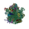
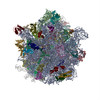
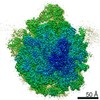
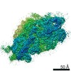
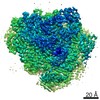
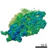
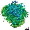
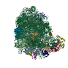
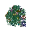
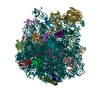
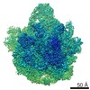
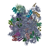
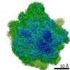
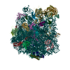
- PDB-6ysi: Acinetobacter baumannii ribosome-tigecycline complex - 50S subunit -
+ Open data

- Basic information
| Entry | Database: PDB / ID: 6ysi | |||||||||
|---|---|---|---|---|---|---|---|---|---|---|
| Title | Acinetobacter baumannii ribosome-tigecycline complex - 50S subunit | |||||||||
 Components Components |
| |||||||||
 Keywords Keywords | RIBOSOME / antibiotic / tigecycline / translation | |||||||||
| Function / homology |  Function and homology information Function and homology informationassembly of large subunit precursor of preribosome / large ribosomal subunit / transferase activity / ribosome binding / 5S rRNA binding / ribosomal large subunit assembly / large ribosomal subunit rRNA binding / cytosolic large ribosomal subunit / cytoplasmic translation / tRNA binding ...assembly of large subunit precursor of preribosome / large ribosomal subunit / transferase activity / ribosome binding / 5S rRNA binding / ribosomal large subunit assembly / large ribosomal subunit rRNA binding / cytosolic large ribosomal subunit / cytoplasmic translation / tRNA binding / negative regulation of translation / rRNA binding / structural constituent of ribosome / ribosome / translation / ribonucleoprotein complex / mRNA binding / metal ion binding / cytoplasm Similarity search - Function | |||||||||
| Biological species |  Acinetobacter baumannii ATCC 19606 = CIP 70.34 = JCM 6841 (bacteria) Acinetobacter baumannii ATCC 19606 = CIP 70.34 = JCM 6841 (bacteria) | |||||||||
| Method | ELECTRON MICROSCOPY / single particle reconstruction / cryo EM / Resolution: 2.5 Å | |||||||||
 Authors Authors | Nicholson, D. / Edwards, T.A. / O'Neill, A.J. / Ranson, N.A. | |||||||||
| Funding support |  United Kingdom, 2items United Kingdom, 2items
| |||||||||
 Citation Citation | Journal: Structure / Year: 2020 Title: Structure of the 70S Ribosome from the Human Pathogen Acinetobacter baumannii in Complex with Clinically Relevant Antibiotics. Authors: David Nicholson / Thomas A Edwards / Alex J O'Neill / Neil A Ranson / Abstract: Acinetobacter baumannii is a Gram-negative bacterium primarily associated with hospital-acquired, often multidrug-resistant (MDR) infections. The ribosome-targeting antibiotics amikacin and ...Acinetobacter baumannii is a Gram-negative bacterium primarily associated with hospital-acquired, often multidrug-resistant (MDR) infections. The ribosome-targeting antibiotics amikacin and tigecycline are among the limited arsenal of drugs available for treatment of such infections. We present high-resolution structures of the 70S ribosome from A. baumannii in complex with these antibiotics, as determined by cryoelectron microscopy. Comparison with the ribosomes of other bacteria reveals several unique structural features at functionally important sites, including around the exit of the polypeptide tunnel and the periphery of the subunit interface. The structures also reveal the mode and site of interaction of these drugs with the ribosome. This work paves the way for the design of new inhibitors of translation to address infections caused by MDR A. baumannii. | |||||||||
| History |
|
- Structure visualization
| Movie |
Movie viewer |
|---|---|
| Structure viewer | Molecule: MolmilJmol/JSmol |
- Downloads & links
-Download
| PDBx/mmCIF format | 6ysi.cif.gz | 1.8 MB | Display | PDBx/mmCIF format |
|---|---|---|---|---|
| PDB format | pdb6ysi.ent.gz | 1.4 MB | Display | PDB format |
| PDBx/mmJSON format | 6ysi.json.gz | Tree view | PDBx/mmJSON format | |
| Others |  Other downloads Other downloads |
-Validation report
| Arichive directory | https://data.pdbj.org/pub/pdb/validation_reports/ys/6ysiftp://data.pdbj.org/pub/pdb/validation_reports/ys/6ysi | HTTPS FTP |
|---|
-Related structure data
| Related structure data | 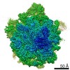 10898MC 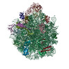 6yhsC 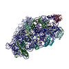 6ypuC 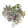 6ys5C 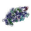 6yt9C 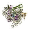 6ytfC M: map data used to model this data C: citing same article ( |
|---|---|
| Similar structure data | |
| EM raw data | EMPIAR-10407 (Title: Motion-corrected micrographs and extracted particle images of the 70S ribosome from the human pathogen Acinetobacter baumannii in complex with tigecycline Data size: 538.5 Data #1: Motion-corrected micrographs of the 70S ribosome from the human pathogen Acinetobacter baumannii in complex with tigecycline [micrographs - single frame] Data #2: Extracted particle images of the 70S ribosome from the human pathogen Acinetobacter baumannii in complex with tigecycline [picked particles - multiframe - processed]) |


-Links
 PDBj
PDBj


- Assembly
| Deposited unit | 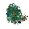
|
|---|---|
| 1 |
|
-Components
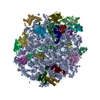
+50S ribosomal protein ... , 28 types, 28 molecules ABCDEFGHIJKLMNOPQRSTUVWXYZab
-RNA chain , 3 types, 4 molecules 1568
| #27: RNA chain | Mass: 940330.125 Da / Num. of mol.: 1 / Source method: isolated from a natural source Source: (natural) Acinetobacter baumannii ATCC 19606 = CIP 70.34 = JCM 6841 (bacteria)References: GenBank: 1188467441 |
|---|---|
| #28: RNA chain | Mass: 37608.344 Da / Num. of mol.: 1 / Source method: isolated from a natural source Source: (natural) Acinetobacter baumannii ATCC 19606 = CIP 70.34 = JCM 6841 (bacteria)References: GenBank: 1560725104 |
| #29: RNA chain | Mass: 24346.498 Da / Num. of mol.: 2 / Source method: isolated from a natural source Details: E. coli fMet-tRNA from PDB 5AFI fitted into EM density - represents a mixture of tRNAs Source: (natural) Acinetobacter baumannii ATCC 19606 = CIP 70.34 = JCM 6841 (bacteria) |
-Non-polymers , 3 types, 166 molecules 
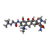

| #32: Chemical | ChemComp-MG /  Mass: 24.305 Da / Num. of mol.: 162 / Source method: obtained synthetically / Formula: Mg Mass: 24.305 Da / Num. of mol.: 162 / Source method: obtained synthetically / Formula: Mg#33: Chemical |  Mass: 587.665 Da / Num. of mol.: 3 / Source method: obtained synthetically / Formula: C29H41N5O8 / Feature type: SUBJECT OF INVESTIGATION / Comment: medication, antibiotic*YM Mass: 587.665 Da / Num. of mol.: 3 / Source method: obtained synthetically / Formula: C29H41N5O8 / Feature type: SUBJECT OF INVESTIGATION / Comment: medication, antibiotic*YM#34: Chemical | ChemComp-ZN / |  Mass: 65.409 Da / Num. of mol.: 1 / Source method: obtained synthetically / Formula: Zn Mass: 65.409 Da / Num. of mol.: 1 / Source method: obtained synthetically / Formula: Zn |
|---|
-Details
| Has ligand of interest | Y |
|---|
-Experimental details
-Experiment
| Experiment | Method: ELECTRON MICROSCOPY |
|---|---|
| EM experiment | Aggregation state: PARTICLE / 3D reconstruction method: single particle reconstruction |
- Sample preparation
| Component | Name: Acinetobacter baumannii ribosome-tigecycline complex - 50S subunit Type: RIBOSOME / Entity ID: #1-#31 / Source: NATURAL |
|---|---|
| Source (natural) | Organism: Acinetobacter baumannii (bacteria) |
| Buffer solution | pH: 7.5 |
| Specimen | Embedding applied: NO / Shadowing applied: NO / Staining applied: NO / Vitrification applied: YES |
| Vitrification | Cryogen name: ETHANE |
- Electron microscopy imaging
| Experimental equipment |  Model: Titan Krios / Image courtesy: FEI Company |
|---|---|
| Microscopy | Model: FEI TITAN KRIOS |
| Electron gun | Electron source:  FIELD EMISSION GUN / Accelerating voltage: 300 kV / Illumination mode: FLOOD BEAM FIELD EMISSION GUN / Accelerating voltage: 300 kV / Illumination mode: FLOOD BEAM |
| Electron lens | Mode: BRIGHT FIELD / Nominal magnification: 75000 X / Nominal defocus max: 2600 nm / Nominal defocus min: 800 nm |
| Image recording | Average exposure time: 1.1 sec. / Electron dose: 62 e/Å2 / Detector mode: INTEGRATING / Film or detector model: FEI FALCON III (4k x 4k) |
| Image scans | Movie frames/image: 50 |
- Processing
| Software |
| ||||||||||||||||||||||||||||
|---|---|---|---|---|---|---|---|---|---|---|---|---|---|---|---|---|---|---|---|---|---|---|---|---|---|---|---|---|---|
| EM software |
| ||||||||||||||||||||||||||||
| CTF correction | Type: PHASE FLIPPING AND AMPLITUDE CORRECTION | ||||||||||||||||||||||||||||
| Symmetry | Point symmetry: C1 (asymmetric) | ||||||||||||||||||||||||||||
| 3D reconstruction | Resolution: 2.5 Å / Resolution method: FSC 0.143 CUT-OFF / Num. of particles: 231159 Details: Multi-body refinement was carried out in RELION 3.0 to obtain the final '50S subunit' reconstruction. The mask used for this procedure is deposited with this entry. Symmetry type: POINT | ||||||||||||||||||||||||||||
| Atomic model building | Protocol: RIGID BODY FIT / Space: REAL / Target criteria: correlation coefficient | ||||||||||||||||||||||||||||
| Atomic model building | 3D fitting-ID: 1 / Source name: PDB / Type: experimental model
| ||||||||||||||||||||||||||||
| Refinement | Cross valid method: NONE Stereochemistry target values: GeoStd + Monomer Library + CDL v1.2 | ||||||||||||||||||||||||||||
| Displacement parameters | Biso mean: 28.7 Å2 | ||||||||||||||||||||||||||||
| Refine LS restraints |
|