Movie
Movie Controller

[English] 日本語
 Yorodumi
- EMDB-10892: Acinetobacter baumannii ribosome-amikacin complex - 30S subunit head -
+ Open data

- Basic information
| Entry | Database: EMDB / ID: EMD-10892 | |||||||||
|---|---|---|---|---|---|---|---|---|---|---|
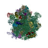
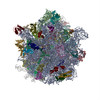
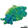
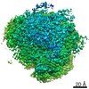
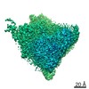
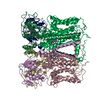
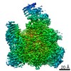
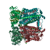
| Title | Acinetobacter baumannii ribosome-amikacin complex - 30S subunit head | |||||||||
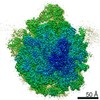
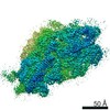
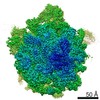
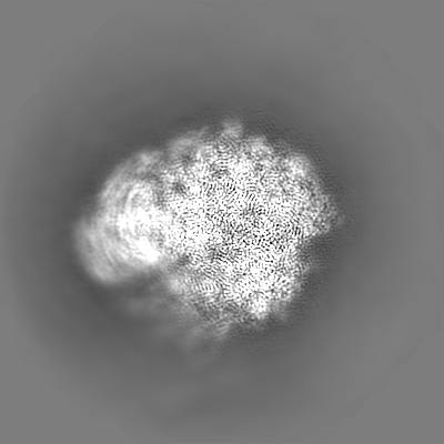
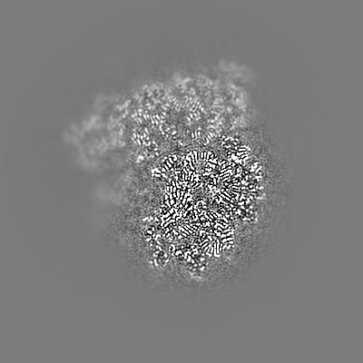
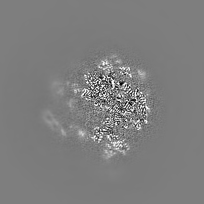
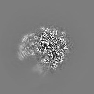
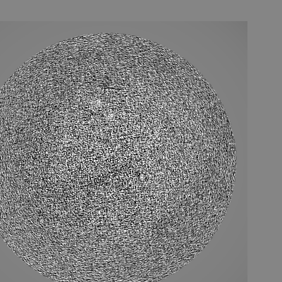
 Map data Map data | Acinetobacter baumannii ribosome-amikacin complex - 30S subunit head, post-processed map | |||||||||
 Sample Sample |
| |||||||||
 Keywords Keywords | antibiotic / amikacin / translation / ribosome | |||||||||
| Function / homology |  Function and homology information Function and homology informationribosomal small subunit assembly / small ribosomal subunit / cytosolic small ribosomal subunit / tRNA binding / rRNA binding / structural constituent of ribosome / ribosome / translation / ribonucleoprotein complex / mRNA binding ...ribosomal small subunit assembly / small ribosomal subunit / cytosolic small ribosomal subunit / tRNA binding / rRNA binding / structural constituent of ribosome / ribosome / translation / ribonucleoprotein complex / mRNA binding / RNA binding / cytoplasm / cytosol Similarity search - Function | |||||||||
| Biological species |  Acinetobacter baumannii ATCC 19606 = CIP 70.34 = JCM 6841 (bacteria) Acinetobacter baumannii ATCC 19606 = CIP 70.34 = JCM 6841 (bacteria) | |||||||||
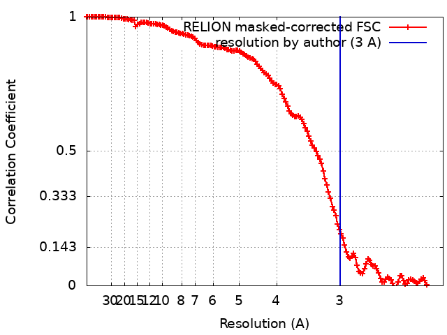
| Method | single particle reconstruction / cryo EM / Resolution: 3.0 Å | |||||||||
 Authors Authors | Nicholson D / Edwards TA | |||||||||
| Funding support |  United Kingdom, 2 items United Kingdom, 2 items
| |||||||||
 Citation Citation | Journal: Structure / Year: 2020 Title: Structure of the 70S Ribosome from the Human Pathogen Acinetobacter baumannii in Complex with Clinically Relevant Antibiotics. Authors: David Nicholson / Thomas A Edwards / Alex J O'Neill / Neil A Ranson / Abstract: Acinetobacter baumannii is a Gram-negative bacterium primarily associated with hospital-acquired, often multidrug-resistant (MDR) infections. The ribosome-targeting antibiotics amikacin and ...Acinetobacter baumannii is a Gram-negative bacterium primarily associated with hospital-acquired, often multidrug-resistant (MDR) infections. The ribosome-targeting antibiotics amikacin and tigecycline are among the limited arsenal of drugs available for treatment of such infections. We present high-resolution structures of the 70S ribosome from A. baumannii in complex with these antibiotics, as determined by cryoelectron microscopy. Comparison with the ribosomes of other bacteria reveals several unique structural features at functionally important sites, including around the exit of the polypeptide tunnel and the periphery of the subunit interface. The structures also reveal the mode and site of interaction of these drugs with the ribosome. This work paves the way for the design of new inhibitors of translation to address infections caused by MDR A. baumannii. | |||||||||
| History |
|
- Structure visualization
| Movie |
Movie viewer |
|---|---|
| Structure viewer | EM map: SurfViewMolmilJmol/JSmol |
| Supplemental images |

- Downloads & links
-EMDB archive
| Map data | emd_10892.map.gz | 14.5 MB | EMDB map data format | |
|---|---|---|---|---|
| Header (meta data) | emd-10892-v30.xmlemd-10892.xml | 29.2 KB 29.2 KB | Display Display | EMDB header |
| FSC (resolution estimation) | emd_10892_fsc.xml | 14.1 KB | Display | FSC data file |
| Images |  emd_10892.png emd_10892.png | 9.7 KB | ||
| Masks | emd_10892_msk_1.map | 244.1 MB | Mask map | |
| Filedesc metadata | emd-10892.cif.gz | 7.5 KB | ||
| Others | emd_10892_additional.map.gzemd_10892_half_map_1.map.gzemd_10892_half_map_2.map.gz | 139.6 MB 137.6 MB 137.6 MB | ||
| Archive directory |  http://ftp.pdbj.org/pub/emdb/structures/EMD-10892ftp://ftp.pdbj.org/pub/emdb/structures/EMD-10892 http://ftp.pdbj.org/pub/emdb/structures/EMD-10892ftp://ftp.pdbj.org/pub/emdb/structures/EMD-10892 | HTTPS FTP |
-Related structure data
| Related structure data | 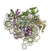 6ys5MC 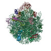 6yhsC 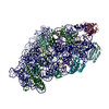 6ypuC 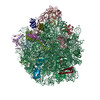 6ysiC 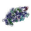 6yt9C 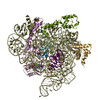 6ytfC C: citing same article ( M: atomic model generated by this map |
|---|---|
| Similar structure data | |
| EM raw data | EMPIAR-10406 (Title: Motion-corrected micrographs and extracted particle images of the 70S ribosome from the human pathogen Acinetobacter baumannii in complex with amikacin Data size: 177.7 Data #1: Motion-corrected micrographs of the 70S ribosome from the human pathogen Acinetobacter baumannii in complex with amikacin [micrographs - single frame] Data #2: Extracted particle images of the 70S ribosome from the human pathogen Acinetobacter baumannii in complex with amikacin [picked particles - multiframe - processed]) |
-Links
| EMDB pages | EMDB (EBI/PDBe) / EMDataResource |
|---|---|
| Related items in Molecule of the Month |


-Map
| File | Download / File: emd_10892.map.gz / Format: CCP4 / Size: 244.1 MB / Type: IMAGE STORED AS FLOATING POINT NUMBER (4 BYTES) | ||||||||||||||||||||||||||||||||||||||||||||||||||||||||||||
|---|---|---|---|---|---|---|---|---|---|---|---|---|---|---|---|---|---|---|---|---|---|---|---|---|---|---|---|---|---|---|---|---|---|---|---|---|---|---|---|---|---|---|---|---|---|---|---|---|---|---|---|---|---|---|---|---|---|---|---|---|---|
| Annotation | Acinetobacter baumannii ribosome-amikacin complex - 30S subunit head, post-processed map | ||||||||||||||||||||||||||||||||||||||||||||||||||||||||||||
| Projections & slices | Image control
Images are generated by Spider. | ||||||||||||||||||||||||||||||||||||||||||||||||||||||||||||
| Voxel size | X=Y=Z: 1.07 Å | ||||||||||||||||||||||||||||||||||||||||||||||||||||||||||||
| Density |
| ||||||||||||||||||||||||||||||||||||||||||||||||||||||||||||
| Symmetry | Space group: 1 | ||||||||||||||||||||||||||||||||||||||||||||||||||||||||||||
| Details | EMDB XML:
CCP4 map header:
| ||||||||||||||||||||||||||||||||||||||||||||||||||||||||||||
 Z (Sec.)
 Y (Row.)
 X (Col.)


-Supplemental data
-Mask #1
| File | emd_10892_msk_1.map | ||||||||||||
|---|---|---|---|---|---|---|---|---|---|---|---|---|---|
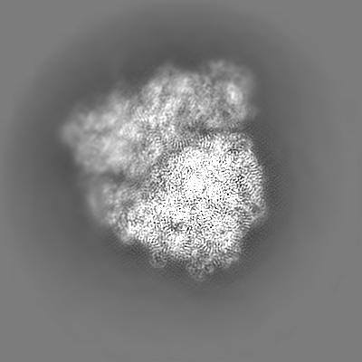
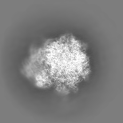
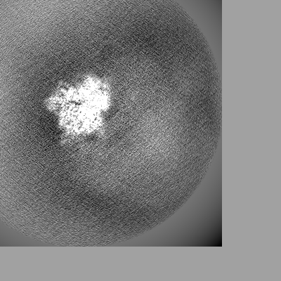
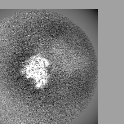
| Projections & Slices |
| ||||||||||||
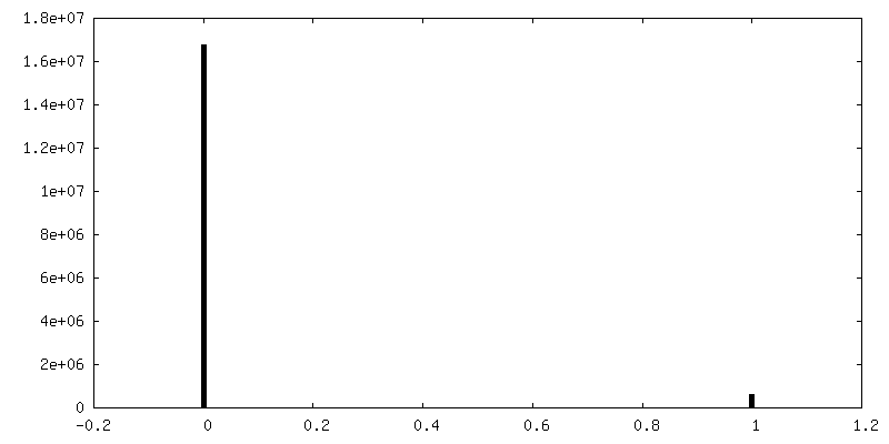
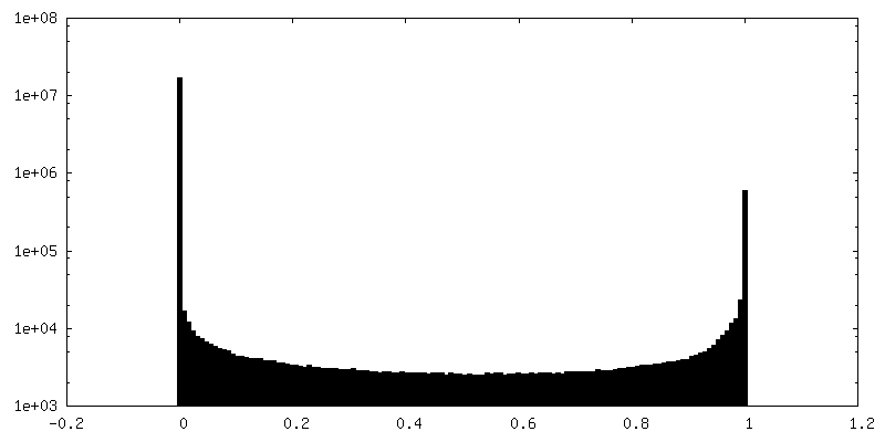
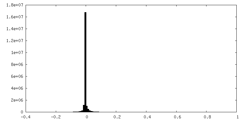
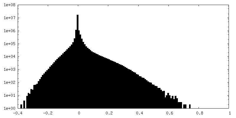
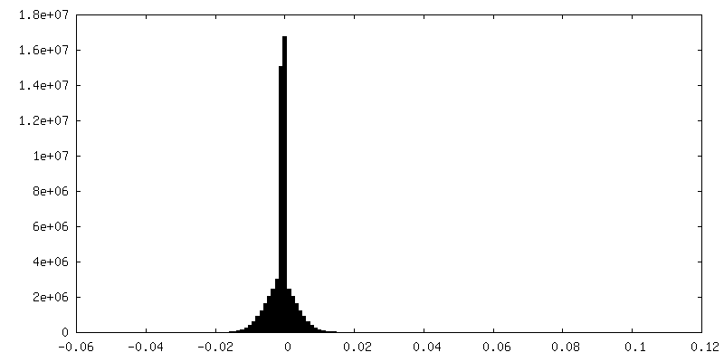
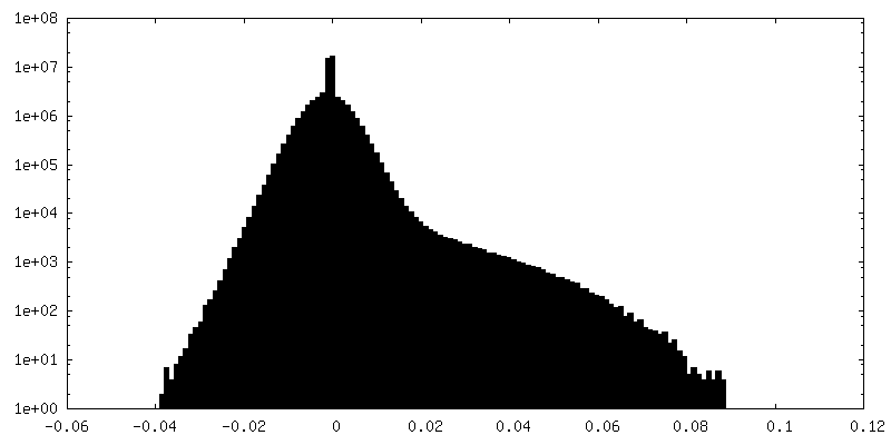
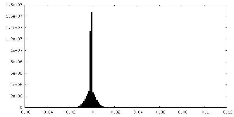
| Density Histograms |


-Additional map: Acinetobacter baumannii ribosome-amikacin complex, consensus map filtered by...
| File | emd_10892_additional.map | ||||||||||||
|---|---|---|---|---|---|---|---|---|---|---|---|---|---|
| Annotation | Acinetobacter baumannii ribosome-amikacin complex, consensus map filtered by local resolution | ||||||||||||
| Projections & Slices |
| ||||||||||||
| Density Histograms |
-Half map: Acinetobacter baumannii ribosome-amikacin complex - 30S subunit head,...
| File | emd_10892_half_map_1.map | ||||||||||||
|---|---|---|---|---|---|---|---|---|---|---|---|---|---|
| Annotation | Acinetobacter baumannii ribosome-amikacin complex - 30S subunit head, half map 1 | ||||||||||||
| Projections & Slices |
| ||||||||||||
| Density Histograms |
-Half map: Acinetobacter baumannii ribosome-amikacin complex - 30S subunit head,...
| File | emd_10892_half_map_2.map | ||||||||||||
|---|---|---|---|---|---|---|---|---|---|---|---|---|---|
| Annotation | Acinetobacter baumannii ribosome-amikacin complex - 30S subunit head, half map 2 | ||||||||||||
| Projections & Slices |
| ||||||||||||
| Density Histograms |


- Sample components
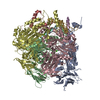
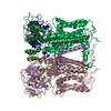
+Entire : Acinetobacter baumannii ribosome-amikacin complex - 30S subunit head
+Supramolecule #1: Acinetobacter baumannii ribosome-amikacin complex - 30S subunit head
+Macromolecule #1: 16S ribosomal RNA
+Macromolecule #2: E-site tRNA
+Macromolecule #3: mRNA
+Macromolecule #4: 30S ribosomal protein S3
+Macromolecule #5: 30S ribosomal protein S7
+Macromolecule #6: 30S ribosomal protein S9
+Macromolecule #7: 30S ribosomal protein S10
+Macromolecule #8: 30S ribosomal protein S13
+Macromolecule #9: 30S ribosomal protein S14
+Macromolecule #10: 30S ribosomal protein S19
+Macromolecule #11: MAGNESIUM ION
-Experimental details
-Structure determination
| Method | cryo EM |
|---|---|
 Processing Processing | single particle reconstruction |
| Aggregation state | particle |
-Sample preparation
| Buffer | pH: 7.5 |
|---|---|
| Vitrification | Cryogen name: ETHANE |
- Electron microscopy
| Microscope | FEI TITAN KRIOS |
|---|---|
| Image recording | Film or detector model: GATAN K2 SUMMIT (4k x 4k) / Detector mode: COUNTING / Average exposure time: 10.0 sec. / Average electron dose: 58.0 e/Å2 |
| Electron beam | Acceleration voltage: 300 kV / Electron source:  FIELD EMISSION GUN FIELD EMISSION GUN |
| Electron optics | Illumination mode: FLOOD BEAM / Imaging mode: BRIGHT FIELD / Nominal defocus max: 2.7 µm / Nominal defocus min: 0.8 µm / Nominal magnification: 130000 |
| Experimental equipment |  Model: Titan Krios / Image courtesy: FEI Company |
+Image processing
-Atomic model buiding 1
| Initial model |
| ||||||
|---|---|---|---|---|---|---|---|
| Refinement | Space: REAL / Protocol: RIGID BODY FIT / Target criteria: correlation coefficient | ||||||
| Output model | PDB-6ys5: |