ムービー
ムービー コントローラー


+ データを開く

- 基本情報
基本情報
| 登録情報 | データベース: EMDB / ID: EMD-10892 | |||||||||
|---|---|---|---|---|---|---|---|---|---|---|
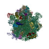
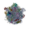
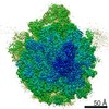
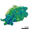
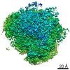
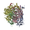
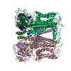
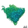
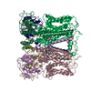
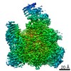
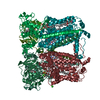
| タイトル | Acinetobacter baumannii ribosome-amikacin complex - 30S subunit head | |||||||||
 マップデータ マップデータ | Acinetobacter baumannii ribosome-amikacin complex - 30S subunit head, post-processed map | |||||||||
 試料 試料 |
| |||||||||
| 機能・相同性 |  機能・相同性情報 機能・相同性情報small ribosomal subunit /  tRNA binding / rRNA binding / リボソーム / structural constituent of ribosome / ribonucleoprotein complex / 翻訳 (生物学) / mRNA binding / 細胞質 tRNA binding / rRNA binding / リボソーム / structural constituent of ribosome / ribonucleoprotein complex / 翻訳 (生物学) / mRNA binding / 細胞質類似検索 - 分子機能 | |||||||||
| 生物種 |  Acinetobacter baumannii ATCC 19606 = CIP 70.34 = JCM 6841 (バクテリア) Acinetobacter baumannii ATCC 19606 = CIP 70.34 = JCM 6841 (バクテリア) | |||||||||
| 手法 | 単粒子再構成法 / クライオ電子顕微鏡法 / 解像度: 3.0 Å | |||||||||
 データ登録者 データ登録者 | Nicholson D / Edwards TA / O'Neill AJ / Ranson NA | |||||||||
| 資金援助 |  英国, 2件 英国, 2件
| |||||||||
 引用 引用 | ジャーナル: Structure / 年: 2020 タイトル: Structure of the 70S Ribosome from the Human Pathogen Acinetobacter baumannii in Complex with Clinically Relevant Antibiotics. 著者: David Nicholson / Thomas A Edwards / Alex J O'Neill / Neil A Ranson / 要旨: Acinetobacter baumannii is a Gram-negative bacterium primarily associated with hospital-acquired, often multidrug-resistant (MDR) infections. The ribosome-targeting antibiotics amikacin and ...Acinetobacter baumannii is a Gram-negative bacterium primarily associated with hospital-acquired, often multidrug-resistant (MDR) infections. The ribosome-targeting antibiotics amikacin and tigecycline are among the limited arsenal of drugs available for treatment of such infections. We present high-resolution structures of the 70S ribosome from A. baumannii in complex with these antibiotics, as determined by cryoelectron microscopy. Comparison with the ribosomes of other bacteria reveals several unique structural features at functionally important sites, including around the exit of the polypeptide tunnel and the periphery of the subunit interface. The structures also reveal the mode and site of interaction of these drugs with the ribosome. This work paves the way for the design of new inhibitors of translation to address infections caused by MDR A. baumannii. | |||||||||
| 履歴 |
|
- 構造の表示
構造の表示
| ムービー |
ムービービューア |
|---|---|
| 構造ビューア | EMマップ: SurfViewMolmilJmol/JSmol |
| 添付画像 |

- ダウンロードとリンク
-EMDBアーカイブ
| マップデータ | emd_10892.map.gz | 14.5 MB | EMDBマップデータ形式 | |
|---|---|---|---|---|
| ヘッダ (付随情報) | emd-10892-v30.xmlemd-10892.xml | 28.9 KB 28.9 KB | 表示 表示 | EMDBヘッダ |
| FSC (解像度算出) | emd_10892_fsc.xml | 14.1 KB | 表示 | FSCデータファイル |
| 画像 |  emd_10892.png emd_10892.png | 9.7 KB | ||
| マスクデータ | emd_10892_msk_1.map | 244.1 MB | マスクマップ | |
| その他 | emd_10892_additional.map.gzemd_10892_half_map_1.map.gzemd_10892_half_map_2.map.gz | 139.6 MB 137.6 MB 137.6 MB | ||
| アーカイブディレクトリ |  http://ftp.pdbj.org/pub/emdb/structures/EMD-10892ftp://ftp.pdbj.org/pub/emdb/structures/EMD-10892 http://ftp.pdbj.org/pub/emdb/structures/EMD-10892ftp://ftp.pdbj.org/pub/emdb/structures/EMD-10892 | HTTPS FTP |
-関連構造データ
| 関連構造データ | 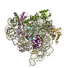 6ys5MC 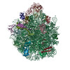 6yhsC 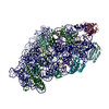 6ypuC 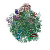 6ysiC 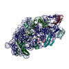 6yt9C 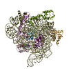 6ytfC C: 同じ文献を引用 ( M: このマップから作成された原子モデル |
|---|---|
| 類似構造データ | |
| 電子顕微鏡画像生データ | EMPIAR-10406 (タイトル: Motion-corrected micrographs and extracted particle images of the 70S ribosome from the human pathogen Acinetobacter baumannii in complex with amikacin Data size: 177.7 Data #1: Motion-corrected micrographs of the 70S ribosome from the human pathogen Acinetobacter baumannii in complex with amikacin [micrographs - single frame] Data #2: Extracted particle images of the 70S ribosome from the human pathogen Acinetobacter baumannii in complex with amikacin [picked particles - multiframe - processed]) |
-リンク
| EMDBのページ | EMDB (EBI/PDBe) / EMDataResource |
|---|---|
| 「今月の分子」の関連する項目 |


-マップ
| ファイル | ダウンロード / ファイル: emd_10892.map.gz / 形式: CCP4 / 大きさ: 244.1 MB / タイプ: IMAGE STORED AS FLOATING POINT NUMBER (4 BYTES) | ||||||||||||||||||||||||||||||||||||||||||||||||||||||||||||
|---|---|---|---|---|---|---|---|---|---|---|---|---|---|---|---|---|---|---|---|---|---|---|---|---|---|---|---|---|---|---|---|---|---|---|---|---|---|---|---|---|---|---|---|---|---|---|---|---|---|---|---|---|---|---|---|---|---|---|---|---|---|
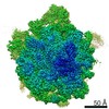
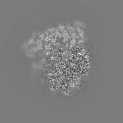
| 注釈 | Acinetobacter baumannii ribosome-amikacin complex - 30S subunit head, post-processed map | ||||||||||||||||||||||||||||||||||||||||||||||||||||||||||||
| ボクセルのサイズ | X=Y=Z: 1.07 Å | ||||||||||||||||||||||||||||||||||||||||||||||||||||||||||||
| 密度 |
| ||||||||||||||||||||||||||||||||||||||||||||||||||||||||||||
| 対称性 | 空間群: 1 | ||||||||||||||||||||||||||||||||||||||||||||||||||||||||||||
| 詳細 | EMDB XML:
CCP4マップ ヘッダ情報:
| ||||||||||||||||||||||||||||||||||||||||||||||||||||||||||||
-添付データ
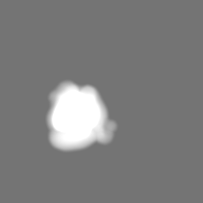
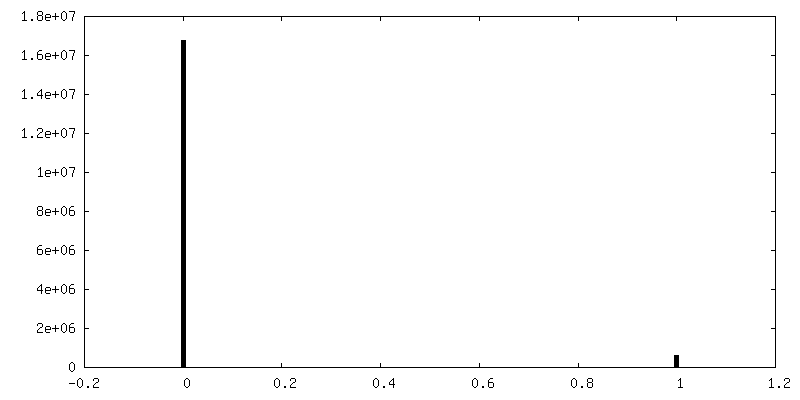
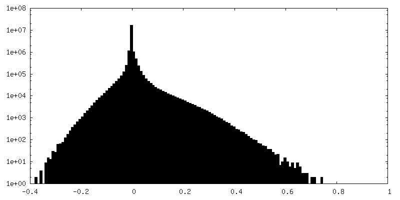
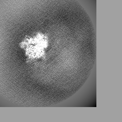
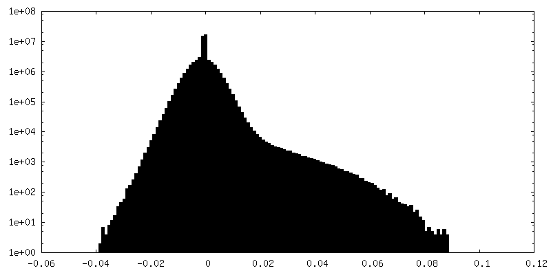
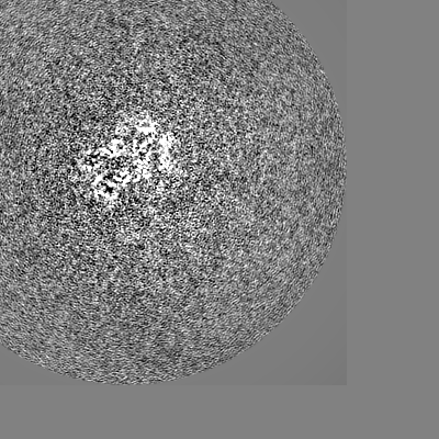
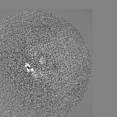
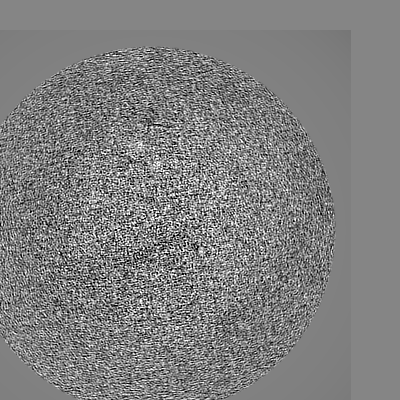
-マスク #1
| ファイル | emd_10892_msk_1.map | ||||||||||||
|---|---|---|---|---|---|---|---|---|---|---|---|---|---|
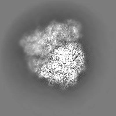
| 投影像・断面図 |
| ||||||||||||
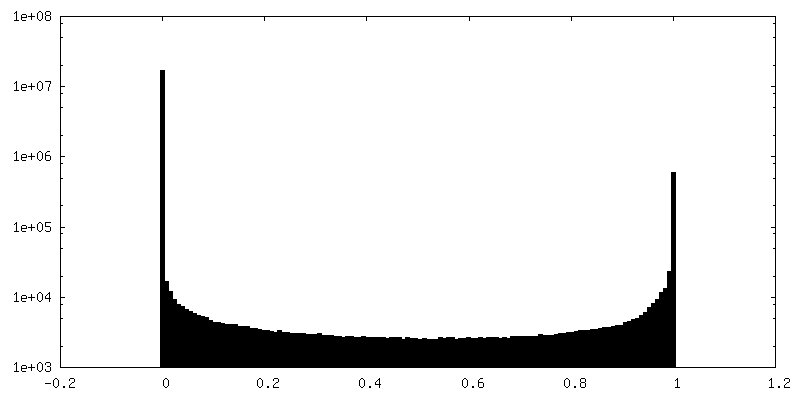
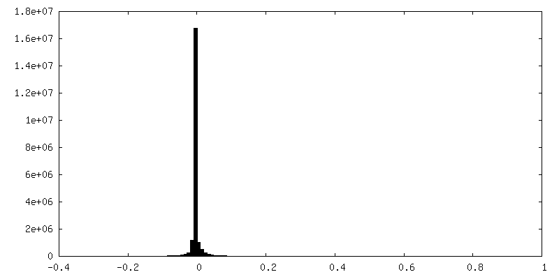
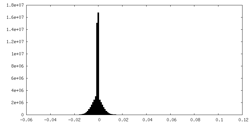
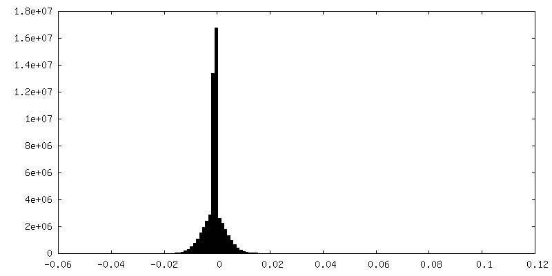
| 密度ヒストグラム |
 Z
Z Y
Y X
X


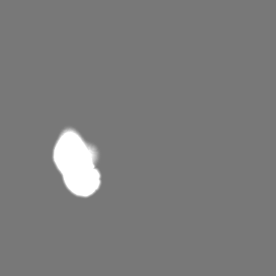

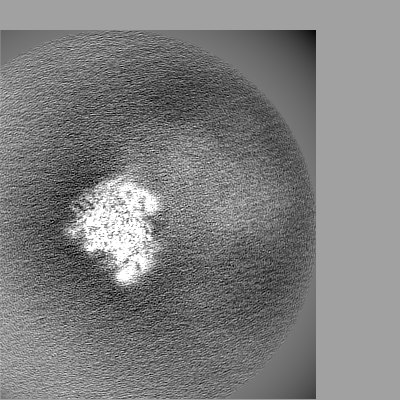
-追加マップ: Acinetobacter baumannii ribosome-amikacin complex, consensus map filtered by...
| ファイル | emd_10892_additional.map | ||||||||||||
|---|---|---|---|---|---|---|---|---|---|---|---|---|---|
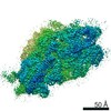
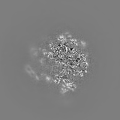
| 注釈 | Acinetobacter baumannii ribosome-amikacin complex, consensus map filtered by local resolution | ||||||||||||
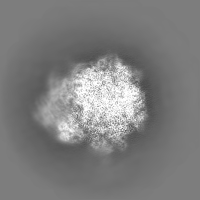
| 投影像・断面図 |
| ||||||||||||
| 密度ヒストグラム |
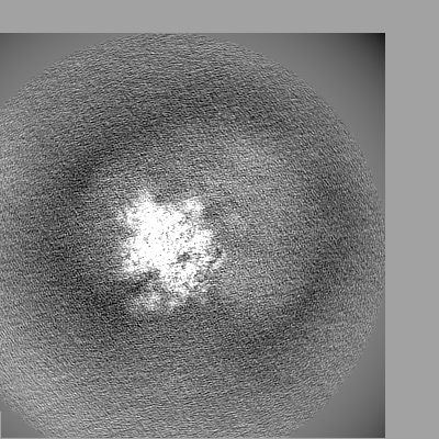
-ハーフマップ: Acinetobacter baumannii ribosome-amikacin complex - 30S subunit head,...
| ファイル | emd_10892_half_map_1.map | ||||||||||||
|---|---|---|---|---|---|---|---|---|---|---|---|---|---|
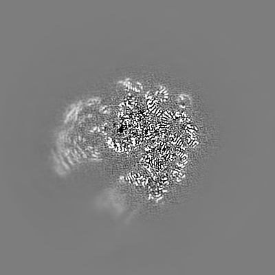
| 注釈 | Acinetobacter baumannii ribosome-amikacin complex - 30S subunit head, half map 1 | ||||||||||||
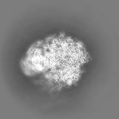
| 投影像・断面図 |
| ||||||||||||
| 密度ヒストグラム |
-ハーフマップ: Acinetobacter baumannii ribosome-amikacin complex - 30S subunit head,...
| ファイル | emd_10892_half_map_2.map | ||||||||||||
|---|---|---|---|---|---|---|---|---|---|---|---|---|---|
| 注釈 | Acinetobacter baumannii ribosome-amikacin complex - 30S subunit head, half map 2 | ||||||||||||
| 投影像・断面図 |
| ||||||||||||
| 密度ヒストグラム |


- 試料の構成要素
+全体 : Acinetobacter baumannii ribosome-amikacin complex - 30S subunit head
+超分子 #1: Acinetobacter baumannii ribosome-amikacin complex - 30S subunit head
+分子 #1: 16S ribosomal RNA
+分子 #2: E-site tRNA
+分子 #3: mRNA
+分子 #4: 30S ribosomal protein S3
+分子 #5: 30S ribosomal protein S7
+分子 #6: 30S ribosomal protein S9
+分子 #7: 30S ribosomal protein S10
+分子 #8: 30S ribosomal protein S13
+分子 #9: 30S ribosomal protein S14
+分子 #10: 30S ribosomal protein S19
+分子 #11: MAGNESIUM ION
-実験情報
-構造解析
| 手法 | クライオ電子顕微鏡法 |
|---|---|
 解析 解析 | 単粒子再構成法 |
| 試料の集合状態 | particle |
-試料調製
| 緩衝液 | pH: 7.5 |
|---|---|
| 凍結 | 凍結剤: ETHANE |
- 電子顕微鏡法
| 顕微鏡 | FEI TITAN KRIOS |
|---|---|
| 電子線 | 加速電圧: 300 kV / 電子線源: FIELD EMISSION GUN |
| 電子光学系 | 照射モード: FLOOD BEAM / 撮影モード: BRIGHT FIELDBright-field microscopy / 最大 デフォーカス(公称値): 2.7 µm / 最小 デフォーカス(公称値): 0.8 µm / 倍率(公称値): 130000 |
| 撮影 | フィルム・検出器のモデル: GATAN K2 SUMMIT (4k x 4k) 検出モード: COUNTING / 平均露光時間: 10.0 sec. / 平均電子線量: 58.0 e/Å2 |
| 実験機器 |  モデル: Titan Krios / 画像提供: FEI Company |
-画像解析
| CTF補正 | ソフトウェア - 名称: Gctf (ver. 1.18) |
|---|---|
| 初期モデル | モデルのタイプ: INSILICO MODEL In silico モデル: Made by stochastic gradient descent in RELION 3.0 |
| 初期 角度割当 | タイプ: MAXIMUM LIKELIHOOD / ソフトウェア - 名称: RELION (ver. 3.0) |
| 最終 角度割当 | タイプ: MAXIMUM LIKELIHOOD / ソフトウェア - 名称: RELION (ver. 3.0) |
| 最終 再構成 | 想定した対称性 - 点群: C1 (非対称) / 解像度のタイプ: BY AUTHOR / 解像度: 3.0 Å / 解像度の算出法: FSC 0.143 CUT-OFF / ソフトウェア - 名称: RELION (ver. 3.0) 詳細: Multi-body refinement was carried out in RELION 3.0 to obtain the final '30S subunit head' reconstruction. The mask used for this procedure is deposited with this entry. 使用した粒子像数: 51958 |
| FSC曲線 (解像度の算出) | 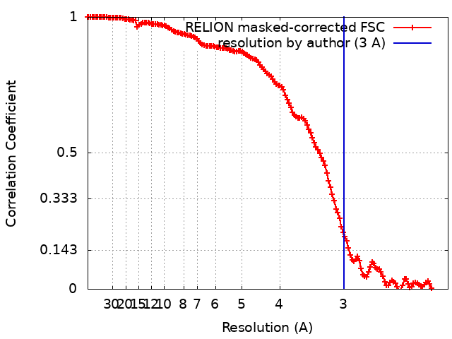 |