[English] 日本語
Yorodumi
- PDB-6qul: Structure of a bacterial 50S ribosomal subunit in complex with th... -

+
Open data


ID or keywords:

Loading...

-
Basic information

Entry
Database: PDB / ID: 6qul
TitleStructure of a bacterial 50S ribosomal subunit in complex with the novel quinoxolidinone antibiotic cadazolid
Components
  • (50S ribosomal protein ...) x 28
  • 23S rRNA23S ribosomal RNA
  • 5S rRNA5S ribosomal RNA
  • P-site fMet-tRNA(fMet)
KeywordsANTIBIOTIC / ribosome / cadazolid / quinoxolidinone / 50S
Function / homology
Function and homology information


transcriptional attenuation / endoribonuclease inhibitor activity / RNA-binding transcription regulator activity / positive regulation of ribosome biogenesis / negative regulation of cytoplasmic translation / DnaA-L2 complex / translation repressor activity / negative regulation of DNA-templated DNA replication initiation / ribosome assembly / mRNA regulatory element binding translation repressor activity ...transcriptional attenuation / endoribonuclease inhibitor activity / RNA-binding transcription regulator activity / positive regulation of ribosome biogenesis / negative regulation of cytoplasmic translation / DnaA-L2 complex / translation repressor activity / negative regulation of DNA-templated DNA replication initiation / ribosome assembly / mRNA regulatory element binding translation repressor activity / response to reactive oxygen species / assembly of large subunit precursor of preribosome / cytosolic ribosome assembly / regulation of cell growth / DNA-templated transcription termination / response to radiation / mRNA 5'-UTR binding / ribosomal large subunit assembly / large ribosomal subunit rRNA binding / ribosome binding / large ribosomal subunit / cytoplasmic translation / 5S rRNA binding / cytosolic large ribosomal subunit / transferase activity / tRNA binding / negative regulation of translation / rRNA binding / ribosome / structural constituent of ribosome / translation / response to antibiotic / mRNA binding / negative regulation of DNA-templated transcription / DNA binding / RNA binding / zinc ion binding / cytosol / cytoplasm
Similarity search - Function
Ribosomal protein L33 / Helix Hairpins - #3980 / Ribosomal protein L17 / 50s Ribosomal Protein L17; Chain: A, / Ribosomal Protein L25; Chain P / Ribosomal protein L2; domain 3 / Ribosomal protein L2, domain 3 / Ribosomal Protein L25; Chain P / Ribosomal protein L30/L7 / Nucleotidyltransferase; domain 5 - #100 ...Ribosomal protein L33 / Helix Hairpins - #3980 / Ribosomal protein L17 / 50s Ribosomal Protein L17; Chain: A, / Ribosomal Protein L25; Chain P / Ribosomal protein L2; domain 3 / Ribosomal protein L2, domain 3 / Ribosomal Protein L25; Chain P / Ribosomal protein L30/L7 / Nucleotidyltransferase; domain 5 - #100 / Ribosomal protein L16/L10 / Ribosomal protein L22/L17 / Ribosomal Protein L14 / Ribosomal protein L14/L23 / Ribosomal Protein L22; Chain A / Ribosomal Protein L30; Chain: A, / Aldehyde Oxidoreductase; domain 3 / Rubrerythrin, domain 2 / RRM (RNA recognition motif) domain / Ribosomal protein L25, short-form / Single Sheet / Nucleic acid-binding proteins / Ribosomal protein L21, conserved site / Ribosomal protein L21 signature. / Ribosomal protein L16 signature 1. / : / Ribosomal protein L6, conserved site / Ribosomal protein L6 signature 1. / Ribosomal protein L16, conserved site / Ribosomal protein L16 signature 2. / Ribosomal protein L17 signature. / Ribosomal protein L9 signature. / Ribosomal protein L9, bacteria/chloroplast / Ribosomal protein L9, C-terminal / Ribosomal protein L9, C-terminal domain / Ribosomal protein L9, C-terminal domain superfamily / Ribosomal L25p family / Ribosomal protein L25 / Ribosomal protein L28/L24 superfamily / Ribosomal protein L36 signature. / Ribosomal protein L25/Gln-tRNA synthetase, N-terminal / Ribosomal protein L32p, bacterial type / Ribosomal protein L25/Gln-tRNA synthetase, anti-codon-binding domain superfamily / Ribosomal protein L9, N-terminal domain superfamily / Ribosomal protein L9 / Ribosomal protein L9, N-terminal / Ribosomal protein L9, N-terminal domain / Ribosomal protein L28 / Ribosomal protein L35, conserved site / Ribosomal protein L35 signature. / Ribosomal protein L33, conserved site / Ribosomal protein L33 signature. / Ribosomal protein L35, non-mitochondrial / Ribosomal protein L5, bacterial-type / Ribosomal protein L6, bacterial-type / Ribosomal protein L18, bacterial-type / Ribosomal protein L19, conserved site / Ribosomal protein L19 signature. / Ribosomal protein L36 / Ribosomal protein L36 superfamily / Ribosomal protein L36 / Ribosomal protein L9/RNase H1, N-terminal / Ribosomal protein L20 signature. / Ribosomal protein L27, conserved site / Ribosomal protein L27 signature. / Ribosomal protein L14P, bacterial-type / Ribosomal protein L34, conserved site / Ribosomal protein L34 signature. / Ribosomal protein L22, bacterial/chloroplast-type / Ribosomal protein L35 / Ribosomal protein L35 superfamily / Ribosomal protein L2, bacterial/organellar-type / Ribosomal protein L35 / Ribosomal L28 family / Ribosomal protein L33 / Ribosomal protein L33 / Ribosomal protein L28/L24 / Ribosomal protein L33 superfamily / : / Ribosomal protein L30, bacterial-type / Ribosomal protein L16 / Ribosomal protein L18 / Ribosomal L18 of archaea, bacteria, mitoch. and chloroplast / L28p-like / Ribosomal protein L20 / Ribosomal protein L20 / Ribosomal protein L20, C-terminal / Ribosomal protein L21 / Ribosomal protein L27 / Ribosomal L27 protein / Ribosomal protein L19 / Ribosomal protein L19 superfamily / Ribosomal protein L19 / Ribosomal proteins 50S L24/mitochondrial 39S L24 / Ribosomal protein L17 / Ribosomal protein L17 superfamily / Ribosomal protein L17 / Ribosomal protein L21-like / L21-like superfamily / Ribosomal prokaryotic L21 protein
Similarity search - Domain/homology
ADENOSINE-5'-MONOPHOSPHATE / N-FORMYLMETHIONINE / cadazolid / : / RNA / RNA (> 10) / RNA (> 100) / RNA (> 1000) / Large ribosomal subunit protein uL15 / Large ribosomal subunit protein bL19 ...ADENOSINE-5'-MONOPHOSPHATE / N-FORMYLMETHIONINE / cadazolid / : / RNA / RNA (> 10) / RNA (> 100) / RNA (> 1000) / Large ribosomal subunit protein uL15 / Large ribosomal subunit protein bL19 / Large ribosomal subunit protein bL20 / Large ribosomal subunit protein bL27 / Large ribosomal subunit protein bL28 / Large ribosomal subunit protein uL29 / Large ribosomal subunit protein bL32 / Large ribosomal subunit protein bL33 / Large ribosomal subunit protein bL34 / Large ribosomal subunit protein bL35 / Large ribosomal subunit protein bL36A / Large ribosomal subunit protein bL9 / Large ribosomal subunit protein uL13 / Large ribosomal subunit protein uL14 / Large ribosomal subunit protein uL16 / Large ribosomal subunit protein uL23 / Large ribosomal subunit protein bL17 / Large ribosomal subunit protein bL21 / Large ribosomal subunit protein uL30 / Large ribosomal subunit protein uL6 / Large ribosomal subunit protein uL18 / Large ribosomal subunit protein uL2 / Large ribosomal subunit protein uL3 / Large ribosomal subunit protein uL24 / Large ribosomal subunit protein uL4 / Large ribosomal subunit protein uL22 / Large ribosomal subunit protein uL5 / Large ribosomal subunit protein bL25
Similarity search - Component
Biological speciesEscherichia coli (E. coli)
MethodELECTRON MICROSCOPY / single particle reconstruction / cryo EM / Resolution: 3 Å
AuthorsScaiola, A. / Leibundgut, M. / Boehringer, D. / Ritz, D.
CitationJournal: Sci Rep / Year: 2019
Title: Structural basis of translation inhibition by cadazolid, a novel quinoxolidinone antibiotic.
Authors: Alain Scaiola / Marc Leibundgut / Daniel Boehringer / Patrick Caspers / Daniel Bur / Hans H Locher / Georg Rueedi / Daniel Ritz /
Abstract: Oxazolidinones are synthetic antibiotics used for treatment of infections caused by Gram-positive bacteria. They target the bacterial protein synthesis machinery by binding to the peptidyl ...Oxazolidinones are synthetic antibiotics used for treatment of infections caused by Gram-positive bacteria. They target the bacterial protein synthesis machinery by binding to the peptidyl transferase centre (PTC) of the ribosome and interfering with the peptidyl transferase reaction. Cadazolid is the first member of quinoxolidinone antibiotics, which are characterized by combining the pharmacophores of oxazolidinones and fluoroquinolones, and it is evaluated for treatment of Clostridium difficile gastrointestinal infections that frequently occur in hospitalized patients. In vitro protein synthesis inhibition by cadazolid was shown in Escherichia coli and Staphylococcus aureus, including an isolate resistant against linezolid, the prototypical oxazolidinone antibiotic. To better understand the mechanism of inhibition, we determined a 3.0 Å cryo-electron microscopy structure of cadazolid bound to the E. coli ribosome in complex with mRNA and initiator tRNA. Here we show that cadazolid binds with its oxazolidinone moiety in a binding pocket in close vicinity of the PTC as observed previously for linezolid, and that it extends its unique fluoroquinolone moiety towards the A-site of the PTC. In this position, the drug inhibits protein synthesis by interfering with the binding of tRNA to the A-site, suggesting that its chemical features also can enable the inhibition of linezolid-resistant strains.
History
DepositionFeb 27, 2019Deposition site: PDBE / Processing site: PDBE
Revision 1.0Apr 10, 2019Provider: repository / Type: Initial release
Revision 1.1Apr 17, 2019Group: Data collection / Database references
Category: citation / citation_author ...citation / citation_author / em_admin / pdbx_database_proc
Item: _citation.page_last / _citation.pdbx_database_id_PubMed ..._citation.page_last / _citation.pdbx_database_id_PubMed / _citation.title / _citation_author.identifier_ORCID / _em_admin.last_update
Revision 1.2Dec 18, 2019Group: Data collection / Other / Category: atom_sites / em_image_scans
Item: _atom_sites.fract_transf_matrix[1][1] / _atom_sites.fract_transf_matrix[2][2] / _atom_sites.fract_transf_matrix[3][3]

-
Structure visualization

Movie
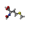
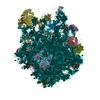
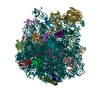
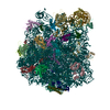
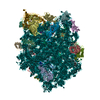
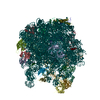
  • Deposited structure unit
  • Imaged by Jmol
  • Download
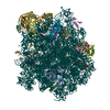
  • Superimposition on EM map
  • EMDB-4638
  • Imaged by UCSF Chimera
  • Download
Movie viewer
Structure viewerMolecule:
MolmilJmol/JSmol

Downloads & links

-
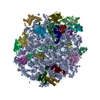
Assembly

Deposited unit
1: P-site fMet-tRNA(fMet)
A: 23S rRNA
B: 5S rRNA
C: 50S ribosomal protein L2
D: 50S ribosomal protein L3
E: 50S ribosomal protein L4
F: 50S ribosomal protein L5
G: 50S ribosomal protein L6
H: 50S ribosomal protein L9
K: 50S ribosomal protein L13
L: 50S ribosomal protein L14
M: 50S ribosomal protein L15
N: 50S ribosomal protein L16
O: 50S ribosomal protein L17
P: 50S ribosomal protein L18
Q: 50S ribosomal protein L19
R: 50S ribosomal protein L20
S: 50S ribosomal protein L21
T: 50S ribosomal protein L22
U: 50S ribosomal protein L23
V: 50S ribosomal protein L24
W: 50S ribosomal protein L25
X: 50S ribosomal protein L27
Y: 50S ribosomal protein L28
Z: 50S ribosomal protein L29
a: 50S ribosomal protein L30
b: 50S ribosomal protein L32
c: 50S ribosomal protein L33
d: 50S ribosomal protein L34
e: 50S ribosomal protein L35
f: 50S ribosomal protein L36
hetero molecules


Theoretical massNumber of molelcules
Total (without water)1,319,248417
Polymers1,308,78831
Non-polymers10,460386
Water0
1


  • Idetical with deposited unit
  • defined by author&software
  • Evidence: microscopy
TypeNameSymmetry operationNumber
identity operation1_555x,y,z1
Buried area192690 Å2
ΔGint-4456 kcal/mol
Surface area456090 Å2
MethodPISA

-
Components

-
RNA chain , 3 types, 3 molecules 1AB

#1: RNA chain P-site fMet-tRNA(fMet)


Mass: 1223.818 Da / Num. of mol.: 1 / Source method: isolated from a natural source / Source: (natural) Escherichia coli (E. coli)
#2: RNA chain 23S rRNA / 23S ribosomal RNA


Mass: 906514.125 Da / Num. of mol.: 1 / Source method: isolated from a natural source / Source: (natural) Escherichia coli (E. coli)
#3: RNA chain 5S rRNA / 5S ribosomal RNA


Mass: 38790.090 Da / Num. of mol.: 1 / Source method: isolated from a natural source / Source: (natural) Escherichia coli (E. coli) / References: GenBank: 1476653947

+
50S ribosomal protein ... , 28 types, 28 molecules CDEFGHKLMNOPQRSTUVWXYZabcdef

#4: Protein 50S ribosomal protein L2 / / ribosomal protein uL2


Mass: 29923.619 Da / Num. of mol.: 1 / Source method: isolated from a natural source / Source: (natural) Escherichia coli (E. coli) / References: UniProt: P60422
#5: Protein 50S ribosomal protein L3 / / ribosomal protein uL3


Mass: 22277.535 Da / Num. of mol.: 1 / Source method: isolated from a natural source / Source: (natural) Escherichia coli (E. coli) / References: UniProt: P60438
#6: Protein 50S ribosomal protein L4 / / ribosomal protein uL4


Mass: 22121.566 Da / Num. of mol.: 1 / Source method: isolated from a natural source / Source: (natural) Escherichia coli (E. coli) / References: UniProt: P60723
#7: Protein 50S ribosomal protein L5 / / ribosomal protein uL5


Mass: 20333.611 Da / Num. of mol.: 1 / Source method: isolated from a natural source / Source: (natural) Escherichia coli (E. coli) / References: UniProt: P62399
#8: Protein 50S ribosomal protein L6 / / ribosomal protein uL6


Mass: 18932.791 Da / Num. of mol.: 1 / Source method: isolated from a natural source / Source: (natural) Escherichia coli (E. coli) / References: UniProt: P0AG55
#9: Protein 50S ribosomal protein L9 / / ribosomal protein bL9


Mass: 15789.020 Da / Num. of mol.: 1 / Source method: isolated from a natural source / Source: (natural) Escherichia coli (E. coli) / References: UniProt: P0A7R1
#10: Protein 50S ribosomal protein L13 / / ribosomal protein uL13


Mass: 16050.606 Da / Num. of mol.: 1 / Source method: isolated from a natural source / Source: (natural) Escherichia coli (E. coli) / References: UniProt: P0AA10
#11: Protein 50S ribosomal protein L14 / / ribosomal protein uL14


Mass: 13565.067 Da / Num. of mol.: 1 / Source method: isolated from a natural source / Source: (natural) Escherichia coli (E. coli) / References: UniProt: P0ADY3
#12: Protein 50S ribosomal protein L15 / / ribosomal protein uL15


Mass: 15008.471 Da / Num. of mol.: 1 / Source method: isolated from a natural source / Source: (natural) Escherichia coli (E. coli) / References: UniProt: P02413
#13: Protein 50S ribosomal protein L16 / / ribosomal protein uL16


Mass: 15312.269 Da / Num. of mol.: 1 / Source method: isolated from a natural source / Source: (natural) Escherichia coli (E. coli) / References: UniProt: P0ADY7
#14: Protein 50S ribosomal protein L17 / / ribosomal protein uL17


Mass: 14393.657 Da / Num. of mol.: 1 / Source method: isolated from a natural source / Source: (natural) Escherichia coli (E. coli) / References: UniProt: P0AG44
#15: Protein 50S ribosomal protein L18 / / ribosomal protein uL18


Mass: 12794.668 Da / Num. of mol.: 1 / Source method: isolated from a natural source / Source: (natural) Escherichia coli (E. coli) / References: UniProt: P0C018
#16: Protein 50S ribosomal protein L19 / / ribosomal protein bL19


Mass: 13159.278 Da / Num. of mol.: 1 / Source method: isolated from a natural source / Source: (natural) Escherichia coli (E. coli) / References: UniProt: P0A7K6
#17: Protein 50S ribosomal protein L20 / / ribosomal protein bL20


Mass: 13528.024 Da / Num. of mol.: 1 / Source method: isolated from a natural source / Source: (natural) Escherichia coli (E. coli) / References: UniProt: P0A7L3
#18: Protein 50S ribosomal protein L21 / / ribosomal protein bL21


Mass: 11586.374 Da / Num. of mol.: 1 / Source method: isolated from a natural source / Source: (natural) Escherichia coli (E. coli) / References: UniProt: P0AG48
#19: Protein 50S ribosomal protein L22 / / ribosomal protein uL22


Mass: 12253.359 Da / Num. of mol.: 1 / Source method: isolated from a natural source / Source: (natural) Escherichia coli (E. coli) / References: UniProt: P61175
#20: Protein 50S ribosomal protein L23 / / ribosomal protein uL23


Mass: 11222.160 Da / Num. of mol.: 1 / Source method: isolated from a natural source / Source: (natural) Escherichia coli (E. coli) / References: UniProt: P0ADZ0
#21: Protein 50S ribosomal protein L24 / / ribosomal protein uL24


Mass: 11339.250 Da / Num. of mol.: 1 / Source method: isolated from a natural source / Source: (natural) Escherichia coli (E. coli) / References: UniProt: P60624
#22: Protein 50S ribosomal protein L25 / / ribosomal protein bL25


Mass: 10713.465 Da / Num. of mol.: 1 / Source method: isolated from a natural source / Source: (natural) Escherichia coli (E. coli) / References: UniProt: P68919
#23: Protein 50S ribosomal protein L27 / / ribosomal protein bL27


Mass: 9146.540 Da / Num. of mol.: 1 / Source method: isolated from a natural source / Source: (natural) Escherichia coli (E. coli) / References: UniProt: P0A7L8
#24: Protein 50S ribosomal protein L28 / / ribosomal protein bL28


Mass: 9027.551 Da / Num. of mol.: 1 / Source method: isolated from a natural source / Source: (natural) Escherichia coli (E. coli) / References: UniProt: P0A7M2
#25: Protein 50S ribosomal protein L29 / / ribosomal protein uL29


Mass: 7286.464 Da / Num. of mol.: 1 / Source method: isolated from a natural source / Source: (natural) Escherichia coli (E. coli) / References: UniProt: P0A7M6
#26: Protein 50S ribosomal protein L30 / / ribosomal protein uL30


Mass: 6554.820 Da / Num. of mol.: 1 / Source method: isolated from a natural source / Source: (natural) Escherichia coli (E. coli) / References: UniProt: P0AG51
#27: Protein 50S ribosomal protein L32 / / ribosomal protein bL32


Mass: 6463.445 Da / Num. of mol.: 1 / Source method: isolated from a natural source / Source: (natural) Escherichia coli (E. coli) / References: UniProt: P0A7N4
#28: Protein 50S ribosomal protein L33 / / ribosomal protein bL33


Mass: 6388.631 Da / Num. of mol.: 1 / Source method: isolated from a natural source / Source: (natural) Escherichia coli (E. coli) / References: UniProt: P0A7N9
#29: Protein/peptide 50S ribosomal protein L34 / / ribosomal protein bL34


Mass: 5397.463 Da / Num. of mol.: 1 / Source method: isolated from a natural source / Source: (natural) Escherichia coli (E. coli) / References: UniProt: P0A7P5
#30: Protein 50S ribosomal protein L35 / / Ribosomal protein A / ribosomal protein bL35


Mass: 7313.032 Da / Num. of mol.: 1 / Source method: isolated from a natural source / Source: (natural) Escherichia coli (E. coli) / References: UniProt: P0A7Q1
#31: Protein/peptide 50S ribosomal protein L36 / / Ribosomal protein B / ribosomal protein bL36


Mass: 4377.390 Da / Num. of mol.: 1 / Source method: isolated from a natural source / Source: (natural) Escherichia coli (E. coli) / References: UniProt: P0A7Q6

-
Non-polymers , 5 types, 386 molecules
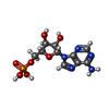
#32: Chemical ChemComp-FME / N-FORMYLMETHIONINE / N-Formylmethionine


Type: L-peptide linking / Mass: 177.221 Da / Num. of mol.: 1 / Source method: obtained synthetically / Formula: C6H11NO3S
#33: Chemical...
ChemComp-MG / MAGNESIUM ION


Mass: 24.305 Da / Num. of mol.: 382 / Source method: obtained synthetically / Formula: Mg
#34: Chemical ChemComp-A / ADENOSINE-5'-MONOPHOSPHATE / Adenosine monophosphate


Type: RNA linking / Mass: 347.221 Da / Num. of mol.: 1 / Source method: obtained synthetically / Formula: C10H14N5O7P
#35: Chemical ChemComp-JJH / cadazolid / Cadazolid


Mass: 585.553 Da / Num. of mol.: 1 / Source method: obtained synthetically / Formula: C29H29F2N3O8 / Feature type: SUBJECT OF INVESTIGATION / Comment: antibiotic*YM
#36: Chemical ChemComp-ZN / ZINC ION


Mass: 65.409 Da / Num. of mol.: 1 / Source method: obtained synthetically / Formula: Zn

-
Experimental details

-
Experiment

ExperimentMethod: ELECTRON MICROSCOPY
EM experimentAggregation state: PARTICLE / 3D reconstruction method: single particle reconstruction

-
Sample preparation

ComponentName: Structure of a bacterial 50S ribosomal subunit in complex with the novel quinoxolidinone antibiotic cadazolid
Type: RIBOSOME / Entity ID: #1-#31 / Source: NATURAL
Molecular weightUnits: MEGADALTONS / Experimental value: NO
Source (natural)Organism: Escherichia coli (E. coli)
Buffer solutionpH: 7.4
SpecimenEmbedding applied: NO / Shadowing applied: NO / Staining applied: NO / Vitrification applied: YES
Specimen supportGrid material: COPPER / Grid type: Quantifoil R2/2
VitrificationInstrument: FEI VITROBOT MARK IV / Cryogen name: ETHANE-PROPANE / Humidity: 100 % / Chamber temperature: 277 K

-
Electron microscopy imaging

Experimental equipment
Model: Titan Krios / Image courtesy: FEI Company
MicroscopyModel: FEI TITAN KRIOS
Electron gunElectron source: FIELD EMISSION GUN / Accelerating voltage: 300 kV / Illumination mode: FLOOD BEAM
Electron lensMode: BRIGHT FIELDBright-field microscopy
Image recordingElectron dose: 1.14 e/Å2 / Detector mode: INTEGRATING / Film or detector model: FEI FALCON II (4k x 4k)

-
Processing

EM software
IDNameVersionCategoryDetails
1EMANparticle selectionbatchboxer
2EPUimage acquisition
4GctfCTF correction
10RELION2initial Euler assignment
11RELION3final Euler assignment
12RELION2classification
13RELION33D reconstruction
CTF correctionType: PHASE FLIPPING AND AMPLITUDE CORRECTION
SymmetryPoint symmetry: C1 (asymmetric)
3D reconstructionResolution: 3 Å / Resolution method: FSC 0.143 CUT-OFF / Num. of particles: 59148 / Symmetry type: POINT

+
About Yorodumi

-
News

-
Feb 9, 2022. New format data for meta-information of EMDB entries

New format data for meta-information of EMDB entries

  • Version 3 of the EMDB header file is now the official format.
  • The previous official version 1.9 will be removed from the archive.

Related info.:EMDB header

External links:wwPDB to switch to version 3 of the EMDB data model

-
Aug 12, 2020. Covid-19 info

Covid-19 info

URL: https://pdbj.org/emnavi/covid19.php

New page: Covid-19 featured information page in EM Navigator.

Related info.:Covid-19 info / Mar 5, 2020. Novel coronavirus structure data

+
Mar 5, 2020. Novel coronavirus structure data

Novel coronavirus structure data

Related info.:Yorodumi Speices / Aug 12, 2020. Covid-19 info

External links:COVID-19 featured content - PDBj / Molecule of the Month (242):Coronavirus Proteases

+
Jan 31, 2019. EMDB accession codes are about to change! (news from PDBe EMDB page)

EMDB accession codes are about to change! (news from PDBe EMDB page)

  • The allocation of 4 digits for EMDB accession codes will soon come to an end. Whilst these codes will remain in use, new EMDB accession codes will include an additional digit and will expand incrementally as the available range of codes is exhausted. The current 4-digit format prefixed with “EMD-” (i.e. EMD-XXXX) will advance to a 5-digit format (i.e. EMD-XXXXX), and so on. It is currently estimated that the 4-digit codes will be depleted around Spring 2019, at which point the 5-digit format will come into force.
  • The EM Navigator/Yorodumi systems omit the EMD- prefix.

Related info.:Q: What is EMD? / ID/Accession-code notation in Yorodumi/EM Navigator

External links:EMDB Accession Codes are Changing Soon! / Contact to PDBj

+
Jul 12, 2017. Major update of PDB

Major update of PDB

  • wwPDB released updated PDB data conforming to the new PDBx/mmCIF dictionary.
  • This is a major update changing the version number from 4 to 5, and with Remediation, in which all the entries are updated.
  • In this update, many items about electron microscopy experimental information are reorganized (e.g. em_software).
  • Now, EM Navigator and Yorodumi are based on the updated data.

External links:wwPDB Remediation / Enriched Model Files Conforming to OneDep Data Standards Now Available in the PDB FTP Archive

-
Yorodumi

Thousand views of thousand structures

  • Yorodumi is a browser for structure data from EMDB, PDB, SASBDB, etc.
  • This page is also the successor to EM Navigator detail page, and also detail information page/front-end page for Omokage search.
  • The word "yorodu" (or yorozu) is an old Japanese word meaning "ten thousand". "mi" (miru) is to see.

Related info.:EMDB / PDB / SASBDB / Comparison of 3 databanks / Yorodumi Search / Aug 31, 2016. New EM Navigator & Yorodumi / Yorodumi Papers / Jmol/JSmol / Function and homology information / Changes in new EM Navigator and Yorodumi

Read more