 Movie
Movie Controller

[English] 日本語
 Yorodumi
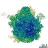
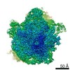
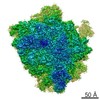
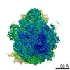
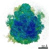
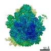
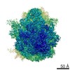
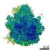
- EMDB-4639: Structure of a bacterial 70S ribosomal subunit in complex with th... -
+ Open data

- Basic information
| Entry | Database: EMDB / ID: EMD-4639 | |||||||||
|---|---|---|---|---|---|---|---|---|---|---|
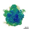
| Title | Structure of a bacterial 70S ribosomal subunit in complex with the novel quinoxolidinone antibiotic cadazolid | |||||||||
 Map data Map data | ||||||||||
 Sample Sample |
| |||||||||
| Biological species |  | |||||||||
| Method | single particle reconstruction / cryo EM / Resolution: 3.0 Å | |||||||||
 Authors Authors | Scaiola A / Leibundgut M / Boehringer D / Ritz D | |||||||||
 Citation Citation | Journal: Sci Rep / Year: 2019 Title: Structural basis of translation inhibition by cadazolid, a novel quinoxolidinone antibiotic. Authors: Alain Scaiola / Marc Leibundgut / Daniel Boehringer / Patrick Caspers / Daniel Bur / Hans H Locher / Georg Rueedi / Daniel Ritz /  Abstract: Oxazolidinones are synthetic antibiotics used for treatment of infections caused by Gram-positive bacteria. They target the bacterial protein synthesis machinery by binding to the peptidyl ...Oxazolidinones are synthetic antibiotics used for treatment of infections caused by Gram-positive bacteria. They target the bacterial protein synthesis machinery by binding to the peptidyl transferase centre (PTC) of the ribosome and interfering with the peptidyl transferase reaction. Cadazolid is the first member of quinoxolidinone antibiotics, which are characterized by combining the pharmacophores of oxazolidinones and fluoroquinolones, and it is evaluated for treatment of Clostridium difficile gastrointestinal infections that frequently occur in hospitalized patients. In vitro protein synthesis inhibition by cadazolid was shown in Escherichia coli and Staphylococcus aureus, including an isolate resistant against linezolid, the prototypical oxazolidinone antibiotic. To better understand the mechanism of inhibition, we determined a 3.0 Å cryo-electron microscopy structure of cadazolid bound to the E. coli ribosome in complex with mRNA and initiator tRNA. Here we show that cadazolid binds with its oxazolidinone moiety in a binding pocket in close vicinity of the PTC as observed previously for linezolid, and that it extends its unique fluoroquinolone moiety towards the A-site of the PTC. In this position, the drug inhibits protein synthesis by interfering with the binding of tRNA to the A-site, suggesting that its chemical features also can enable the inhibition of linezolid-resistant strains. | |||||||||
| History |
|
- Structure visualization
| Movie |
 Movie viewer Movie viewer |
|---|---|
| Structure viewer | EM map: SurfViewMolmilJmol/JSmol |
| Supplemental images |

- Downloads & links
-EMDB archive
| Map data | emd_4639.map.gz | 21.3 MB | EMDB map data format | |
|---|---|---|---|---|
| Header (meta data) | emd-4639-v30.xmlemd-4639.xml | 17.3 KB 17.3 KB | Display Display | EMDB header |
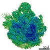
| Images | 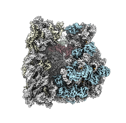 emd_4639.png emd_4639.png | 169.5 KB | ||
| Others | emd_4639_half_map_1.map.gzemd_4639_half_map_2.map.gz | 193.3 MB 193.1 MB | ||
| Archive directory |  http://ftp.pdbj.org/pub/emdb/structures/EMD-4639ftp://ftp.pdbj.org/pub/emdb/structures/EMD-4639 http://ftp.pdbj.org/pub/emdb/structures/EMD-4639ftp://ftp.pdbj.org/pub/emdb/structures/EMD-4639 | HTTPS FTP |
-Related structure data
-Links
| EMDB pages | EMDB (EBI/PDBe) / EMDataResource |
|---|---|
| Related items in Molecule of the Month |

-Map
| File | Download / File: emd_4639.map.gz / Format: CCP4 / Size: 89.8 MB / Type: IMAGE STORED AS FLOATING POINT NUMBER (4 BYTES) | ||||||||||||||||||||||||||||||||||||||||||||||||||||||||||||||||||||
|---|---|---|---|---|---|---|---|---|---|---|---|---|---|---|---|---|---|---|---|---|---|---|---|---|---|---|---|---|---|---|---|---|---|---|---|---|---|---|---|---|---|---|---|---|---|---|---|---|---|---|---|---|---|---|---|---|---|---|---|---|---|---|---|---|---|---|---|---|---|
| Projections & slices | Image control
Images are generated by Spider. generated in cubic-lattice coordinate | ||||||||||||||||||||||||||||||||||||||||||||||||||||||||||||||||||||
| Voxel size | X=Y=Z: 1.079 Å | ||||||||||||||||||||||||||||||||||||||||||||||||||||||||||||||||||||
| Density |
| ||||||||||||||||||||||||||||||||||||||||||||||||||||||||||||||||||||
| Symmetry | Space group: 1 | ||||||||||||||||||||||||||||||||||||||||||||||||||||||||||||||||||||
| Details | EMDB XML:
CCP4 map header:
| ||||||||||||||||||||||||||||||||||||||||||||||||||||||||||||||||||||
 Z (Sec.)
 Y (Row.)
 X (Col.)


-Supplemental data
-Half map: #2
| File | emd_4639_half_map_1.map | ||||||||||||
|---|---|---|---|---|---|---|---|---|---|---|---|---|---|
| Projections & Slices |
| ||||||||||||
| Density Histograms |


-Half map: #1
| File | emd_4639_half_map_2.map | ||||||||||||
|---|---|---|---|---|---|---|---|---|---|---|---|---|---|
| Projections & Slices |
| ||||||||||||
| Density Histograms |


- Sample components
-Entire : Structure of a bacterial 70S ribosomal subunit in complex with th...
| Entire | Name: Structure of a bacterial 70S ribosomal subunit in complex with the novel quinoxolidinone antibiotic cadazolid |
|---|---|
| Components |
|
-Supramolecule #1: Structure of a bacterial 70S ribosomal subunit in complex with th...
| Supramolecule | Name: Structure of a bacterial 70S ribosomal subunit in complex with the novel quinoxolidinone antibiotic cadazolid type: complex / ID: 1 / Parent: 0 / Macromolecule list: #1-#31 |
|---|---|
| Source (natural) | Organism: |
-Experimental details
-Structure determination
| Method | cryo EM |
|---|---|
 Processing Processing | single particle reconstruction |
| Aggregation state | particle |
-Sample preparation
| Buffer | pH: 7.4 |
|---|---|
| Vitrification | Cryogen name: ETHANE-PROPANE / Chamber humidity: 100 % / Chamber temperature: 277 K / Instrument: FEI VITROBOT MARK IV |
- Electron microscopy
| Microscope | FEI TITAN KRIOS |
|---|---|
| Image recording | Film or detector model: FEI FALCON II (4k x 4k) / Detector mode: INTEGRATING / Average electron dose: 1.14 e/Å2 |
| Electron beam | Acceleration voltage: 300 kV / Electron source:  FIELD EMISSION GUN FIELD EMISSION GUN |
| Electron optics | Illumination mode: FLOOD BEAM / Imaging mode: BRIGHT FIELD |
| Experimental equipment |  Model: Titan Krios / Image courtesy: FEI Company |

