 Movie
Movie Controller

[English] 日本語
 Yorodumi
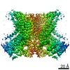
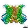
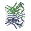
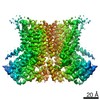
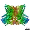
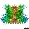
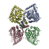
- PDB-6e1o: afTMEM16 reconstituted in nanodiscs in the presence of Ca2+ and c... -
+ Open data

- Basic information
| Entry | Database: PDB / ID: 6e1o | ||||||
|---|---|---|---|---|---|---|---|
| Title | afTMEM16 reconstituted in nanodiscs in the presence of Ca2+ and ceramide 24:0 | ||||||
 Components Components | Plasma membrane channel protein (Aqy1), putative | ||||||
 Keywords Keywords | LIPID TRANSPORT / scramblase / Ca2+-activated / membrane-reorganization | ||||||
| Function / homology |  Function and homology information Function and homology informationphospholipid scramblase activity / cortical endoplasmic reticulum / phospholipid translocation / chloride channel activity / voltage-gated calcium channel activity / chloride transmembrane transport / monoatomic ion transmembrane transport / membrane Similarity search - Function | ||||||
| Biological species |  | ||||||
| Method | ELECTRON MICROSCOPY / single particle reconstruction / cryo EM / Resolution: 3.59 Å | ||||||
 Authors Authors | Falzone, M.E. / Accardi, A. | ||||||
| Funding support |  United States, 1items United States, 1items
| ||||||
 Citation Citation | Journal: Elife / Year: 2019 Title: Structural basis of Ca-dependent activation and lipid transport by a TMEM16 scramblase. Authors: Maria E Falzone / Jan Rheinberger / Byoung-Cheol Lee / Thasin Peyear / Linda Sasset / Ashleigh M Raczkowski / Edward T Eng / Annarita Di Lorenzo / Olaf S Andersen / Crina M Nimigean / Alessio Accardi /  Abstract: The lipid distribution of plasma membranes of eukaryotic cells is asymmetric and phospholipid scramblases disrupt this asymmetry by mediating the rapid, nonselective transport of lipids down their ...The lipid distribution of plasma membranes of eukaryotic cells is asymmetric and phospholipid scramblases disrupt this asymmetry by mediating the rapid, nonselective transport of lipids down their concentration gradients. As a result, phosphatidylserine is exposed to the outer leaflet of membrane, an important step in extracellular signaling networks controlling processes such as apoptosis, blood coagulation, membrane fusion and repair. Several TMEM16 family members have been identified as Ca-activated scramblases, but the mechanisms underlying their Ca-dependent gating and their effects on the surrounding lipid bilayer remain poorly understood. Here, we describe three high-resolution cryo-electron microscopy structures of a fungal scramblase from , afTMEM16, reconstituted in lipid nanodiscs. These structures reveal that Ca-dependent activation of the scramblase entails global rearrangement of the transmembrane and cytosolic domains. These structures, together with functional experiments, suggest that activation of the protein thins the membrane near the transport pathway to facilitate rapid transbilayer lipid movement. | ||||||
| History |
|
- Structure visualization
| Movie |
Movie viewer |
|---|---|
| Structure viewer | Molecule: MolmilJmol/JSmol |
- Downloads & links
-Download
| PDBx/mmCIF format | 6e1o.cif.gz | 227.7 KB | Display | PDBx/mmCIF format |
|---|---|---|---|---|
| PDB format | pdb6e1o.ent.gz | 179.4 KB | Display | PDB format |
| PDBx/mmJSON format | 6e1o.json.gz | Tree view | PDBx/mmJSON format | |
| Others |  Other downloads Other downloads |
-Validation report
| Arichive directory | https://data.pdbj.org/pub/pdb/validation_reports/e1/6e1oftp://data.pdbj.org/pub/pdb/validation_reports/e1/6e1o | HTTPS FTP |
|---|
-Related structure data
| Related structure data | 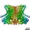 8959MC 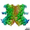 8931C 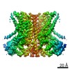 8948C 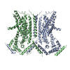 6dz7C 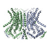 6e0hC C: citing same article ( M: map data used to model this data |
|---|---|
| Similar structure data | |
| EM raw data | EMPIAR-10240 (Title: afTMEM16/nanodisc complex in the presence of Ca2+ and 5mol% Ceramide 24:0 Data size: 113.8 Data #1: motion corrected 2D micrographs from 45 frames of afTMEM16/nanodisc complex in the presence of Ca2+ and 5 mol% ceramide 24:0 [micrographs - single frame]) |

-Links
 PDBj
PDBj- Assembly
| Deposited unit | 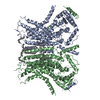
|
|---|---|
| 1 |
|
| 2 |
|
-Components
-Protein , 1 types, 2 molecules BA
| #1: Protein | Mass: 84616.859 Da / Num. of mol.: 2 Source method: isolated from a genetically manipulated source Source: (gene. exp.) Strain: ATCC MYA-4609 / Af293 / CBS 101355 / FGSC A1100 / Gene: AFUA_4G02970 / Production host:  |
|---|
-Non-polymers , 5 types, 18 molecules 


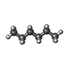
| #2: Chemical | ChemComp-CA /  Mass: 40.078 Da / Num. of mol.: 4 / Source method: obtained synthetically / Formula: Ca Mass: 40.078 Da / Num. of mol.: 4 / Source method: obtained synthetically / Formula: Ca#3: Chemical | ChemComp-D12 /  Mass: 170.335 Da / Num. of mol.: 8 / Source method: obtained synthetically / Formula: C12H26 Mass: 170.335 Da / Num. of mol.: 8 / Source method: obtained synthetically / Formula: C12H26#4: Chemical |  Mass: 142.282 Da / Num. of mol.: 2 / Source method: obtained synthetically / Formula: C10H22 Mass: 142.282 Da / Num. of mol.: 2 / Source method: obtained synthetically / Formula: C10H22#5: Chemical |  Mass: 254.494 Da / Num. of mol.: 2 / Source method: obtained synthetically / Formula: C18H38 Mass: 254.494 Da / Num. of mol.: 2 / Source method: obtained synthetically / Formula: C18H38#6: Chemical |  Mass: 86.175 Da / Num. of mol.: 2 / Source method: obtained synthetically / Formula: C6H14 Mass: 86.175 Da / Num. of mol.: 2 / Source method: obtained synthetically / Formula: C6H14 |
|---|
-Experimental details
-Experiment
| Experiment | Method: ELECTRON MICROSCOPY |
|---|---|
| EM experiment | Aggregation state: PARTICLE / 3D reconstruction method: single particle reconstruction |
- Sample preparation
| Component | Name: afTMEM16 reconstituted in nanodiscs in the presence of Ca2+ and ceramide 24:0 Type: COMPLEX / Entity ID: #1 / Source: RECOMBINANT |
|---|---|
| Molecular weight | Value: 168 kDa/nm / Experimental value: NO |
| Source (natural) | Organism: |
| Source (recombinant) | Organism: |
| Buffer solution | pH: 8 |
| Specimen | Conc.: 7 mg/ml / Embedding applied: NO / Shadowing applied: NO / Staining applied: NO / Vitrification applied: YES Details: afTMEM16 reconstituted in nanodiscs in the absence of Ca2+ |
| Vitrification | Instrument: FEI VITROBOT MARK IV / Cryogen name: ETHANE / Humidity: 100 % / Chamber temperature: 288 K |
- Electron microscopy imaging
| Experimental equipment |  Model: Titan Krios / Image courtesy: FEI Company |
|---|---|
| Microscopy | Model: FEI TITAN KRIOS |
| Electron gun | Electron source:  FIELD EMISSION GUN / Accelerating voltage: 300 kV / Illumination mode: OTHER FIELD EMISSION GUN / Accelerating voltage: 300 kV / Illumination mode: OTHER |
| Electron lens | Mode: BRIGHT FIELD |
| Image recording | Electron dose: 62.75 e/Å2 / Detector mode: COUNTING / Film or detector model: GATAN K2 SUMMIT (4k x 4k) / Num. of grids imaged: 1 |
- Processing
| Software |
| ||||||||||||||||||||||||
|---|---|---|---|---|---|---|---|---|---|---|---|---|---|---|---|---|---|---|---|---|---|---|---|---|---|
| EM software |
| ||||||||||||||||||||||||
| CTF correction | Type: PHASE FLIPPING AND AMPLITUDE CORRECTION | ||||||||||||||||||||||||
| 3D reconstruction | Resolution: 3.59 Å / Resolution method: FSC 0.143 CUT-OFF / Num. of particles: 24602 / Symmetry type: POINT | ||||||||||||||||||||||||
| Refinement | Stereochemistry target values: GeoStd + Monomer Library | ||||||||||||||||||||||||
| Refine LS restraints |
|
