Mass: 18.015 Da / Num. of mol.: 2 / Source method: isolated from a natural source / Formula: H2O
-
Details
Sequence details
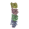
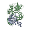
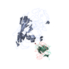
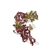
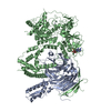
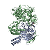
The sequence of kinesin (chain K) included mutations. The construct is known as cys-lite. for ...The sequence of kinesin (chain K) included mutations. The construct is known as cys-lite. for tubulin the bovine brain tubulin sequence was used for refinement because the sequence of ovine brain tubulin is not available. for alpha-tubulin (chain A) The alpha 1B isotype sequence (NCBI NP_001108328.1) was used. For beta-tubulin (chain B) the beta 2B isotype sequence (NCBI NP_001003900.1) was used.
-
Experimental details
-
Experiment
Experiment
Method: X-RAY DIFFRACTION / Number of used crystals: 3
-
Sample preparation
Crystal
Density Matthews: 3.44 Å3/Da / Density % sol: 64 % Description: DUE TO THE ANISOTROPIC NATURE OF THE DIFFRACTION, THE DATA WERE PROCESSED IN TWO DIFFERENT WAYS: 1) BY APPLYING AN ANISOTROPIC CORRECTION TO THE DATA, AND 2) WITHOUT ANY ANISOTROPY ...Description: DUE TO THE ANISOTROPIC NATURE OF THE DIFFRACTION, THE DATA WERE PROCESSED IN TWO DIFFERENT WAYS: 1) BY APPLYING AN ANISOTROPIC CORRECTION TO THE DATA, AND 2) WITHOUT ANY ANISOTROPY CORRECTION. STRUCTURE FACTORS FOR BOTH PROCESSING HAVE BEEN DEPOSITED WITH THE PDB. THE COORDINATES AND STATISTICS REPORTED HERE ARE THOSE OBTAINED WITH THE DATA CORRECTED FOR ANISOTROPY.
Crystal grow
Temperature: 293 K / Method: vapor diffusion, hanging drop / pH: 6.8 Details: PEG, PIPES BUFFER, PH 6.8, 0.8 MM ALCL3, 4 MM NAF, VAPOR DIFFUSION, HANGING DROP, temperature 293K
Resolution: 3.19→59.14 Å / Cor.coef. Fo:Fc: 0.9004 / Cor.coef. Fo:Fc free: 0.8708 / Cross valid method: THROUGHOUT / σ(F): 0 / Stereochemistry target values: Engh & Huber Details: BEFORE REFINEMENT, THE REFLECTION DATA WERE ANISOTROPICALLY SCALED AND TRUNCATED USING THE ANISOTROPY SERVER AT HTTP://SERVICES.MBI.UCLA.EDU/ANISOSCALE/
Rfactor
Num. reflection
% reflection
Selection details
Rfree
0.2113
1519
5.12 %
RANDOM
Rwork
0.1767
-
-
-
obs
0.1784
29641
80.25 %
-
Displacement parameters
Biso mean: 80.26 Å2
Baniso -1
Baniso -2
Baniso -3
1-
-15.3645 Å2
0 Å2
0 Å2
2-
-
5.305 Å2
0 Å2
3-
-
-
10.0595 Å2
Refine analyze
Luzzati coordinate error obs: 0.656 Å
Refinement step
Cycle: LAST / Resolution: 3.19→59.14 Å
Protein
Nucleic acid
Ligand
Solvent
Total
Num. atoms
10505
0
100
2
10607
Refine LS restraints
Refine-ID
Type
Dev ideal
Number
Restraint function
Weight
X-RAY DIFFRACTION
t_bond_d
0.01
10811
HARMONIC
2
X-RAY DIFFRACTION
t_angle_deg
1.23
14660
HARMONIC
2
X-RAY DIFFRACTION
t_dihedral_angle_d
3763
SINUSOIDAL
2
X-RAY DIFFRACTION
t_incorr_chiral_ct
X-RAY DIFFRACTION
t_pseud_angle
X-RAY DIFFRACTION
t_trig_c_planes
297
HARMONIC
2
X-RAY DIFFRACTION
t_gen_planes
1603
HARMONIC
5
X-RAY DIFFRACTION
t_it
10811
HARMONIC
20
X-RAY DIFFRACTION
t_nbd
4
SEMIHARMONIC
5
X-RAY DIFFRACTION
t_omega_torsion
2.8
X-RAY DIFFRACTION
t_other_torsion
20.06
X-RAY DIFFRACTION
t_improper_torsion
X-RAY DIFFRACTION
t_chiral_improper_torsion
1427
SEMIHARMONIC
5
X-RAY DIFFRACTION
t_sum_occupancies
X-RAY DIFFRACTION
t_utility_distance
X-RAY DIFFRACTION
t_utility_angle
X-RAY DIFFRACTION
t_utility_torsion
X-RAY DIFFRACTION
t_ideal_dist_contact
12228
SEMIHARMONIC
4
LS refinement shell
Resolution: 3.19→3.3 Å / Total num. of bins used: 15
Rfactor
Num. reflection
% reflection
Rfree
0.3416
39
4.56 %
Rwork
0.2246
817
-
all
0.2293
856
-
obs
-
-
80.25 %
Refinement TLS params.
Method: refined / Refine-ID: X-RAY DIFFRACTION
ID
L11 (°2)
L12 (°2)
L13 (°2)
L22 (°2)
L23 (°2)
L33 (°2)
S11 (Å °)
S12 (Å °)
S13 (Å °)
S21 (Å °)
S22 (Å °)
S23 (Å °)
S31 (Å °)
S32 (Å °)
S33 (Å °)
T11 (Å2)
T12 (Å2)
T13 (Å2)
T22 (Å2)
T23 (Å2)
T33 (Å2)
Origin x (Å)
Origin y (Å)
Origin z (Å)
1
1.665
0.2009
-0.9889
3.7782
-0.8879
3.3673
-0.0114
-0.382
-0.2481
0.6472
0.0058
0.517
0.0338
0.0163
0.0056
0.0256
-0.0706
0.304
-0.0641
0.0614
-0.057
-4.693
42.3853
38.2622
2
1.2759
0.5028
0.1213
2.5439
-0.9504
2.5562
-0.0593
0.0329
0.0939
-0.3715
0.1001
0.5085
-0.089
-0.3304
-0.0408
0.0612
0.0051
0.081
-0.0234
-0.0752
0.0624
-5.606
59.4465
-0.1471
3
3.8594
2.1391
1.2584
8.9352
1.6709
5.656
-0.0133
0.2682
-0.1428
0.1478
0.048
0.316
0.2501
-0.4047
-0.0347
0.066
-0.073
-0.0391
0.0326
0.0931
-0.2222
5.3775
71.6902
-31.922
4
1.8431
0.3535
0.4223
2.7785
-0.2326
4.0753
0.0783
0.018
0.0691
-0.0303
-0.0713
0.0629
-0.1438
0.1825
-0.0071
-0.175
-0.1473
0.1699
-0.3268
-0.1012
-0.3344
19.1965
83.9067
24.1645
Refinement TLS group
ID
Refine-ID
Refine TLS-ID
Selection details
Auth asym-ID
Auth seq-ID
1
X-RAY DIFFRACTION
1
{ A|* }
A
1 - 601
2
X-RAY DIFFRACTION
2
{ B|* }
B
1 - 650
3
X-RAY DIFFRACTION
3
{ D|* }
D
11 - 169
4
X-RAY DIFFRACTION
4
{ K|* }
K
4 - 650
+
About Yorodumi
-
News
-
Feb 9, 2022. New format data for meta-information of EMDB entries
New format data for meta-information of EMDB entries
Version 3 of the EMDB header file is now the official format.
The previous official version 1.9 will be removed from the archive.
In the structure databanks used in Yorodumi, some data are registered as the other names, "COVID-19 virus" and "2019-nCoV". Here are the details of the virus and the list of structure data.
Jan 31, 2019. EMDB accession codes are about to change! (news from PDBe EMDB page)
EMDB accession codes are about to change! (news from PDBe EMDB page)
The allocation of 4 digits for EMDB accession codes will soon come to an end. Whilst these codes will remain in use, new EMDB accession codes will include an additional digit and will expand incrementally as the available range of codes is exhausted. The current 4-digit format prefixed with “EMD-” (i.e. EMD-XXXX) will advance to a 5-digit format (i.e. EMD-XXXXX), and so on. It is currently estimated that the 4-digit codes will be depleted around Spring 2019, at which point the 5-digit format will come into force.
The EM Navigator/Yorodumi systems omit the EMD- prefix.
Related info.:Q: What is EMD? / ID/Accession-code notation in Yorodumi/EM Navigator
Yorodumi is a browser for structure data from EMDB, PDB, SASBDB, etc.
This page is also the successor to EM Navigator detail page, and also detail information page/front-end page for Omokage search.
The word "yorodu" (or yorozu) is an old Japanese word meaning "ten thousand". "mi" (miru) is to see.
Related info.:EMDB / PDB / SASBDB / Comparison of 3 databanks / Yorodumi Search / Aug 31, 2016. New EM Navigator & Yorodumi / Yorodumi Papers / Jmol/JSmol / Function and homology information / Changes in new EM Navigator and Yorodumi
 Movie
Movie Controller

 Yorodumi
 Open data
 Basic information
 Components
 Keywords
 Function and homology information
 Homo sapiens (human)
 X-RAY DIFFRACTION /
  Authors
 Citation
 Structure visualization
 Downloads & links
 Other downloads
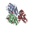


 PDBj
PDBj


 Assembly


 Mass: 523.180 Da / Num. of mol.: 1 / Source method: obtained synthetically / Formula: C10H16N5O14P3 / Comment: GTP, energy-carrying molecule*YM
 Mass: 24.305 Da / Num. of mol.: 3 / Source method: obtained synthetically / Formula: Mg
 Type: RNA linking / Mass: 443.201 Da / Num. of mol.: 1 / Source method: obtained synthetically / Formula: C10H15N5O11P2 / Comment: GDP, energy-carrying molecule*YM
 Mass: 102.975 Da / Num. of mol.: 2 / Source method: obtained synthetically / Formula: AlF4
 Mass: 427.201 Da / Num. of mol.: 1 / Source method: obtained synthetically / Formula: C10H15N5O10P2 / Comment: ADP, energy-carrying molecule*YM
 Sample preparation
 / Beamline: ID29 / Wavelength: 0.979 / Wavelength: 0.979 Å
 Processing