 Movie
Movie Controller

[English] 日本語
 Yorodumi
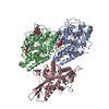
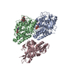
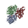
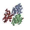
- PDB-2p4n: Human Monomeric Kinesin (1BG2) and Bovine Tubulin (1JFF) Docked i... -
+ Open data

- Basic information
| Entry | Database: PDB / ID: 2p4n | ||||||
|---|---|---|---|---|---|---|---|
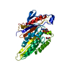
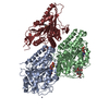
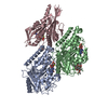
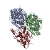
| Title | Human Monomeric Kinesin (1BG2) and Bovine Tubulin (1JFF) Docked into the 9-Angstrom Cryo-EM Map of Nucleotide-Free Kinesin Complexed to the Microtubule | ||||||
 Components Components |
| ||||||
 Keywords Keywords | TRANSPORT PROTEIN / Motor protein / ATPase | ||||||
| Function / homology |  Function and homology information Function and homology informationregulation of modification of synapse structure, modulating synaptic transmission / plus-end-directed vesicle transport along microtubule / cytoplasm organization / cytolytic granule membrane / mitocytosis / anterograde dendritic transport of neurotransmitter receptor complex / anterograde axonal protein transport / anterograde neuronal dense core vesicle transport / retrograde neuronal dense core vesicle transport / ciliary rootlet ...regulation of modification of synapse structure, modulating synaptic transmission / plus-end-directed vesicle transport along microtubule / cytoplasm organization / cytolytic granule membrane / mitocytosis / anterograde dendritic transport of neurotransmitter receptor complex / anterograde axonal protein transport / anterograde neuronal dense core vesicle transport / retrograde neuronal dense core vesicle transport / ciliary rootlet / lysosome localization / positive regulation of potassium ion transport / plus-end-directed microtubule motor activity / RHO GTPases activate KTN1 / Kinesins / positive regulation of axon guidance / vesicle transport along microtubule / centrosome localization / natural killer cell mediated cytotoxicity / microtubule motor activity / kinesin complex / mitochondrion transport along microtubule / COPI-dependent Golgi-to-ER retrograde traffic / microtubule-based movement / stress granule disassembly / Insulin processing / synaptic vesicle transport / centriolar satellite / postsynaptic cytosol / microtubule-based process / phagocytic vesicle / axon cytoplasm / MHC class II antigen presentation / axon guidance / dendrite cytoplasm / positive regulation of synaptic transmission, GABAergic / positive regulation of protein localization to plasma membrane / sperm end piece / regulation of membrane potential / neuron migration / cellular response to type II interferon / microtubule cytoskeleton organization / structural constituent of cytoskeleton / neuron differentiation / Signaling by ALK fusions and activated point mutants / microtubule cytoskeleton / mitotic cell cycle / nuclear membrane / microtubule binding / vesicle / microtubule / Hydrolases; Acting on acid anhydrides; Acting on GTP to facilitate cellular and subcellular movement / cadherin binding / protein heterodimerization activity / hydrolase activity / GTPase activity / GTP binding / protein-containing complex binding / perinuclear region of cytoplasm / ATP hydrolysis activity / mitochondrion / ATP binding / membrane / metal ion binding / identical protein binding / cytosol / cytoplasm Similarity search - Function | ||||||
| Biological species |  Homo sapiens (human) Homo sapiens (human) | ||||||
| Method | ELECTRON MICROSCOPY / single particle reconstruction / cryo EM / Resolution: 9 Å | ||||||
 Authors Authors | Sindelar, C.V. / Downing, K.H. | ||||||
 Citation Citation | Journal: J Cell Biol / Year: 2007 Title: The beginning of kinesin's force-generating cycle visualized at 9-A resolution. Authors: Charles V Sindelar / Kenneth H Downing /  Abstract: We have used cryo-electron microscopy of kinesin-decorated microtubules to resolve the structure of the motor protein kinesin's crucial nucleotide response elements, switch I and the switch II helix, ...We have used cryo-electron microscopy of kinesin-decorated microtubules to resolve the structure of the motor protein kinesin's crucial nucleotide response elements, switch I and the switch II helix, in kinesin's poorly understood nucleotide-free state. Both of the switch elements undergo conformational change relative to the microtubule-free state. The changes in switch I suggest a role for it in "ejecting" adenosine diphosphate when kinesin initially binds to the microtubule. The switch II helix has an N-terminal extension, apparently stabilized by conserved microtubule contacts, implying a microtubule activation mechanism that could convey the state of the bound nucleotide to kinesin's putative force-delivering element (the "neck linker"). In deriving this structure, we have adapted an image-processing technique, single-particle reconstruction, for analyzing decorated microtubules. The resulting reconstruction visualizes the asymmetric seam present in native, 13-protofilament microtubules, and this method will provide an avenue to higher-resolution characterization of a variety of microtubule- binding proteins, as well as the microtubule itself. | ||||||
| History |
|
- Structure visualization
| Movie |
Movie viewer |
|---|---|
| Structure viewer | Molecule: MolmilJmol/JSmol |
 UCSF Chimera
- Downloads & links
-Download
| PDBx/mmCIF format | 2p4n.cif.gz | 249.4 KB | Display | PDBx/mmCIF format |
|---|---|---|---|---|
| PDB format | pdb2p4n.ent.gz | 193.1 KB | Display | PDB format |
| PDBx/mmJSON format | 2p4n.json.gz | Tree view | PDBx/mmJSON format | |
| Others |  Other downloads Other downloads |
-Validation report
| Arichive directory | https://data.pdbj.org/pub/pdb/validation_reports/p4/2p4nftp://data.pdbj.org/pub/pdb/validation_reports/p4/2p4n | HTTPS FTP |
|---|
-Related structure data
| Related structure data | 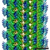 1340MC M: map data used to model this data C: citing same article ( |
|---|---|
| Similar structure data |


-Links
 PDBj
PDBj


- Assembly
| Deposited unit | 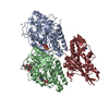
|
|---|---|
| 1 |
|
-Components
-Protein , 3 types, 3 molecules KAB
| #1: Protein | Mass: 36405.070 Da / Num. of mol.: 1 / Fragment: K349 Construct of Human Kinesin / Source method: isolated from a natural source Details: The actual construct used in the EM studies is a mutant protein (called cys-lite) Source: (natural) Homo sapiens (human) / References: UniProt: P33176*PLUS |
|---|---|
| #2: Protein | Mass: 50121.266 Da / Num. of mol.: 1 / Source method: isolated from a natural source / Source: (natural) |
| #3: Protein | Mass: 49907.770 Da / Num. of mol.: 1 / Source method: isolated from a natural source / Source: (natural) |
-Non-polymers , 6 types, 7 molecules 


| #4: Chemical |  Mass: 24.305 Da / Num. of mol.: 2 / Source method: obtained synthetically / Formula: Mg Mass: 24.305 Da / Num. of mol.: 2 / Source method: obtained synthetically / Formula: Mg#5: Chemical | ChemComp-ADP / |  Mass: 427.201 Da / Num. of mol.: 1 / Source method: obtained synthetically / Formula: C10H15N5O10P2 / Comment: ADP, energy-carrying molecule*YM Mass: 427.201 Da / Num. of mol.: 1 / Source method: obtained synthetically / Formula: C10H15N5O10P2 / Comment: ADP, energy-carrying molecule*YM#6: Chemical | ChemComp-ZN / |  Mass: 65.409 Da / Num. of mol.: 1 / Source method: obtained synthetically / Formula: Zn Mass: 65.409 Da / Num. of mol.: 1 / Source method: obtained synthetically / Formula: Zn#7: Chemical | ChemComp-GTP / |  Mass: 523.180 Da / Num. of mol.: 1 / Source method: obtained synthetically / Formula: C10H16N5O14P3 / Comment: GTP, energy-carrying molecule*YM Mass: 523.180 Da / Num. of mol.: 1 / Source method: obtained synthetically / Formula: C10H16N5O14P3 / Comment: GTP, energy-carrying molecule*YM#8: Chemical | ChemComp-GDP / |  Type: RNA linking / Mass: 443.201 Da / Num. of mol.: 1 / Source method: obtained synthetically / Formula: C10H15N5O11P2 / Comment: GDP, energy-carrying molecule*YM Type: RNA linking / Mass: 443.201 Da / Num. of mol.: 1 / Source method: obtained synthetically / Formula: C10H15N5O11P2 / Comment: GDP, energy-carrying molecule*YM#9: Chemical | ChemComp-TA1 / |  Mass: 853.906 Da / Num. of mol.: 1 / Source method: obtained synthetically / Formula: C47H51NO14 / Comment: medication, chemotherapy*YM Mass: 853.906 Da / Num. of mol.: 1 / Source method: obtained synthetically / Formula: C47H51NO14 / Comment: medication, chemotherapy*YM |
|---|
-Details
| Sequence details | THE SEQUENCE OF KINESIN (CHAIN K) IN THE SAMPLE INCLUDED MUTATIONS.THE CONSTRUCT IS KNOWN AS CYS- ...THE SEQUENCE OF KINESIN (CHAIN K) IN THE SAMPLE INCLUDED MUTATIONS.THE CONSTRUCT IS KNOWN AS CYS-LITE. MODEL CRYSTAL STRUCTURE FOR FITTING THE MAP WAS THE NATIVE SEQUENCE. IN THE CASE OF TUBULIN A AND B (CHAINS A AND B), THE SEQUENCE IN THE SAMPLE WAS DERIVED FROM COW, WHILE THE MODEL USED FOR FITTING THE MAP WAS A PIG PROTEIN. |
|---|
-Experimental details
-Experiment
| Experiment | Method: ELECTRON MICROSCOPY |
|---|---|
| EM experiment | Aggregation state: FILAMENT / 3D reconstruction method: single particle reconstruction |
- Sample preparation
| Component |
| |||||||||||||||||||||||||
|---|---|---|---|---|---|---|---|---|---|---|---|---|---|---|---|---|---|---|---|---|---|---|---|---|---|---|
| Buffer solution | pH: 6.8 | |||||||||||||||||||||||||
| Specimen | Embedding applied: YES / Shadowing applied: NO / Staining applied: NO / Vitrification applied: YES | |||||||||||||||||||||||||
| Specimen support | Details: 300 mesh copper grid | |||||||||||||||||||||||||
| Vitrification | Instrument: HOMEMADE PLUNGER / Cryogen name: ETHANE / Details: ETHANE |
- Electron microscopy imaging
| Microscopy | Model: JEOL 4000 |
|---|---|
| Electron gun | Electron source: LAB6 / Accelerating voltage: 400 kV / Illumination mode: FLOOD BEAM |
| Electron lens | Mode: BRIGHT FIELD / Nominal magnification: 60000 X / Nominal defocus max: 1500 nm / Nominal defocus min: 700 nm / Cs: 4.1 mm |
| Specimen holder | Temperature: 103.15 K / Tilt angle max: 0 ° / Tilt angle min: 0 ° |
| Image recording | Electron dose: 16 e/Å2 / Film or detector model: KODAK SO-163 FILM |
- Processing
| EM software |
| |||||||||||||||||||||
|---|---|---|---|---|---|---|---|---|---|---|---|---|---|---|---|---|---|---|---|---|---|---|
| CTF correction | Details: CTF correction was integrated into the back projection process with a customized C program | |||||||||||||||||||||
| 3D reconstruction | Method: Back projection with integrated CTF correction / Resolution: 9 Å / Num. of particles: 150000 / Nominal pixel size: 1 Å / Actual pixel size: 0.98 Å Magnification calibration: The magnification was calibrated by assuming a microtubule dimer spacing of 80.0 Angstroms. Details: Single-particle analysis was employed. / Symmetry type: HELICAL | |||||||||||||||||||||
| Atomic model building | Space: RECIPROCAL | |||||||||||||||||||||
| Atomic model building |
| |||||||||||||||||||||
| Refinement step | Cycle: LAST
|
