Movie
Movie Controller

[English] 日本語
 Yorodumi
- EMDB-1340: The beginning of kinesin's force-generating cycle visualized at 9... -
+ Open data

- Basic information
| Entry | Database: EMDB / ID: EMD-1340 | |||||||||
|---|---|---|---|---|---|---|---|---|---|---|
| Title | The beginning of kinesin's force-generating cycle visualized at 9-A resolution. | |||||||||
 Map data Map data | 9-Angstrom map of nucleotide-free kinesin complexed to the microtubule | |||||||||
 Sample Sample |
| |||||||||
| Function / homology |  Function and homology information Function and homology informationregulation of modification of synapse structure, modulating synaptic transmission / plus-end-directed vesicle transport along microtubule / cytoplasm organization / cytolytic granule membrane / anterograde dendritic transport of neurotransmitter receptor complex / mitocytosis / anterograde axonal protein transport / anterograde neuronal dense core vesicle transport / retrograde neuronal dense core vesicle transport / ciliary rootlet ...regulation of modification of synapse structure, modulating synaptic transmission / plus-end-directed vesicle transport along microtubule / cytoplasm organization / cytolytic granule membrane / anterograde dendritic transport of neurotransmitter receptor complex / mitocytosis / anterograde axonal protein transport / anterograde neuronal dense core vesicle transport / retrograde neuronal dense core vesicle transport / ciliary rootlet / lysosome localization / positive regulation of potassium ion transport / plus-end-directed microtubule motor activity / RHO GTPases activate KTN1 / Kinesins / positive regulation of axon guidance / vesicle transport along microtubule / centrosome localization / microtubule motor activity / kinesin complex / mitochondrion transport along microtubule / COPI-dependent Golgi-to-ER retrograde traffic / natural killer cell mediated cytotoxicity / microtubule-based movement / stress granule disassembly / Insulin processing / synaptic vesicle transport / postsynaptic cytosol / microtubule-based process / phagocytic vesicle / axon cytoplasm / MHC class II antigen presentation / dendrite cytoplasm / axon guidance / positive regulation of synaptic transmission, GABAergic / positive regulation of protein localization to plasma membrane / sperm end piece / regulation of membrane potential / structural constituent of cytoskeleton / cellular response to type II interferon / microtubule cytoskeleton organization / neuron migration / centriolar satellite / Signaling by ALK fusions and activated point mutants / mitotic cell cycle / microtubule cytoskeleton / nuclear membrane / microtubule binding / vesicle / microtubule / Hydrolases; Acting on acid anhydrides; Acting on GTP to facilitate cellular and subcellular movement / cadherin binding / protein heterodimerization activity / hydrolase activity / GTPase activity / GTP binding / protein-containing complex binding / perinuclear region of cytoplasm / ATP hydrolysis activity / mitochondrion / ATP binding / membrane / metal ion binding / identical protein binding / cytoplasm / cytosol Similarity search - Function | |||||||||
| Biological species |  Homo sapiens (human) Homo sapiens (human) | |||||||||
| Method | single particle reconstruction / cryo EM / Resolution: 9.0 Å | |||||||||
 Authors Authors | Sindelar CV / Downing KH | |||||||||
 Citation Citation | Journal: J Cell Biol / Year: 2007 Title: The beginning of kinesin's force-generating cycle visualized at 9-A resolution. Authors: Charles V Sindelar / Kenneth H Downing /  Abstract: We have used cryo-electron microscopy of kinesin-decorated microtubules to resolve the structure of the motor protein kinesin's crucial nucleotide response elements, switch I and the switch II helix, ...We have used cryo-electron microscopy of kinesin-decorated microtubules to resolve the structure of the motor protein kinesin's crucial nucleotide response elements, switch I and the switch II helix, in kinesin's poorly understood nucleotide-free state. Both of the switch elements undergo conformational change relative to the microtubule-free state. The changes in switch I suggest a role for it in "ejecting" adenosine diphosphate when kinesin initially binds to the microtubule. The switch II helix has an N-terminal extension, apparently stabilized by conserved microtubule contacts, implying a microtubule activation mechanism that could convey the state of the bound nucleotide to kinesin's putative force-delivering element (the "neck linker"). In deriving this structure, we have adapted an image-processing technique, single-particle reconstruction, for analyzing decorated microtubules. The resulting reconstruction visualizes the asymmetric seam present in native, 13-protofilament microtubules, and this method will provide an avenue to higher-resolution characterization of a variety of microtubule- binding proteins, as well as the microtubule itself. | |||||||||
| History |
|
- Structure visualization
| Movie |
Movie viewer |
|---|---|
| Structure viewer | EM map: SurfViewMolmilJmol/JSmol |
| Supplemental images |
 UCSF Chimera
- Downloads & links
-EMDB archive
| Map data | emd_1340.map.gz | 15.4 MB | EMDB map data format | |
|---|---|---|---|---|
| Header (meta data) | emd-1340-v30.xmlemd-1340.xml | 12.5 KB 12.5 KB | Display Display | EMDB header |
| Images | 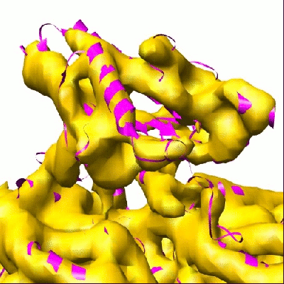 1340.gif 1340.gif | 90.8 KB | ||
| Archive directory |  http://ftp.pdbj.org/pub/emdb/structures/EMD-1340ftp://ftp.pdbj.org/pub/emdb/structures/EMD-1340 http://ftp.pdbj.org/pub/emdb/structures/EMD-1340ftp://ftp.pdbj.org/pub/emdb/structures/EMD-1340 | HTTPS FTP |
-Related structure data
| Related structure data | 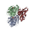 2p4nMC M: atomic model generated by this map C: citing same article ( |
|---|---|
| Similar structure data |


-Links
| EMDB pages | EMDB (EBI/PDBe) / EMDataResource |
|---|---|
| Related items in Molecule of the Month |


-Map
| File | Download / File: emd_1340.map.gz / Format: CCP4 / Size: 21.7 MB / Type: IMAGE STORED AS FLOATING POINT NUMBER (4 BYTES) | ||||||||||||||||||||||||||||||||||||||||||||||||||||||||||||||||||||
|---|---|---|---|---|---|---|---|---|---|---|---|---|---|---|---|---|---|---|---|---|---|---|---|---|---|---|---|---|---|---|---|---|---|---|---|---|---|---|---|---|---|---|---|---|---|---|---|---|---|---|---|---|---|---|---|---|---|---|---|---|---|---|---|---|---|---|---|---|---|
| Annotation | 9-Angstrom map of nucleotide-free kinesin complexed to the microtubule | ||||||||||||||||||||||||||||||||||||||||||||||||||||||||||||||||||||
| Projections & slices | Image control
Images are generated by Spider. | ||||||||||||||||||||||||||||||||||||||||||||||||||||||||||||||||||||
| Voxel size | X=Y=Z: 2 Å | ||||||||||||||||||||||||||||||||||||||||||||||||||||||||||||||||||||
| Density |
| ||||||||||||||||||||||||||||||||||||||||||||||||||||||||||||||||||||
| Symmetry | Space group: 1 | ||||||||||||||||||||||||||||||||||||||||||||||||||||||||||||||||||||
| Details | EMDB XML:
CCP4 map header:
| ||||||||||||||||||||||||||||||||||||||||||||||||||||||||||||||||||||
 Z (Sec.)
 Y (Row.)
 X (Col.)


-Supplemental data
- Sample components
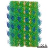
-Entire : Nucleotide-free kinesin bound to a 13-protofilament microtubule
| Entire | Name: Nucleotide-free kinesin bound to a 13-protofilament microtubule |
|---|---|
| Components |
|
-Supramolecule #1000: Nucleotide-free kinesin bound to a 13-protofilament microtubule
| Supramolecule | Name: Nucleotide-free kinesin bound to a 13-protofilament microtubule type: sample / ID: 1000 Oligomeric state: The 13-protofilament microtubule forms a pseudo helix interrupted by a single seam Number unique components: 3 |
|---|
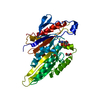
-Macromolecule #1: Kinesin
| Macromolecule | Name: Kinesin / type: protein_or_peptide / ID: 1 / Name.synonym: K349 Details: C220 "cys-lite" K349 construct: 6 of 8 native cysteines eliminated, introduced cysteine at position 220 Number of copies: 1 / Oligomeric state: Monomer / Recombinant expression: Yes |
|---|---|
| Source (natural) | Organism: Homo sapiens (human) / synonym: Human |
| Molecular weight | Experimental: 36.4134 KDa |
| Recombinant expression | Organism:  |
-Macromolecule #2: Alpha-tubulin
| Macromolecule | Name: Alpha-tubulin / type: protein_or_peptide / ID: 2 / Details: Glycerol-free tubulin purchased from Cytoskeleton / Number of copies: 1 / Oligomeric state: Dimer / Recombinant expression: No / Database: NCBI |
|---|---|
| Source (natural) | Organism: Homo sapiens (human) / synonym: Human / Tissue: Brain / Location in cell: Brain |
| Molecular weight | Experimental: 50.1279 KDa |
-Macromolecule #3: Beta-tubulin
| Macromolecule | Name: Beta-tubulin / type: protein_or_peptide / ID: 3 / Details: Glycerol-free tubulin purchased from Cytoskeleton / Number of copies: 1 / Oligomeric state: Dimer / Recombinant expression: No / Database: NCBI |
|---|---|
| Source (natural) | Organism: Homo sapiens (human) / synonym: Human / Tissue: Brain |
| Molecular weight | Experimental: 49.9283 KDa |
-Experimental details
-Structure determination
| Method | cryo EM |
|---|---|
 Processing Processing | single particle reconstruction |
| Aggregation state | particle |
-Sample preparation
| Buffer | pH: 6.8 / Details: 25mM Pipes, 25mM NaCl, 2mM MgCl2, 1mM EGTA |
|---|---|
| Grid | Details: 300 mesh copper grid |
| Vitrification | Cryogen name: ETHANE / Chamber temperature: 93 K / Instrument: HOMEMADE PLUNGER / Details: Vitrification instrument: Home-built Method: Grids were not glow discharged. Liquid was almost entirely wicked away prior to blotting. Blotting time was less than 0.5 sec. |
- Electron microscopy
| Microscope | JEOL 4000EX |
|---|---|
| Temperature | Average: 105 K |
| Alignment procedure | Legacy - Astigmatism: e.g objective lens astigmatism was corrected at 400,000 times magnification |
| Image recording | Category: FILM / Film or detector model: KODAK SO-163 FILM / Digitization - Scanner: OTHER / Digitization - Sampling interval: 6.3 µm / Number real images: 350 / Average electron dose: 16 e/Å2 / Bits/pixel: 16 |
| Tilt angle min | 0 |
| Tilt angle max | 0 |
| Electron beam | Acceleration voltage: 400 kV / Electron source: LAB6 |
| Electron optics | Illumination mode: FLOOD BEAM / Imaging mode: BRIGHT FIELD / Cs: 4.1 mm / Nominal defocus max: 1.5 µm / Nominal defocus min: 0.8 µm / Nominal magnification: 60000 |
| Sample stage | Specimen holder: Eucentric / Specimen holder model: GATAN LIQUID NITROGEN |
-Image processing
| CTF correction | Details: CTF correction was integrated into the back-projection process |
|---|---|
| Final reconstruction | Algorithm: OTHER / Resolution.type: BY AUTHOR / Resolution: 9.0 Å / Resolution method: FSC 0.5 CUT-OFF / Software - Name: SPIDER Details: Data from 188 microtubules were incorporated into the final reconstruction Number images used: 150000 |
-Atomic model buiding 1
| Initial model | (PDB ID: , ) |
|---|---|
| Software | Name: SITUS |
| Details | Protocol: Rigid body. Kinesin and tubulin were separately fitted into the final map using the program SITUS. |
| Refinement | Space: REAL / Protocol: RIGID BODY FIT / Target criteria: Cross-correlation |
| Output model | PDB-2p4n: |