 Movie
Movie Controller

[English] 日本語
 Yorodumi
- PDB-5vn1: horse liver alcohol dehydrogenae complexed with NADH (R,S)-N-1-me... -
+ Open data

- Basic information
| Entry | Database: PDB / ID: 5vn1 | |||||||||
|---|---|---|---|---|---|---|---|---|---|---|
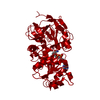
| Title | horse liver alcohol dehydrogenae complexed with NADH (R,S)-N-1-methylhexylformamide | |||||||||
 Components Components | Alcohol dehydrogenase E chain | |||||||||
 Keywords Keywords | OXIDOREDUCTASE / alcohol dehydrogenase / horse liver / NADH N-1-methylhexylformamide | |||||||||
| Function / homology |  Function and homology information Function and homology informationall-trans-retinol dehydrogenase (NAD+) activity / alcohol dehydrogenase / retinoic acid metabolic process / retinol metabolic process / zinc ion binding / cytosol Similarity search - Function | |||||||||
| Biological species |  | |||||||||
| Method |  X-RAY DIFFRACTION / SYNCHROTRON / MOLECULAR REPLACEMENT / Resolution: 1.25 Å X-RAY DIFFRACTION / SYNCHROTRON / MOLECULAR REPLACEMENT / Resolution: 1.25 Å | |||||||||
 Authors Authors | Plapp, B.V. / Ramaswamy, S. / Ferraro, D.J. / Baskar Raj, S. | |||||||||
| Funding support |  United States, 2items United States, 2items
| |||||||||
 Citation Citation | Journal: Biochemistry / Year: 2017 Title: Horse Liver Alcohol Dehydrogenase: Zinc Coordination and Catalysis. Authors: Plapp, B.V. / Savarimuthu, B.R. / Ferraro, D.J. / Rubach, J.K. / Brown, E.N. / Ramaswamy, S. #1: Journal: Biochemistry / Year: 2012Title: Atomic-resolution structures of horse liver alcohol dehydrogenase with NAD(+) and fluoroalcohols define strained Michaelis complexes. Authors: Plapp, B.V. / Ramaswamy, S. #2: Journal: J. Biol. Chem. / Year: 2003Title: Formamides mimic aldehydes and inhibit liver alcohol dehydrogenases and ethanol metabolism. Authors: Venkataramaiah, T.H. / Plapp, B.V. #3: Journal: Biochemistry / Year: 1997Title: Flexibility of liver alcohol dehydrogenase in stereoselective binding of 3-butylthiolane 1-oxides. Authors: Cho, H. / Ramaswamy, S. / Plapp, B.V. #4: Journal: Biochemistry / Year: 1997Title: Binding of formamides to liver alcohol dehydrogenase. Authors: Ramaswamy, S. / Scholze, M. / Plapp, B.V. | |||||||||
| History |
|
- Structure visualization
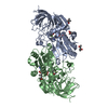
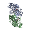
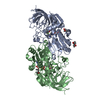
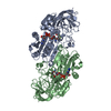
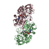
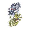
| Structure viewer | Molecule: MolmilJmol/JSmol |
|---|
- Downloads & links
-Download
| PDBx/mmCIF format | 5vn1.cif.gz | 621.6 KB | Display | PDBx/mmCIF format |
|---|---|---|---|---|
| PDB format | pdb5vn1.ent.gz | 511.5 KB | Display | PDB format |
| PDBx/mmJSON format | 5vn1.json.gz | Tree view | PDBx/mmJSON format | |
| Others |  Other downloads Other downloads |
-Validation report
| Arichive directory | https://data.pdbj.org/pub/pdb/validation_reports/vn/5vn1ftp://data.pdbj.org/pub/pdb/validation_reports/vn/5vn1 | HTTPS FTP |
|---|
-Related structure data
| Related structure data | 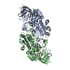 5vj5C 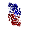 5vjgC 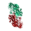 5vkrC 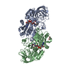 5vl0C  1p1rS S: Starting model for refinement C: citing same article ( |
|---|---|
| Similar structure data |


-Links
 PDBj
PDBj


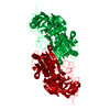
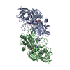
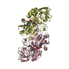
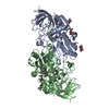
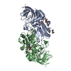
- Assembly
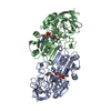
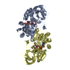
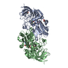
| Deposited unit | 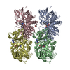
| ||||||||
|---|---|---|---|---|---|---|---|---|---|
| 1 | 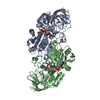
| ||||||||
| 2 | 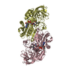
| ||||||||
| Unit cell |
|
-Components
-Protein , 1 types, 4 molecules ABCD
| #1: Protein | Mass: 39853.273 Da / Num. of mol.: 4 / Source method: isolated from a natural source / Source: (natural) |
|---|
-Non-polymers , 6 types, 1340 molecules 

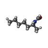

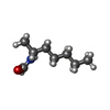

| #2: Chemical | ChemComp-ZN /  Mass: 65.409 Da / Num. of mol.: 8 / Source method: obtained synthetically / Formula: Zn Mass: 65.409 Da / Num. of mol.: 8 / Source method: obtained synthetically / Formula: Zn#3: Chemical | ChemComp-NAI /  Mass: 665.441 Da / Num. of mol.: 4 / Source method: obtained synthetically / Formula: C21H29N7O14P2 Mass: 665.441 Da / Num. of mol.: 4 / Source method: obtained synthetically / Formula: C21H29N7O14P2#4: Chemical | ChemComp-NWH /  Mass: 143.227 Da / Num. of mol.: 4 / Source method: obtained synthetically / Formula: C8H17NO Mass: 143.227 Da / Num. of mol.: 4 / Source method: obtained synthetically / Formula: C8H17NO#5: Chemical |  Mass: 118.174 Da / Num. of mol.: 2 / Source method: obtained synthetically / Formula: C6H14O2 / Comment: precipitant*YM Mass: 118.174 Da / Num. of mol.: 2 / Source method: obtained synthetically / Formula: C6H14O2 / Comment: precipitant*YM#6: Chemical | ChemComp-NMH / ( |  Mass: 143.227 Da / Num. of mol.: 1 / Source method: obtained synthetically / Formula: C8H17NO Mass: 143.227 Da / Num. of mol.: 1 / Source method: obtained synthetically / Formula: C8H17NO#7: Water | ChemComp-HOH / | Mass: 18.015 Da / Num. of mol.: 1321 / Source method: isolated from a natural source / Formula: H2O |
|---|
-Experimental details
-Experiment
| Experiment | Method: X-RAY DIFFRACTION / Number of used crystals: 1 |
|---|
- Sample preparation
| Crystal | Density Matthews: 2.38 Å3/Da / Density % sol: 48.22 % / Description: long block |
|---|---|
| Crystal grow | Temperature: 278 K / Method: microdialysis / pH: 7 Details: 10 mg protein/ml dialyzed against 50 mM ammonium N-[tris(hydroxymethyl)methyl]-2-aminoethanesulfonate buffer, pH 7 (pH 6.7 at 25 deg C) with 1 mM NADH and 10 mM (racemic) (R,S)-N-1- ...Details: 10 mg protein/ml dialyzed against 50 mM ammonium N-[tris(hydroxymethyl)methyl]-2-aminoethanesulfonate buffer, pH 7 (pH 6.7 at 25 deg C) with 1 mM NADH and 10 mM (racemic) (R,S)-N-1-methylhexylformamide as the concentration of 2-methyl-2,4-pentanediol was raised to 25%. Crystal on loop plunged into liquid N2. |
-Data collection
| Diffraction | Mean temperature: 100 K | ||||||||||||||||||||||||||||||||||||||||||||||||||||||||||||||||||||||||||||||||||||
|---|---|---|---|---|---|---|---|---|---|---|---|---|---|---|---|---|---|---|---|---|---|---|---|---|---|---|---|---|---|---|---|---|---|---|---|---|---|---|---|---|---|---|---|---|---|---|---|---|---|---|---|---|---|---|---|---|---|---|---|---|---|---|---|---|---|---|---|---|---|---|---|---|---|---|---|---|---|---|---|---|---|---|---|---|---|
| Diffraction source | Source: SYNCHROTRON / Site: ESRF  / Beamline: ID14-4 / Wavelength: 0.936 Å / Beamline: ID14-4 / Wavelength: 0.936 Å | ||||||||||||||||||||||||||||||||||||||||||||||||||||||||||||||||||||||||||||||||||||
| Detector | Type: MAR CCD 165 mm / Detector: CCD / Date: Mar 1, 1999 | ||||||||||||||||||||||||||||||||||||||||||||||||||||||||||||||||||||||||||||||||||||
| Radiation | Protocol: SINGLE WAVELENGTH / Monochromatic (M) / Laue (L): M / Scattering type: x-ray | ||||||||||||||||||||||||||||||||||||||||||||||||||||||||||||||||||||||||||||||||||||
| Radiation wavelength | Wavelength: 0.936 Å / Relative weight: 1 | ||||||||||||||||||||||||||||||||||||||||||||||||||||||||||||||||||||||||||||||||||||
| Reflection | Resolution: 1.2→20 Å / Num. obs: 386016 / % possible obs: 83.8 % / Observed criterion σ(I): -3 / Redundancy: 1.762 % / Rmerge F obs: 0.13 / Rmerge(I) obs: 0.122 / Rrim(I) all: 0.151 / Net I/σ(I): 5.77 / Num. measured all: 680160 / Scaling rejects: 57 | ||||||||||||||||||||||||||||||||||||||||||||||||||||||||||||||||||||||||||||||||||||
| Reflection shell | Diffraction-ID: 1
|
- Processing
| Software |
| |||||||||||||||||||||||||||||||||||||||||||||||||||||||||||||||||||||||||||
|---|---|---|---|---|---|---|---|---|---|---|---|---|---|---|---|---|---|---|---|---|---|---|---|---|---|---|---|---|---|---|---|---|---|---|---|---|---|---|---|---|---|---|---|---|---|---|---|---|---|---|---|---|---|---|---|---|---|---|---|---|---|---|---|---|---|---|---|---|---|---|---|---|---|---|---|---|
| Refinement | Method to determine structure: MOLECULAR REPLACEMENT Starting model: 1p1r Resolution: 1.25→20 Å / Cor.coef. Fo:Fc: 0.977 / Cor.coef. Fo:Fc free: 0.969 / SU B: 1.431 / SU ML: 0.027 / Cross valid method: THROUGHOUT / σ(F): 0 / ESU R: 0.05 / ESU R Free: 0.05 Details: HYDROGENS HAVE BEEN ADDED IN THE RIDING POSITIONS U VALUES : REFINED INDIVIDUALLY
| |||||||||||||||||||||||||||||||||||||||||||||||||||||||||||||||||||||||||||
| Solvent computation | Ion probe radii: 0.8 Å / Shrinkage radii: 0.8 Å / VDW probe radii: 1.2 Å | |||||||||||||||||||||||||||||||||||||||||||||||||||||||||||||||||||||||||||
| Displacement parameters | Biso max: 85.95 Å2 / Biso mean: 25.461 Å2 / Biso min: 15.64 Å2
| |||||||||||||||||||||||||||||||||||||||||||||||||||||||||||||||||||||||||||
| Refinement step | Cycle: final / Resolution: 1.25→20 Å
| |||||||||||||||||||||||||||||||||||||||||||||||||||||||||||||||||||||||||||
| Refine LS restraints |
| |||||||||||||||||||||||||||||||||||||||||||||||||||||||||||||||||||||||||||
| LS refinement shell | Resolution: 1.25→1.282 Å / Rfactor Rfree error: 0 / Total num. of bins used: 20
|
