 Movie
Movie Controller

[English] 日本語
 Yorodumi
- PDB-1qa7: CRYSTAL COMPLEX OF THE 3C PROTEINASE FROM HEPATITIS A VIRUS WITH ... -
+ Open data

- Basic information
| Entry | Database: PDB / ID: 1qa7 | ||||||
|---|---|---|---|---|---|---|---|
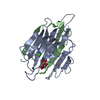
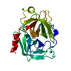
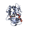
| Title | CRYSTAL COMPLEX OF THE 3C PROTEINASE FROM HEPATITIS A VIRUS WITH ITS INHIBITOR AND IMPLICATIONS FOR THE POLYPROTEIN PROCESSING IN HAV | ||||||
 Components Components | HAV 3C PROTEINASE | ||||||
 Keywords Keywords | hydrolase/hydrolase inhibitor / CHYMOTRYPSIN-LIKE CYSTEINE PROTEINASE VIRAL PROTEASE P'-SITE INHIBITOR / HYDROLASE / hydrolase-hydrolase inhibitor complex | ||||||
| Function / homology |  Function and homology information Function and homology informationhost cell mitochondrial outer membrane / symbiont-mediated suppression of host cytoplasmic pattern recognition receptor signaling pathway via inhibition of MAVS activity / picornain 3C / T=pseudo3 icosahedral viral capsid / host cell cytoplasmic vesicle membrane / host multivesicular body / ribonucleoside triphosphate phosphatase activity / nucleoside-triphosphate phosphatase / channel activity / monoatomic ion transmembrane transport ...host cell mitochondrial outer membrane / symbiont-mediated suppression of host cytoplasmic pattern recognition receptor signaling pathway via inhibition of MAVS activity / picornain 3C / T=pseudo3 icosahedral viral capsid / host cell cytoplasmic vesicle membrane / host multivesicular body / ribonucleoside triphosphate phosphatase activity / nucleoside-triphosphate phosphatase / channel activity / monoatomic ion transmembrane transport / RNA helicase activity / RNA-directed RNA polymerase / cysteine-type endopeptidase activity / viral RNA genome replication / RNA-directed RNA polymerase activity / symbiont entry into host cell / virion attachment to host cell / structural molecule activity / DNA-templated transcription / proteolysis / RNA binding / ATP binding Similarity search - Function | ||||||
| Biological species |  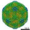 Hepatitis A virus Hepatitis A virus | ||||||
| Method |  X-RAY DIFFRACTION / SYNCHROTRON / MOLECULAR REPLACEMENT / Resolution: 1.9 Å X-RAY DIFFRACTION / SYNCHROTRON / MOLECULAR REPLACEMENT / Resolution: 1.9 Å | ||||||
 Authors Authors | Bergmann, E.M. / Cherney, M.M. / Mckendrick, J. / Vederas, J.C. / James, M.N.G. | ||||||
 Citation Citation | Journal: Virology / Year: 1999 Title: Crystal structure of an inhibitor complex of the 3C proteinase from hepatitis A virus (HAV) and implications for the polyprotein processing in HAV. Authors: Bergmann, E.M. / Cherney, M.M. / Mckendrick, J. / Frormann, S. / Luo, C. / Malcolm, B.A. / Vederas, J.C. / James, M.N. #1: Journal: J.Virol. / Year: 1997Title: The refined crystal structure of the 3C gene product from hepatitis A virus: specific proteinase activity and RNA recognition Authors: Bergmann, E.M. / Mosimann, S.C. / Chernaia, M.M. / Malcolm, B.A. / James, M.N.G. #2: Journal: Handbook of Proteolytic Enzymes / Year: 1998Title: Hepatitis A virus picornain 3C Authors: Bergmann, E.M. #3: Journal: Handbook of Exp. Pharmacol., Vol. Proteases as Targets for TherapyYear: 1999 Title: The 3C proteinases of picornaviruses and other positive-sense, single-stranded RNA viruses Authors: Bergmann, E.M. / James, M.N.G. #4: Journal: Biochemistry / Year: 1992Title: Expression and characterization of recombinant hepatitis A virus 3C proteinase Authors: Malcolm, B.A. / Chin, S.M. / Jewell, D.A. / Stratton-Thomas, J.R. / Thudium, K. | ||||||
| History |
|
- Structure visualization
| Structure viewer | Molecule: MolmilJmol/JSmol |
|---|
- Downloads & links
-Download
| PDBx/mmCIF format | 1qa7.cif.gz | 188.6 KB | Display | PDBx/mmCIF format |
|---|---|---|---|---|
| PDB format | pdb1qa7.ent.gz | 150.7 KB | Display | PDB format |
| PDBx/mmJSON format | 1qa7.json.gz | Tree view | PDBx/mmJSON format | |
| Others |  Other downloads Other downloads |
-Validation report
| Arichive directory | https://data.pdbj.org/pub/pdb/validation_reports/qa/1qa7ftp://data.pdbj.org/pub/pdb/validation_reports/qa/1qa7 | HTTPS FTP |
|---|
-Related structure data
| Related structure data | 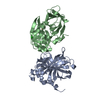 1havS S: Starting model for refinement |
|---|---|
| Similar structure data |


-Links
 PDBj
PDBj

- Assembly
| Deposited unit | 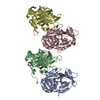
| ||||||||
|---|---|---|---|---|---|---|---|---|---|
| 1 | 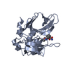
| ||||||||
| 2 | 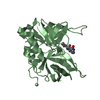
| ||||||||
| 3 | 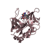
| ||||||||
| 4 | 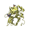
| ||||||||
| 5 |
| ||||||||
| 6 | 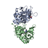
| ||||||||
| 7 | 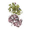
| ||||||||
| Unit cell |
|
-Components
| #1: Protein | Mass: 23828.469 Da / Num. of mol.: 4 / Fragment: HAV 3C PROTEINASE / Mutation: C24S, F82A Source method: isolated from a genetically manipulated source Details: PROTEINASE CHEMICALLY BONDED TO INHIBITOR ACE-VAL-NFA. IT WAS CHEMICALLY SYNTHESIZED AS IODOACETYL-VALYL-PHENYLALANYL AMIDE Source: (gene. exp.) Hepatitis A virus / Genus: Hepatovirus / Plasmid: PHAV3-CEX / Production host:  #2: Chemical | ChemComp-IVF /   Type: peptide-like / Mass: 431.269 Da / Num. of mol.: 4 / Source method: obtained synthetically / Formula: C16H22IN3O3 Type: peptide-like / Mass: 431.269 Da / Num. of mol.: 4 / Source method: obtained synthetically / Formula: C16H22IN3O3#3: Chemical | ChemComp-DMS / |   Mass: 78.133 Da / Num. of mol.: 1 / Source method: obtained synthetically / Formula: C2H6OS / Comment: DMSO, precipitant*YM Mass: 78.133 Da / Num. of mol.: 1 / Source method: obtained synthetically / Formula: C2H6OS / Comment: DMSO, precipitant*YM#4: Chemical | ChemComp-GOL / |   Mass: 92.094 Da / Num. of mol.: 1 / Source method: obtained synthetically / Formula: C3H8O3 Mass: 92.094 Da / Num. of mol.: 1 / Source method: obtained synthetically / Formula: C3H8O3#5: Water | ChemComp-HOH / |  Mass: 18.015 Da / Num. of mol.: 514 / Source method: isolated from a natural source / Formula: H2O Mass: 18.015 Da / Num. of mol.: 514 / Source method: isolated from a natural source / Formula: H2OHas protein modification | Y | |
|---|
-Experimental details
-Experiment
| Experiment | Method: X-RAY DIFFRACTION / Number of used crystals: 1 |
|---|
- Sample preparation
| Crystal | Density Matthews: 2.17 Å3/Da / Density % sol: 43.29 % | ||||||||||||||||||||||||||||||
|---|---|---|---|---|---|---|---|---|---|---|---|---|---|---|---|---|---|---|---|---|---|---|---|---|---|---|---|---|---|---|---|
| Crystal grow | Temperature: 298 K / Method: vapor diffusion, hanging drop / pH: 8.5 Details: 100mM TRIS, 5% DMSO, 18% PEG 8000 , pH 8.5, VAPOR DIFFUSION, HANGING DROP, temperature 298K | ||||||||||||||||||||||||||||||
| Crystal grow | *PLUS pH: 7.5 | ||||||||||||||||||||||||||||||
| Components of the solutions | *PLUS
|
-Data collection
| Diffraction | Mean temperature: 100 K |
|---|---|
| Diffraction source | Source: SYNCHROTRON / Site: SSRL  / Beamline: BL7-1 / Wavelength: 1 / Beamline: BL7-1 / Wavelength: 1 |
| Detector | Type: MARRESEARCH / Detector: IMAGE PLATE / Date: Dec 10, 1997 |
| Radiation | Protocol: SINGLE WAVELENGTH / Monochromatic (M) / Laue (L): M / Scattering type: x-ray |
| Radiation wavelength | Wavelength: 1 Å / Relative weight: 1 |
| Reflection | Resolution: 1.9→15 Å / Num. all: 64826 / Num. obs: 59384 / % possible obs: 92.3 % / Observed criterion σ(F): 0 / Observed criterion σ(I): -3 / Redundancy: 3 % / Biso Wilson estimate: 32 Å2 / Rmerge(I) obs: 0.033 / Net I/σ(I): 4.8 |
| Reflection shell | Resolution: 1.9→1.96 Å / Redundancy: 1.6 % / Rmerge(I) obs: 0.157 / Num. unique all: 4001 / % possible all: 74.7 |
| Reflection | *PLUS Num. measured all: 494113 |
| Reflection shell | *PLUS % possible obs: 74.7 % / Num. unique obs: 4001 / Mean I/σ(I) obs: 1.8 |
- Processing
| Software |
| |||||||||||||||||||||||||
|---|---|---|---|---|---|---|---|---|---|---|---|---|---|---|---|---|---|---|---|---|---|---|---|---|---|---|
| Refinement | Method to determine structure: MOLECULAR REPLACEMENT Starting model: PDB ID 1hav Resolution: 1.9→15 Å / Cross valid method: THROUGHOUT / σ(F): 0 / σ(I): 0 / Stereochemistry target values: Engh & Huber / Details: no non-crystallographic restraints
| |||||||||||||||||||||||||
| Refinement step | Cycle: LAST / Resolution: 1.9→15 Å
| |||||||||||||||||||||||||
| Refine LS restraints |
| |||||||||||||||||||||||||
| Software | *PLUS Name: TNT / Version: 5E / Classification: refinement | |||||||||||||||||||||||||
| Refinement | *PLUS Highest resolution: 1.9 Å / σ(F): 0 / % reflection Rfree: 6.5 % | |||||||||||||||||||||||||
| Solvent computation | *PLUS | |||||||||||||||||||||||||
| Displacement parameters | *PLUS | |||||||||||||||||||||||||
| Refine LS restraints | *PLUS
|
