 Movie
Movie Controller


+ Open data

- Basic information
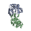
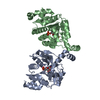
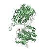
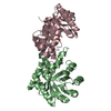
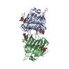
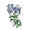
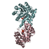
| Entry | Database: PDB / ID: 1hav | ||||||
|---|---|---|---|---|---|---|---|
| Title | HEPATITIS A VIRUS 3C PROTEINASE | ||||||
 Components Components | (HEPATITIS A VIRUS 3C PROTEINASE) x 2 | ||||||
 Keywords Keywords | HYDROLASE / POLYPROTEIN / COAT PROTEIN / CORE PROTEIN / RNA-DIRECTED RNA POLYMERASE / THIOL PROTEASE | ||||||
| Function / homology |  Function and homology information Function and homology informationhost cell mitochondrial outer membrane / symbiont-mediated suppression of host cytoplasmic pattern recognition receptor signaling pathway via inhibition of MAVS activity / picornain 3C / T=pseudo3 icosahedral viral capsid / host cell cytoplasmic vesicle membrane / host multivesicular body / ribonucleoside triphosphate phosphatase activity / nucleoside-triphosphate phosphatase / channel activity / monoatomic ion transmembrane transport ...host cell mitochondrial outer membrane / symbiont-mediated suppression of host cytoplasmic pattern recognition receptor signaling pathway via inhibition of MAVS activity / picornain 3C / T=pseudo3 icosahedral viral capsid / host cell cytoplasmic vesicle membrane / host multivesicular body / ribonucleoside triphosphate phosphatase activity / nucleoside-triphosphate phosphatase / channel activity / monoatomic ion transmembrane transport / RNA helicase activity / RNA-directed RNA polymerase / cysteine-type endopeptidase activity / viral RNA genome replication / RNA-directed RNA polymerase activity / symbiont entry into host cell / virion attachment to host cell / DNA-templated transcription / structural molecule activity / proteolysis / RNA binding / ATP binding Similarity search - Function | ||||||
| Biological species |  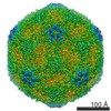 Hepatitis A virus Hepatitis A virus | ||||||
| Method |  X-RAY DIFFRACTION / MOLECULAR REPLACEMENT / Resolution: 2 Å X-RAY DIFFRACTION / MOLECULAR REPLACEMENT / Resolution: 2 Å | ||||||
 Authors Authors | Bergmann, E.M. / James, M.N.G. | ||||||
 Citation Citation | Journal: J.Virol. / Year: 1997 Title: The refined crystal structure of the 3C gene product from hepatitis A virus: specific proteinase activity and RNA recognition. Authors: Bergmann, E.M. / Mosimann, S.C. / Chernaia, M.M. / Malcolm, B.A. / James, M.N. #2: Journal: Nature / Year: 1994Title: Picornaviral 3C Cysteine Proteinases Have a Fold Similar to Chymotrypsin-Like Serine Proteinases Authors: Allaire, M. / Chernaia, M.M. / Malcolm, B.A. / James, M.N. #3: Journal: Biochemistry / Year: 1992Title: Expression and Characterization of Recombinant Hepatitis a Virus 3C Proteinase Authors: Malcolm, B.A. / Chin, S.M. / Jewell, D.A. / Stratton-Thomas, J.R. / Thudium, K.B. / Ralston, R. / Rosenberg, S. | ||||||
| History |
|
- Structure visualization
| Structure viewer | Molecule: MolmilJmol/JSmol |
|---|
- Downloads & links
-Download
| PDBx/mmCIF format | 1hav.cif.gz | 96.3 KB | Display | PDBx/mmCIF format |
|---|---|---|---|---|
| PDB format | pdb1hav.ent.gz | 73.2 KB | Display | PDB format |
| PDBx/mmJSON format | 1hav.json.gz | Tree view | PDBx/mmJSON format | |
| Others |  Other downloads Other downloads |
-Validation report
| Arichive directory | https://data.pdbj.org/pub/pdb/validation_reports/ha/1havftp://data.pdbj.org/pub/pdb/validation_reports/ha/1hav | HTTPS FTP |
|---|
-Related structure data
| Similar structure data |
|---|


-Links
 PDBj
PDBj

- Assembly
| Deposited unit | 
| ||||||||
|---|---|---|---|---|---|---|---|---|---|
| 1 |
| ||||||||
| 2 | 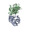
| ||||||||
| 3 | 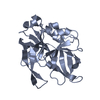
| ||||||||
| 4 | 
| ||||||||
| Unit cell |
| ||||||||
| Noncrystallographic symmetry (NCS) | NCS oper: (Code: given Matrix: (0.8808, -0.4661, -0.0832), Vector: |
-Components
| #1: Protein | Mass: 23904.564 Da / Num. of mol.: 1 / Mutation: C24S Source method: isolated from a genetically manipulated source Source: (gene. exp.) Hepatitis A virus / Genus: Hepatovirus / Gene: 3C / Plasmid: PHAV-3CEX / Gene (production host): 3C / Production host:  |
|---|---|
| #2: Protein | Mass: 23952.562 Da / Num. of mol.: 1 / Mutation: C24S Source method: isolated from a genetically manipulated source Source: (gene. exp.) Hepatitis A virus / Genus: Hepatovirus / Gene: 3C / Plasmid: PHAV-3CEX / Gene (production host): 3C / Production host: |
| #3: Chemical | ChemComp-CL /   Mass: 35.453 Da / Num. of mol.: 1 / Source method: obtained synthetically / Formula: Cl Mass: 35.453 Da / Num. of mol.: 1 / Source method: obtained synthetically / Formula: Cl |
| #4: Water | ChemComp-HOH /  Mass: 18.015 Da / Num. of mol.: 107 / Source method: isolated from a natural source / Formula: H2O Mass: 18.015 Da / Num. of mol.: 107 / Source method: isolated from a natural source / Formula: H2O |
| Compound details | ACTIVE SITE CYSTEINE 172 IS OXIDIZED IN MOLECULE B. MOLECULE A RESEMBLES ACTIVE PROTEINASE |
-Experimental details
-Experiment
| Experiment | Method: X-RAY DIFFRACTION / Number of used crystals: 1 |
|---|
- Sample preparation
| Crystal | Density Matthews: 2.21 Å3/Da / Density % sol: 44.36 % | ||||||||||||||||||||||||||||||||||||||||||
|---|---|---|---|---|---|---|---|---|---|---|---|---|---|---|---|---|---|---|---|---|---|---|---|---|---|---|---|---|---|---|---|---|---|---|---|---|---|---|---|---|---|---|---|
| Crystal grow | pH: 8.5 / Details: pH 8.5 | ||||||||||||||||||||||||||||||||||||||||||
| Crystal grow | *PLUS Method: vapor diffusion, hanging drop / Details: used to seeding | ||||||||||||||||||||||||||||||||||||||||||
| Components of the solutions | *PLUS
|
-Data collection
| Diffraction | Mean temperature: 295 K |
|---|---|
| Diffraction source | Source: ROTATING ANODE / Type: RIGAKU RU200 / Wavelength: 1.5418 |
| Detector | Type: XUONG-HAMLIN MULTIWIRE / Detector: AREA DETECTOR / Date: Oct 30, 1994 |
| Radiation | Monochromator: GRAPHITE(002) / Monochromatic (M) / Laue (L): M / Scattering type: x-ray |
| Radiation wavelength | Wavelength: 1.5418 Å / Relative weight: 1 |
| Reflection | Resolution: 2→20 Å / Num. obs: 25299 / % possible obs: 87 % / Redundancy: 2.4 % / Rmerge(I) obs: 0.0673 / Net I/σ(I): 9.1 |
| Reflection shell | Resolution: 2→2.15 Å / Redundancy: 1.5 % / Rmerge(I) obs: 0.2713 / Mean I/σ(I) obs: 1.4 / % possible all: 67 |
| Reflection | *PLUS Num. measured all: 64410 |
| Reflection shell | *PLUS % possible obs: 67.2 % / Num. unique obs: 3841 / Num. measured obs: 5802 |
- Processing
| Software |
| ||||||||||||||||||||||||||||||||||||||||||||||||||||||||||||
|---|---|---|---|---|---|---|---|---|---|---|---|---|---|---|---|---|---|---|---|---|---|---|---|---|---|---|---|---|---|---|---|---|---|---|---|---|---|---|---|---|---|---|---|---|---|---|---|---|---|---|---|---|---|---|---|---|---|---|---|---|---|
| Refinement | Method to determine structure: MOLECULAR REPLACEMENT Starting model: INACTIVE DOUBLE MUTANT OF ALLAIRE ET AL. (1994), SEE REFERENCE 2. Resolution: 2→20 Å / σ(F): 2 Details: MAXIMUM LIKELIHOOD VERSION OF X-PLOR USED INSTEAD OF CRYSTALLOGRAPHIC RESIDUAL, SEE PANNU N.S. AND READ R.J. (1996) ACTA CRYST, VOL.A50, PP.659-668 RESIDUES 147 - 151 IN BOTH MOLECULES, A ...Details: MAXIMUM LIKELIHOOD VERSION OF X-PLOR USED INSTEAD OF CRYSTALLOGRAPHIC RESIDUAL, SEE PANNU N.S. AND READ R.J. (1996) ACTA CRYST, VOL.A50, PP.659-668 RESIDUES 147 - 151 IN BOTH MOLECULES, A AND B, ARE POORLY DEFINED AND PRESUMABLY FLEXIBLE. RESIDUES 147 - 151 IN BOTH MOLECULES, A AND B, ARE POORLY DEFINED AND PRESUMABLY FLEXIBLE. ASP A 36 AND ASP B 36: UNUSUAL MAIN CHAIN CONFORMATIONAL ANGLES RESIDUE I+1 OF II' REVERSE TURN AND WELL-DEFINED IN ELECTRON DENSITY DESPITE BEING FLAGGED AN OUTLIER IN A RAMACHANDRAN PLOT.
| ||||||||||||||||||||||||||||||||||||||||||||||||||||||||||||
| Displacement parameters | Biso mean: 21 Å2 | ||||||||||||||||||||||||||||||||||||||||||||||||||||||||||||
| Refine analyze | Luzzati coordinate error obs: 0.25 Å / Luzzati d res low obs: 6 Å / Luzzati sigma a obs: 0.26 Å | ||||||||||||||||||||||||||||||||||||||||||||||||||||||||||||
| Refinement step | Cycle: LAST / Resolution: 2→20 Å
| ||||||||||||||||||||||||||||||||||||||||||||||||||||||||||||
| Refine LS restraints |
| ||||||||||||||||||||||||||||||||||||||||||||||||||||||||||||
| LS refinement shell | Resolution: 2→2.13 Å / Total num. of bins used: 6
| ||||||||||||||||||||||||||||||||||||||||||||||||||||||||||||
| Software | *PLUS Name: X-PLOR / Version: 3.1 / Classification: refinement | ||||||||||||||||||||||||||||||||||||||||||||||||||||||||||||
| Refine LS restraints | *PLUS
|
