 Movie
Movie Controller

[English] 日本語
 Yorodumi
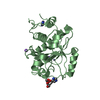
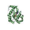
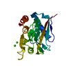
- PDB-6jfr: K3U bound crystal structure of class II peptide deformylase from ... -
+ Open data

- Basic information
| Entry | Database: PDB / ID: 6jfr | ||||||
|---|---|---|---|---|---|---|---|
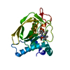
| Title | K3U bound crystal structure of class II peptide deformylase from methicillin resistant Staphylococcus aureus | ||||||
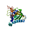
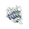
 Components Components | Peptide deformylase | ||||||
 Keywords Keywords | HYDROLASE / peptide deformylase | ||||||
| Function / homology |  Function and homology information Function and homology informationpeptide deformylase / peptide deformylase activity / translation / metal ion binding Similarity search - Function | ||||||
| Biological species |   Staphylococcus aureus (bacteria) Staphylococcus aureus (bacteria) | ||||||
| Method |  X-RAY DIFFRACTION / SYNCHROTRON / MOLECULAR REPLACEMENT / Resolution: 2.4 Å X-RAY DIFFRACTION / SYNCHROTRON / MOLECULAR REPLACEMENT / Resolution: 2.4 Å | ||||||
 Authors Authors | Lee, I.H. / Ho, T.H. / Kang, L.W. | ||||||
 Citation Citation | Journal: To be published Title: K3U bound crystal structure of class II peptide deformylase from methicillin resistant Staphylococcus aureus Authors: Lee, I.H. / Ho, T.H. / Kang, L.W. | ||||||
| History |
|
- Structure visualization
| Structure viewer | Molecule: MolmilJmol/JSmol |
|---|
- Downloads & links
-Download
| PDBx/mmCIF format | 6jfr.cif.gz | 52 KB | Display | PDBx/mmCIF format |
|---|---|---|---|---|
| PDB format | pdb6jfr.ent.gz | 34.4 KB | Display | PDB format |
| PDBx/mmJSON format | 6jfr.json.gz | Tree view | PDBx/mmJSON format | |
| Others |  Other downloads Other downloads |
-Validation report
| Arichive directory | https://data.pdbj.org/pub/pdb/validation_reports/jf/6jfrftp://data.pdbj.org/pub/pdb/validation_reports/jf/6jfr | HTTPS FTP |
|---|
-Related structure data
| Related structure data | 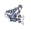 1lm4S S: Starting model for refinement |
|---|---|
| Similar structure data |


-Links
 PDBj
PDBj- Assembly
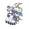
| Deposited unit | 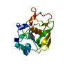
| ||||||||
|---|---|---|---|---|---|---|---|---|---|
| 1 |
| ||||||||
| Unit cell |
|
-Components
| #1: Protein | Mass: 20588.576 Da / Num. of mol.: 1 Source method: isolated from a genetically manipulated source Source: (gene. exp.) Staphylococcus aureus (bacteria) / Strain: CCARM3089 / Gene: def, def1, pdf1 / Plasmid: pET29b / Production host: | ||||
|---|---|---|---|---|---|
| #2: Chemical |   Mass: 58.693 Da / Num. of mol.: 2 / Source method: obtained synthetically / Formula: Ni Mass: 58.693 Da / Num. of mol.: 2 / Source method: obtained synthetically / Formula: Ni#3: Chemical | ChemComp-K3U / | 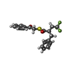  Mass: 366.397 Da / Num. of mol.: 1 / Source method: obtained synthetically / Formula: C19H17F3O2S / Feature type: SUBJECT OF INVESTIGATION Mass: 366.397 Da / Num. of mol.: 1 / Source method: obtained synthetically / Formula: C19H17F3O2S / Feature type: SUBJECT OF INVESTIGATION#4: Water | ChemComp-HOH / |  Mass: 18.015 Da / Num. of mol.: 18 / Source method: isolated from a natural source / Formula: H2O Mass: 18.015 Da / Num. of mol.: 18 / Source method: isolated from a natural source / Formula: H2O |
-Experimental details
-Experiment
| Experiment | Method: X-RAY DIFFRACTION / Number of used crystals: 1 |
|---|
- Sample preparation
| Crystal | Density Matthews: 3.32 Å3/Da / Density % sol: 62.94 % |
|---|---|
| Crystal grow | Temperature: 287 K / Method: evaporation / pH: 8.5 Details: 0.02M CaCl2, 0.1M MgCl2, 15% (v/v) Glycerol, 25% (w/v) PEG 4K, 0.05M Tris pH 8.5 |
-Data collection
| Diffraction | Mean temperature: 100 K / Serial crystal experiment: N | |||||||||||||||||||||||||||||||||||||||||||||||||||||||||||||||||||||||||||||||||||||||||||||||||||||||||||||||||||||||||||||||||||||||||||||||||||||||||||||||||||||||||||||||||||||||||||||
|---|---|---|---|---|---|---|---|---|---|---|---|---|---|---|---|---|---|---|---|---|---|---|---|---|---|---|---|---|---|---|---|---|---|---|---|---|---|---|---|---|---|---|---|---|---|---|---|---|---|---|---|---|---|---|---|---|---|---|---|---|---|---|---|---|---|---|---|---|---|---|---|---|---|---|---|---|---|---|---|---|---|---|---|---|---|---|---|---|---|---|---|---|---|---|---|---|---|---|---|---|---|---|---|---|---|---|---|---|---|---|---|---|---|---|---|---|---|---|---|---|---|---|---|---|---|---|---|---|---|---|---|---|---|---|---|---|---|---|---|---|---|---|---|---|---|---|---|---|---|---|---|---|---|---|---|---|---|---|---|---|---|---|---|---|---|---|---|---|---|---|---|---|---|---|---|---|---|---|---|---|---|---|---|---|---|---|---|---|---|---|
| Diffraction source | Source: SYNCHROTRON / Site: PAL/PLS  / Beamline: 5C (4A) / Wavelength: 0.9794 Å / Beamline: 5C (4A) / Wavelength: 0.9794 Å | |||||||||||||||||||||||||||||||||||||||||||||||||||||||||||||||||||||||||||||||||||||||||||||||||||||||||||||||||||||||||||||||||||||||||||||||||||||||||||||||||||||||||||||||||||||||||||||
| Detector | Type: ADSC QUANTUM 315r / Detector: CCD / Date: Mar 9, 2018 | |||||||||||||||||||||||||||||||||||||||||||||||||||||||||||||||||||||||||||||||||||||||||||||||||||||||||||||||||||||||||||||||||||||||||||||||||||||||||||||||||||||||||||||||||||||||||||||
| Radiation | Protocol: SINGLE WAVELENGTH / Monochromatic (M) / Laue (L): M / Scattering type: x-ray | |||||||||||||||||||||||||||||||||||||||||||||||||||||||||||||||||||||||||||||||||||||||||||||||||||||||||||||||||||||||||||||||||||||||||||||||||||||||||||||||||||||||||||||||||||||||||||||
| Radiation wavelength | Wavelength: 0.9794 Å / Relative weight: 1 | |||||||||||||||||||||||||||||||||||||||||||||||||||||||||||||||||||||||||||||||||||||||||||||||||||||||||||||||||||||||||||||||||||||||||||||||||||||||||||||||||||||||||||||||||||||||||||||
| Reflection | Resolution: 2.4→50 Å / Num. obs: 10475 / % possible obs: 94.7 % / Redundancy: 4.8 % / Rmerge(I) obs: 0.12 / Rpim(I) all: 0.052 / Rrim(I) all: 0.132 / Χ2: 6.366 / Net I/σ(I): 16.4 | |||||||||||||||||||||||||||||||||||||||||||||||||||||||||||||||||||||||||||||||||||||||||||||||||||||||||||||||||||||||||||||||||||||||||||||||||||||||||||||||||||||||||||||||||||||||||||||
| Reflection shell | Diffraction-ID: 1
|
- Processing
| Software |
| ||||||||||||||||||||||||||||||||||||||||||||||||||||||||||||
|---|---|---|---|---|---|---|---|---|---|---|---|---|---|---|---|---|---|---|---|---|---|---|---|---|---|---|---|---|---|---|---|---|---|---|---|---|---|---|---|---|---|---|---|---|---|---|---|---|---|---|---|---|---|---|---|---|---|---|---|---|---|
| Refinement | Method to determine structure: MOLECULAR REPLACEMENT Starting model: 1LM4 Resolution: 2.4→49.99 Å / Cor.coef. Fo:Fc: 0.933 / Cor.coef. Fo:Fc free: 0.886 / SU B: 8.215 / SU ML: 0.181 / SU R Cruickshank DPI: 0.296 / Cross valid method: THROUGHOUT / σ(F): 0 / ESU R: 0.296 / ESU R Free: 0.242 Details: HYDROGENS HAVE BEEN ADDED IN THE RIDING POSITIONS U VALUES : REFINED INDIVIDUALLY
| ||||||||||||||||||||||||||||||||||||||||||||||||||||||||||||
| Solvent computation | Ion probe radii: 0.8 Å / Shrinkage radii: 0.8 Å / VDW probe radii: 1.2 Å | ||||||||||||||||||||||||||||||||||||||||||||||||||||||||||||
| Displacement parameters | Biso max: 100.3 Å2 / Biso mean: 35.356 Å2 / Biso min: 17.22 Å2
| ||||||||||||||||||||||||||||||||||||||||||||||||||||||||||||
| Refinement step | Cycle: final / Resolution: 2.4→49.99 Å
| ||||||||||||||||||||||||||||||||||||||||||||||||||||||||||||
| Refine LS restraints |
| ||||||||||||||||||||||||||||||||||||||||||||||||||||||||||||
| LS refinement shell | Resolution: 2.402→2.465 Å / Rfactor Rfree error: 0 / Total num. of bins used: 20
|
