 Movie
Movie Controller

[English] 日本語
 Yorodumi
- PDB-2ltn: DESIGN, EXPRESSION, AND CRYSTALLIZATION OF RECOMBINANT LECTIN FRO... -
+ Open data

- Basic information
| Entry | Database: PDB / ID: 2ltn | ||||||
|---|---|---|---|---|---|---|---|
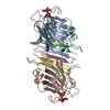
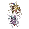
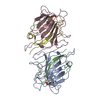
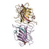
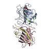
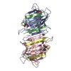
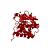
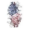
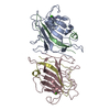
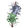
| Title | DESIGN, EXPRESSION, AND CRYSTALLIZATION OF RECOMBINANT LECTIN FROM THE GARDEN PEA (PISUM SATIVUM) | ||||||
 Components Components |
| ||||||
 Keywords Keywords | LECTIN | ||||||
| Function / homology |  Function and homology information Function and homology information | ||||||
| Biological species |  Pisum sativum (garden pea) Pisum sativum (garden pea) | ||||||
| Method |  X-RAY DIFFRACTION / Resolution: 1.7 Å X-RAY DIFFRACTION / Resolution: 1.7 Å | ||||||
 Authors Authors | Suddath, F.L. / Phillips, S.R. / Einspahr, H. | ||||||
 Citation Citation | Journal: J.Biol.Chem. / Year: 1989 Title: Design, expression, and crystallization of recombinant lectin from the garden pea (Pisum sativum). Authors: Prasthofer, T. / Phillips, S.R. / Suddath, F.L. / Engler, J.A. #1: Journal: J.Biol.Chem. / Year: 1986Title: The Crystal Structure of Pea Lectin at 3.0-Angstroms Resolution Authors: Einspahr, H. / Parks, E.H. / Suguna, K. / Subramanian, E. / Suddath, F.L. #2: Journal: Acta Crystallogr.,Sect.B / Year: 1985Title: The Location of Manganese and Calcium Ion Cofactors in Pea Lectin Crystals by Use of Anomalous Dispersion and Tuneable Synchrotron X-Radiation Authors: Einspahr, H. / Suguna, K. / Suddath, F.L. / Ellis, G. / Helliwell, J.R. / Papiz, M.Z. #3: Journal: J.Biol.Chem. / Year: 1982Title: The Crystal Structure of Pea Lectin at 6-Angstroms Resolution Authors: Meehanjunior, E.J. / Mcduffie, J. / Einspahr, H. / Bugg, C.E. / Suddath, F.L. | ||||||
| History |
|
- Structure visualization
| Structure viewer | Molecule: MolmilJmol/JSmol |
|---|
- Downloads & links
-Download
| PDBx/mmCIF format | 2ltn.cif.gz | 107.3 KB | Display | PDBx/mmCIF format |
|---|---|---|---|---|
| PDB format | pdb2ltn.ent.gz | 82.7 KB | Display | PDB format |
| PDBx/mmJSON format | 2ltn.json.gz | Tree view | PDBx/mmJSON format | |
| Others |  Other downloads Other downloads |
-Validation report
| Arichive directory | https://data.pdbj.org/pub/pdb/validation_reports/lt/2ltnftp://data.pdbj.org/pub/pdb/validation_reports/lt/2ltn | HTTPS FTP |
|---|
-Related structure data
| Similar structure data |
|---|
-Links
 PDBj
PDBj

- Assembly
| Deposited unit | 
| ||||||||
|---|---|---|---|---|---|---|---|---|---|
| 1 |
| ||||||||
| 2 | 
| ||||||||
| 3 | 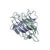
| ||||||||
| Unit cell |
| ||||||||
| Atom site foot note | 1: THE PEPTIDE BOND BETWEEN ALA A 80 AND ASP A 81 AND THE PEPTIDE BOND BETWEEN ALA C 80 AND ASP C 81 ARE BOTH IN THE CIS CONFORMATION. |
-Components
| #1: Protein | Mass: 20001.076 Da / Num. of mol.: 2 Source method: isolated from a genetically manipulated source Source: (gene. exp.) Pisum sativum (garden pea) / Production host:  #2: Protein | Mass: 5596.086 Da / Num. of mol.: 2 Source method: isolated from a genetically manipulated source Source: (gene. exp.) Pisum sativum (garden pea) / References: UniProt: P02867#3: Chemical |   Mass: 54.938 Da / Num. of mol.: 2 / Source method: obtained synthetically / Formula: Mn Mass: 54.938 Da / Num. of mol.: 2 / Source method: obtained synthetically / Formula: Mn#4: Chemical |   Mass: 40.078 Da / Num. of mol.: 2 / Source method: obtained synthetically / Formula: Ca Mass: 40.078 Da / Num. of mol.: 2 / Source method: obtained synthetically / Formula: Ca#5: Water | ChemComp-HOH / |  Mass: 18.015 Da / Num. of mol.: 294 / Source method: isolated from a natural source / Formula: H2O Mass: 18.015 Da / Num. of mol.: 294 / Source method: isolated from a natural source / Formula: H2O |
|---|
-Experimental details
-Experiment
| Experiment | Method: X-RAY DIFFRACTION |
|---|
- Sample preparation
| Crystal | Density Matthews: 2.07 Å3/Da / Density % sol: 40.55 % | ||||||||||||||||||||||||||||||
|---|---|---|---|---|---|---|---|---|---|---|---|---|---|---|---|---|---|---|---|---|---|---|---|---|---|---|---|---|---|---|---|
| Crystal grow | *PLUS Temperature: 4 ℃ / pH: 5 / Method: vapor diffusion, hanging drop | ||||||||||||||||||||||||||||||
| Components of the solutions | *PLUS
|
-Data collection
| Reflection | *PLUS |
|---|
- Processing
| Software | Name: PROLSQ / Classification: refinement | ||||||||||||||||||||||||||||||||||||||||||||||||||||||||||||||||||||||||||||||||||||
|---|---|---|---|---|---|---|---|---|---|---|---|---|---|---|---|---|---|---|---|---|---|---|---|---|---|---|---|---|---|---|---|---|---|---|---|---|---|---|---|---|---|---|---|---|---|---|---|---|---|---|---|---|---|---|---|---|---|---|---|---|---|---|---|---|---|---|---|---|---|---|---|---|---|---|---|---|---|---|---|---|---|---|---|---|---|
| Refinement | Rfactor obs: 0.177 / Highest resolution: 1.7 Å | ||||||||||||||||||||||||||||||||||||||||||||||||||||||||||||||||||||||||||||||||||||
| Refinement step | Cycle: LAST / Highest resolution: 1.7 Å
| ||||||||||||||||||||||||||||||||||||||||||||||||||||||||||||||||||||||||||||||||||||
| Refine LS restraints |
| ||||||||||||||||||||||||||||||||||||||||||||||||||||||||||||||||||||||||||||||||||||
| Refinement | *PLUS Highest resolution: 1.7 Å / Rfactor obs: 0.177 | ||||||||||||||||||||||||||||||||||||||||||||||||||||||||||||||||||||||||||||||||||||
| Solvent computation | *PLUS | ||||||||||||||||||||||||||||||||||||||||||||||||||||||||||||||||||||||||||||||||||||
| Displacement parameters | *PLUS |
