 Movie
Movie Controller

[English] 日本語
 Yorodumi
- PDB-1loe: X-RAY CRYSTAL STRUCTURE DETERMINATION AND REFINEMENT AT 1.9 ANGST... -
+ Open data

- Basic information
| Entry | Database: PDB / ID: 1loe | ||||||
|---|---|---|---|---|---|---|---|
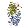
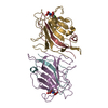
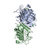
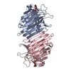
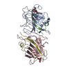
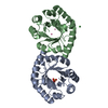
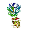
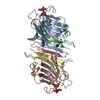
| Title | X-RAY CRYSTAL STRUCTURE DETERMINATION AND REFINEMENT AT 1.9 ANGSTROMS RESOLUTION OF ISOLECTIN I FROM THE SEEDS OF LATHYRUS OCHRUS | ||||||
 Components Components |
| ||||||
 Keywords Keywords | LECTIN | ||||||
| Function / homology |  Function and homology information Function and homology information | ||||||
| Biological species |  Lathyrus ochrus (yellow-flowered pea) Lathyrus ochrus (yellow-flowered pea) | ||||||
| Method |  X-RAY DIFFRACTION / Resolution: 1.9 Å X-RAY DIFFRACTION / Resolution: 1.9 Å | ||||||
 Authors Authors | Bourne, Y. / Cambillau, C. | ||||||
 Citation Citation | Journal: J.Mol.Biol. / Year: 1990 Title: X-ray crystal structure determination and refinement at 1.9 A resolution of isolectin I from the seeds of Lathyrus ochrus. Authors: Bourne, Y. / Abergel, C. / Cambillau, C. / Frey, M. / Rouge, P. / Fontecilla-Camps, J.C. | ||||||
| History |
|
- Structure visualization
| Structure viewer | Molecule: MolmilJmol/JSmol |
|---|
- Downloads & links
-Download
| PDBx/mmCIF format | 1loe.cif.gz | 104 KB | Display | PDBx/mmCIF format |
|---|---|---|---|---|
| PDB format | pdb1loe.ent.gz | 80.6 KB | Display | PDB format |
| PDBx/mmJSON format | 1loe.json.gz | Tree view | PDBx/mmJSON format | |
| Others |  Other downloads Other downloads |
-Validation report
| Arichive directory | https://data.pdbj.org/pub/pdb/validation_reports/lo/1loeftp://data.pdbj.org/pub/pdb/validation_reports/lo/1loe | HTTPS FTP |
|---|
-Related structure data
| Similar structure data |
|---|


-Links
 PDBj
PDBj

- Assembly
| Deposited unit | 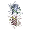
| ||||||||
|---|---|---|---|---|---|---|---|---|---|
| 1 |
| ||||||||
| 2 | 
| ||||||||
| 3 | 
| ||||||||
| Unit cell |
| ||||||||
| Atom site foot note | 1: ALA A 80 - ASP A 81 OMEGA =350.16 PEPTIDE BOND DEVIATES SIGNIFICANTLY FROM TRANS CONFORMATION 2: ALA C 80 - ASP C 81 OMEGA =339.46 PEPTIDE BOND DEVIATES SIGNIFICANTLY FROM TRANS CONFORMATION |
-Components
| #1: Protein | Mass: 19847.857 Da / Num. of mol.: 2 Source method: isolated from a genetically manipulated source Source: (gene. exp.) Lathyrus ochrus (yellow-flowered pea) / Organ: SEED / References: UniProt: P04122#2: Protein | Mass: 5783.322 Da / Num. of mol.: 2 Source method: isolated from a genetically manipulated source Source: (gene. exp.) Lathyrus ochrus (yellow-flowered pea) / Organ: SEED / References: UniProt: P12306#3: Chemical |   Mass: 40.078 Da / Num. of mol.: 2 / Source method: obtained synthetically / Formula: Ca Mass: 40.078 Da / Num. of mol.: 2 / Source method: obtained synthetically / Formula: Ca#4: Chemical |   Mass: 54.938 Da / Num. of mol.: 2 / Source method: obtained synthetically / Formula: Mn Mass: 54.938 Da / Num. of mol.: 2 / Source method: obtained synthetically / Formula: Mn#5: Water | ChemComp-HOH / |  Mass: 18.015 Da / Num. of mol.: 217 / Source method: isolated from a natural source / Formula: H2O Mass: 18.015 Da / Num. of mol.: 217 / Source method: isolated from a natural source / Formula: H2OCompound details | THE AMINO-ACID SEQUENCE OF ISOLECTIN I SHOWS 85 PER CENT HOMOLOGY WITH THAT OF PEA LECTIN. | Sequence details | SEQUENCE ADVISORY NOTICE: DIFFERENCE BETWEEN SWISS-PROT AND PDB SEQUENCE. SWISS-PROT ENTRY NAME: ...SEQUENCE ADVISORY NOTICE: DIFFERENCE | |
|---|
-Experimental details
-Experiment
| Experiment | Method: X-RAY DIFFRACTION |
|---|
- Sample preparation
| Crystal | Density Matthews: 2.28 Å3/Da / Density % sol: 46.05 % | ||||||||||||||||||||||||||||||||||||
|---|---|---|---|---|---|---|---|---|---|---|---|---|---|---|---|---|---|---|---|---|---|---|---|---|---|---|---|---|---|---|---|---|---|---|---|---|---|
| Crystal grow | *PLUS pH: 7.5 / Method: vapor diffusion | ||||||||||||||||||||||||||||||||||||
| Components of the solutions | *PLUS
|
-Data collection
| Radiation | Scattering type: x-ray |
|---|---|
| Radiation wavelength | Relative weight: 1 |
| Reflection | *PLUS Highest resolution: 1.9 Å / Lowest resolution: 9999 Å / Num. obs: 27484 / Observed criterion σ(I): 4.4 / Rmerge(I) obs: 0.066 |
| Reflection shell | *PLUS Highest resolution: 1.9 Å / Lowest resolution: 2 Å / Num. unique obs: 2212 / Rmerge(I) obs: 0.255 |
- Processing
| Software |
| ||||||||||||||||||||||||||||||||||||||||||||||||||||||||||||
|---|---|---|---|---|---|---|---|---|---|---|---|---|---|---|---|---|---|---|---|---|---|---|---|---|---|---|---|---|---|---|---|---|---|---|---|---|---|---|---|---|---|---|---|---|---|---|---|---|---|---|---|---|---|---|---|---|---|---|---|---|---|
| Refinement | Resolution: 1.9→8 Å / Rfactor Rwork: 0.185 / Rfactor obs: 0.185 / σ(F): 1 | ||||||||||||||||||||||||||||||||||||||||||||||||||||||||||||
| Refinement step | Cycle: LAST / Resolution: 1.9→8 Å
| ||||||||||||||||||||||||||||||||||||||||||||||||||||||||||||
| Refine LS restraints |
| ||||||||||||||||||||||||||||||||||||||||||||||||||||||||||||
| Software | *PLUS Name: X-PLOR / Classification: refinement | ||||||||||||||||||||||||||||||||||||||||||||||||||||||||||||
| Refinement | *PLUS Num. reflection obs: 25932 / Rfactor obs: 0.185 / Rfactor Rwork: 0.185 | ||||||||||||||||||||||||||||||||||||||||||||||||||||||||||||
| Solvent computation | *PLUS | ||||||||||||||||||||||||||||||||||||||||||||||||||||||||||||
| Displacement parameters | *PLUS Biso mean: 15.5 Å2 | ||||||||||||||||||||||||||||||||||||||||||||||||||||||||||||
| Refine LS restraints | *PLUS Type: x_angle_d / Dev ideal: 3 |
