 Movie
Movie Controller


+ Open data

- Basic information
| Entry | Database: PDB / ID: 2b7y | |||||||||
|---|---|---|---|---|---|---|---|---|---|---|
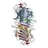
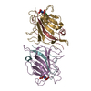
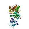
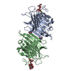
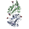
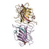
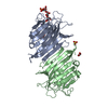
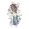
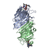
| Title | Fava Bean Lectin-Glucose Complex | |||||||||
 Components Components |
| |||||||||
 Keywords Keywords | LECTIN / lectin-glucose complex / plant lectin / glycoprotein / carbohydrate binding protein / d-glucose / protein-carbohydrate complex | |||||||||
| Function / homology |  Function and homology information Function and homology information | |||||||||
| Biological species |   Vicia faba (fava bean) Vicia faba (fava bean) | |||||||||
| Method |  X-RAY DIFFRACTION / MOLECULAR REPLACEMENT / molecular replacement / Resolution: 3 Å X-RAY DIFFRACTION / MOLECULAR REPLACEMENT / molecular replacement / Resolution: 3 Å | |||||||||
 Authors Authors | Reeke Jr., G.N. / Becker, J.W. | |||||||||
 Citation Citation | Journal: Science / Year: 1986 Title: Three-dimensional structure of favin: saccharide binding-cyclic permutation in leguminous lectins Authors: Reeke Jr., G.N. / Becker, J.W. #1: Journal: J.Mol.Biol. / Year: 1974Title: Favin, a crystalline lectin from Vicia faba Authors: Wang, J.L. / Becker, J.W. / Edelman, G.M. / Reeke Jr., G.N. #2: Journal: J.Biol.Chem. / Year: 1979Title: The chemical characterization of favin, a lectin isolated from Vicia faba Authors: Hemperly, J.J. / Hopp, T.P. / Becker, J.W. / Cunningham, B.A. #3: Journal: J.Biol.Chem. / Year: 1982Title: Amino acid sequence and variant forms of favin, a lectin from Vicia faba Authors: Hopp, T.P. / Hemperly, J.J. / Cunningham, B.A. | |||||||||
| History |
|
- Structure visualization
| Structure viewer | Molecule: MolmilJmol/JSmol |
|---|
- Downloads & links
-Download
| PDBx/mmCIF format | 2b7y.cif.gz | 103.4 KB | Display | PDBx/mmCIF format |
|---|---|---|---|---|
| PDB format | pdb2b7y.ent.gz | 80 KB | Display | PDB format |
| PDBx/mmJSON format | 2b7y.json.gz | Tree view | PDBx/mmJSON format | |
| Others |  Other downloads Other downloads |
-Validation report
| Arichive directory | https://data.pdbj.org/pub/pdb/validation_reports/b7/2b7yftp://data.pdbj.org/pub/pdb/validation_reports/b7/2b7y | HTTPS FTP |
|---|
-Related structure data
| Similar structure data |
|---|

-Links
 PDBj
PDBj


- Assembly
| Deposited unit | 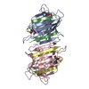
| ||||||||
|---|---|---|---|---|---|---|---|---|---|
| 1 |
| ||||||||
| Unit cell |
| ||||||||
| Details | Asymmetric unit contains biological assembly, an alpha(2)-beta(2) tetramer |
-Components
-Protein , 2 types, 4 molecules ACBD
| #1: Protein | Mass: 19973.172 Da / Num. of mol.: 2 / Fragment: residues 1-182 / Source method: isolated from a natural source / Details: extracted from seeds / Source: (natural) Vicia faba (fava bean) / References: UniProt: P02871#2: Protein | Mass: 5573.199 Da / Num. of mol.: 2 / Fragment: residues 183-233 / Source method: isolated from a natural source / Details: extracted from seeds / Source: (natural) Vicia faba (fava bean) / References: UniProt: P02871 |
|---|
-Sugars , 2 types, 4 molecules 

| #3: Sugar |  Type: D-saccharide, alpha linking / Mass: 180.156 Da / Num. of mol.: 2 Type: D-saccharide, alpha linking / Mass: 180.156 Da / Num. of mol.: 2Source method: isolated from a genetically manipulated source Formula: C6H12O6 #4: Sugar |  Type: D-saccharide, beta linking / Mass: 221.208 Da / Num. of mol.: 2 Type: D-saccharide, beta linking / Mass: 221.208 Da / Num. of mol.: 2Source method: isolated from a genetically manipulated source Formula: C8H15NO6 |
|---|
-Non-polymers , 3 types, 12 molecules 


| #5: Chemical |  Mass: 54.938 Da / Num. of mol.: 2 / Source method: obtained synthetically / Formula: Mn Mass: 54.938 Da / Num. of mol.: 2 / Source method: obtained synthetically / Formula: Mn#6: Chemical |  Mass: 40.078 Da / Num. of mol.: 2 / Source method: obtained synthetically / Formula: Ca Mass: 40.078 Da / Num. of mol.: 2 / Source method: obtained synthetically / Formula: Ca#7: Water | ChemComp-HOH / | Mass: 18.015 Da / Num. of mol.: 8 / Source method: isolated from a natural source / Formula: H2O |
|---|
-Details
| Has protein modification | Y |
|---|
-Experimental details
-Experiment
| Experiment | Method: X-RAY DIFFRACTION |
|---|
- Sample preparation
| Crystal | Density Matthews: 2.7 Å3/Da / Density % sol: 54.2 % Description: The data were collected in 1974, the exact date is not available. |
|---|---|
| Crystal grow | Temperature: 277 K / Method: microdialysis / pH: 6.5 Details: 1 M glucose, 0.01 M sodium phosphate, pH 6.5, MICRODIALYSIS, temperature 277K |
-Data collection
| Diffraction | Mean temperature: 293 K |
|---|---|
| Diffraction source | Source: ROTATING ANODE / Type: ELLIOTT GX-6 / Wavelength: 1.5418 |
| Detector | Type: FILM / Detector: FILM |
| Radiation | Monochromator: Graphite / Protocol: SINGLE WAVELENGTH / Monochromatic (M) / Laue (L): M / Scattering type: x-ray |
| Radiation wavelength | Wavelength: 1.5418 Å / Relative weight: 1 |
| Reflection | Resolution: 3→46.18 Å / Num. all: 10599 / Num. obs: 10599 / % possible obs: 93 % / Observed criterion σ(F): 0 / Observed criterion σ(I): 0 / Biso Wilson estimate: 53.6 Å2 |
-Phasing
| Phasing | Method: molecular replacement |
|---|
- Processing
| Software |
| |||||||||||||||||||||||||||||||||||||||||||||||||||||||||||||||||||||||||||||||||||||||||||||||
|---|---|---|---|---|---|---|---|---|---|---|---|---|---|---|---|---|---|---|---|---|---|---|---|---|---|---|---|---|---|---|---|---|---|---|---|---|---|---|---|---|---|---|---|---|---|---|---|---|---|---|---|---|---|---|---|---|---|---|---|---|---|---|---|---|---|---|---|---|---|---|---|---|---|---|---|---|---|---|---|---|---|---|---|---|---|---|---|---|---|---|---|---|---|---|---|---|
| Refinement | Method to determine structure: MOLECULAR REPLACEMENT Starting model: Pea lectin. Einspahr et al. (1986) J.Biol.Chem. 261:16518-16527. Resolution: 3→46.18 Å / Cor.coef. Fo:Fc: 0.903 / Cor.coef. Fo:Fc free: 0.836 / SU B: 21.043 / SU ML: 0.387 / Cross valid method: THROUGHOUT / σ(F): 0 / ESU R Free: 0.563 / Stereochemistry target values: MAXIMUM LIKELIHOOD Details: HYDROGENS HAVE BEEN ADDED IN THE RIDING POSITIONS. EARLY REFINEMENT USED PROLSQ (AUTHORS: KONNERT AND HENDRICKSON) FOLLOWED BY TORSION-ANGLE MOLECULAR DYNAMICS REFINEMENT USING CNX (AUTHORS: ...Details: HYDROGENS HAVE BEEN ADDED IN THE RIDING POSITIONS. EARLY REFINEMENT USED PROLSQ (AUTHORS: KONNERT AND HENDRICKSON) FOLLOWED BY TORSION-ANGLE MOLECULAR DYNAMICS REFINEMENT USING CNX (AUTHORS: BRUNGER, ADAMS, CLORE, DELANO, GROS, GROSSE-KUNSTLEVE, JIANG, KUSZEWSKI, NILGES, PANNU, READ, RICE, SIMONSON, WARREN).
| |||||||||||||||||||||||||||||||||||||||||||||||||||||||||||||||||||||||||||||||||||||||||||||||
| Solvent computation | Ion probe radii: 0.8 Å / Shrinkage radii: 0.8 Å / VDW probe radii: 1.2 Å / Solvent model: BABINET MODEL WITH MASK | |||||||||||||||||||||||||||||||||||||||||||||||||||||||||||||||||||||||||||||||||||||||||||||||
| Displacement parameters | Biso mean: 25.733 Å2
| |||||||||||||||||||||||||||||||||||||||||||||||||||||||||||||||||||||||||||||||||||||||||||||||
| Refine analyze | Luzzati coordinate error obs: 0.346 Å | |||||||||||||||||||||||||||||||||||||||||||||||||||||||||||||||||||||||||||||||||||||||||||||||
| Refinement step | Cycle: LAST / Resolution: 3→46.18 Å
| |||||||||||||||||||||||||||||||||||||||||||||||||||||||||||||||||||||||||||||||||||||||||||||||
| Refine LS restraints |
| |||||||||||||||||||||||||||||||||||||||||||||||||||||||||||||||||||||||||||||||||||||||||||||||
| LS refinement shell | Resolution: 3→3.071 Å / Total num. of bins used: 20
|
